
Wild rats are associated with transmitting various deadly pathogens with significant zoonotic potential, including blood protozoans. The present study aimed to determine the prevalence of blood protozoan parasites in peri-domestic wild rats in Dhaka City, Bangladesh. A total of 80 wild rats belonging to four species, Rattus norvegicus (n=28), R. rattus (n=14), Bandicota bengalensis (n=30), and M. musculus (n=8), were captured. The wild rats were trapped alive in grocery shops (n=25), rental houses (n=20), slum areas (n=15), and fish markets (n=20). Five blood protozoa, Plasmodium sp. (31.25%), A. marginale (21.25%), A. centrale (17.5%), Babesia sp. (10.0%), and Trypanosoma sp. (3.75%) were detected in this study. The overall prevalence of infection was 50.0%, with the highest infection rate in R. norvegicus (57.14%), followed by B. bengalensis (53.33%), M. musculus (37.5%), and R. rattus (35.71%). In respect to locations, the highest prevalence was found in slum areas (66.67%), followed by fish markets (50%), grocery shops (48%), and rental houses (40%). Both the single and mixed (Co-infection) were observed. This study revealed that the wild rats act as the reservoir of multiple blood protozoan parasites of animals and humans. Therefore, our study underscores an integrated control program against wild rats to prevent rodent-borne protozoan zoonosis in Bangladesh.
Research Article · Volume 12, Pages 33–40, 4 November 2026 · Open Access
Prevalence of Blood Protozoan Parasites of Peri-Domestic Wild Rats in Dhaka City, Bangladesh
· · · · · · · · · · · ·
Affiliations & Notes ⌄
- 1Department of Microbiology and Parasitology, Faculty of Animal Science and Veterinary Medicine, Sher-e-Bangla Agricultural University, Sher-e-Bangla Nagar, Dhaka-1207, Bangladesh
- *Correspondence | Uday Kumar Mohanta; Email: ukmohanta.mipa@sau.edu.bd
Article Info ⌄
- Journal:
- The Journal of Advances in Parasitology
- Submitted Date:
- 14 July 2026
- Accepted Date:
- 29 August 2026
- Published Date:
- 4 November 2026
Abstract
Nitol Chandra Das and Al Wasef contributed equally for this study.
INTRODUCTION
Wild rats, belonging to the order Rodentia, constitute one of the largest groups of mammals on earth. Muridae, being the largest family, comprises over 1,383 species, including mice and wild rats, collectively known as murid rats; they are nocturnal and omnivorous (Britannica, 2020). In Bangladesh, common wild rats species include Rattus rattus (Indur), Bandicoota indica (Dhadi indur), B. bengalensis (Metho indur), R. Norvegicus (Badami indur), Cannomys badius (Bamboo rat), and Suncus murinus Linnaeus (Chika, chuchoo) etc. ; . Wild rats bear economic and public health importance due to their ability to cause substantial losses by feeding on stored food and destroying crops. Moreover, rats play a vital role as a reservoir and definitive hosts for numerous infectious zoonotic organisms (Gratz, 1998) and, therefore, pose health risks by transmitting various infectious agents to humans and animals . Increasing cases of parasitic zoonoses have been reported globally .
Urban areas, particularly cities like Dhaka, are of particular concern due to the emergence of zoonotic diseases facilitated by the favorable habitats for wild rats, which frequently come into close contact with humans . Rat species such as house mice (M. musculus) and wild rats (R. rattus, R. norvegicus) are more prevalent in urban areas and are becoming problematic due to their high reproductive capacity, zoonotic potentials, and close association with humans ; . Over 20 established diseases are directly transmitted from rats to humans, typically via blood-feeding vectors such as fleas, ticks, and mites ; ; . Wild rats harbor a wide range of zoonotic diseases caused by protozoa (e.g. toxoplasmosis, babesiosis, trypanosomiasis, leishmaniasis), helminths (e.g. hymenolepiasis, trichinellosis, echinococcosis, and capillariasis), viruses (e.g. Lassa fever, Hantavirus diseases, tick-borne encephalitis), and bacteria (e.g. plague, leptospirosis, lyme disease, and relapsing fevers) ; . Among these, blood-borne protozoan diseases are transmitted by rats include leishmaniasis (Leishmania major, L. infantum); trypanosomiasis (Trypanosoma lewisi); babesiosis (Babesia microti); malaria (Plasmodium berghei), anaplasmosis (A. marginale, A. centrale) ; ; . In Spain, L. infantum DNA was detected in 27 % of rats , while Plasmodium berghei is commonly studied as a popular organism for human malaria in Africa . In Iran, 11.54% of rats were found infected with Trypanosoma lewisi, and in the USA and Japan, human babesiosis caused by B. microti, transmitted via rats, has been documented ; ; . In Bangladesh, a lower incidence of anaplasmosis (7.5%) and Babesiosis (4.7%) has been reported previously .
Wild rats are known carriers of numerous zoonotic pathogens, facilitated by increased interaction with people due to urbanization and their close proximity to human settlements such as slums, rented houses, and shops. Despite the global public health significance of rodent-borne protozoan diseases, very few studies have been conducted in Bangladesh, even in Southeast Asia. Furthermore, Dhaka, as a densely populated city with a substantial rat population, has lacked previous research attention. Therefore, the aim of this study was to assess the prevalence of blood protozoan parasites in peri-domestic wild rats in Dhaka City.
MATERIALS AND METHODS
The study area and wild rats catching
The research was conducted in Dhaka, situated on the eastern banks of the Buriganga River. The city experiences a distinct monsoon season and has an annual average temperature of 26 °C. Dhaka is one of the most densely populated cities globally, characterized by a mix of multistoried buildings, skyscrapers, and slums. These demographic, topographic, and climatic conditions make Dhaka an ideal habitat for rats. The wild rats were captured from various locations, including grocery shops, vegetable and fish markets, general households, and slum areas in Agargaon, as well as West Kafrul within the Dhaka North City Corporation. A total of 80 wild rats were trapped from different locations using iron stick traps (27 cm × 13 cm × 13 cm). The traps baited and set with care before sunset were collected in the following morning. The captured wild rats were then taken to the Microbiology and Parasitology Laboratory, Sher-e-Bangla Agricultural University, Dhaka, for further studies.
Rodent identification
Each wild rat was placed individually in a glass flask and anesthetized with a cotton plug soaked in chloroform. Then, the rats were identified, classified, and categorized based on their species, sex, site of collection, and date according to the keys and description of Aplin et al. (2003). Species identification involved observation of coat color, size, shape, and appearance, as well as head-body length and specific tail length. Sex was determined by examining the distinct sex organs.
Blood collection and preservation
In this study, blood was collected via cardiac puncture. After identifying the heart, a 1.0 ml or 3.0 ml syringe was used to draw the blood. Then, the collected blood was preserved in a collecting tube containing EDTA (purple tip) for further analysis.
Preparation of thin blood smears, giemsa staining, and microscopic examination
A thin blood smear was prepared by placing a small drop of blood on a clean, grease-free, pre-labeled glass slide. A second slide was set at a 30-45º angle to the drop, allowing the drop to spread along the edge before quickly pushing it to the end of the slide. Three slides were prepared per sample. The smears were air-dried for a few minutes and then fixed in absolute methanol for 2-3 minutes using a Coplin jar. The fixed slides were stained by flooding them with Giemsa working solution (pH 7.1) for 35-40 minutes . After staining, the slides were rinsed with running tap water and left to air dry. The prepared slides were examined under a microscope (100X magnification) using immersion oil. The morphological identification of blood protozoa was conducted according to the keys and description of Urquhart et al. (1996).
Statistical analysis
The data, including locations, rat species and sex, niches, parasite species, and infection nature (single and mixed), were considered as independent variables. Data with low detection rates were excluded. Pearson’s chi-square and Fisher’s exact test were used to analyze the association of parasite detection rates across the different locations, species, sexes, niches, and infection types using GraphPad Prism 8 (GraphPad Software, San Diego, CA, USA). A p value of <0.05 was considered significant.
RESULTS
Morphological identification
Trypanosoma sp.: This species exhibited a classical slender form with a long posterior end. It had a sub-terminal oval kinetoplast and the nucleus located in the anterior part of the body. The flagellum was free (Figure 1).
A. centrale and A. marginale: A. centrale was found to have a round inclusion body in the central part of the erythrocyte (Figure 2A). Whereas A. marginale appeared as an intracellular basophilic, spherical inclusion body located near the margin of erythrocytes (Figure 2B).
Babesia sp.: In rat blood, B. microti is commonly found. In Babesia infection, infected red blood cells (RBCs) showing pleomorphic shapes and sizes which were elliptical (Figure 3A), classic tetrad-forms (Figure 3B), comma (Figure 3C), pleomorphic ring (Figure 3D) or vacuolated (Figure 3E).
Plasmodium sp.: The trophozoites had a compact cytoplasm and a large chromatin dot. Occasional band forms and/or “basket” forms with coarse, dark-brown pigment were also observed (Figure 4A). The ring form had large chromatin dot inside the cytoplasm (Figure 4B). Schizonts containing 6-12 merozoites with large nuclei clustered around a mass of coarse, dark-brown pigment giving an appearance of rosette (Figure 4C). The gametocytes were round to oval with scattered brown pigment and almost filled the infected red blood cell. The shape of gametocytes was bean-shaped (Figure 4D).
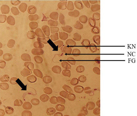
Trypanosoma sp. highlighting the KN=Kinetoplast, NC=Nucleus and FG= Flagellum (100X).
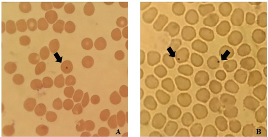
Anaplasma centrale: indicates the inclusion body at the central part of the erythrocyte (A). Anaplasma marginale: indicates the inclusion body near the margin of the erythrocytes (B) (100X).
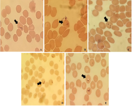
Babesia sp. (100X), showing pleomorphic shapes and sizes including, elliptical shape (A), maltase cross/tetrad (B), comma shape (C), pleomorphic ring (D), and vacuole (E).
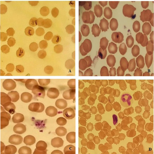
Plasmodium sp. (100X), highlighting the trophozoite stage (A), the ring-shaped trophozoite stage with a large chromatin dot (B), the schizont stage containing 6 to 12 merozoites with large nuclei clustered around a mass of coarse, dark-brown pigment (C), bean-shaped gametocyte (D).
Overall prevalence
The study was carried out with a total of 80 wild rats collected from different locations in Dhaka City, Bangladesh. Out of 80, 40 (50%) rodents were found to be infected with different blood protozoan parasites (Table 1). Five species of blood protozoa were identified in this study. Among the detected blood protozoan parasites, Plasmodium sp. had the highest prevalence (31.25%), followed by A. marginale (21.25%), A. centrale (17.50%), Babesia sp. (10.00%), and Trypanosoma sp. (3.75%).
Table 1: Prevalence of blood parasite infection by wild rat species.
| Rat species | No. of rats examined | No. of infected rats (%) | p-value |
| R. norvegicus | 28 | 16 (57.14%) | |
| B. bengalensis | 30 | 16 (53.33%) | 0.5035 |
| M. musculus | 08 | 3 (37.50%) | |
| R. rattus | 14 | 5 (35.71%) | |
| Total | 80 | 40 (50.00%) |
Prevalence of blood protozoan infection by wild rat species
Four wild rat species were identified among the captured samples. The highest number of rats belonged to Bandicota bengalensis (30), followed by R. norvegicus (28), R. rattus (14), and M. musculus (08). Among these species, R. norvegicus had the highest prevalence of protozoan infection (57.14%, 16/28), followed by B. bengalensis (53.33%, 16/30), M. musculus (37.5%, 3/8), and R. rattus (35.71%, 5/14) (Table 1).
Prevalence of blood protozoan infection by sex of wild rats
Both male and female wild rats from all four species were found to be infected. The prevalence of infection was higher in female rats across all species. In R. norvegicus, the prevalence was 50% in males and 66.67% in females, respectively. Similar observations were observed in B. bengalensis (52.38% in males and 55.55% in females), R. rattus (33.33% in males and 40% in females), and M. musculus (28.57% in males and 100% in females) (Table 2). However, Anaplasma sp. had a higher prevalence in females (44.45%) compared to males (24.52%), similar to Plasmodium sp. Conversely, Babesia sp. and Trypanosoma sp. showed a higher prevalence in males than in females (Table 3).
Table 2: Prevalence of blood parasite infection by sex of wild rats.
| Rat species | Sex of rats | No. of rats | No. of infected rats (%) | p-value |
| R. norvegicus | Male | 16 | 8 (50.00%) | 0.4589 |
| Female | 12 | 8 (66.67%) | ||
| Total | 28 | 16 (57.14%) | ||
| B. bengalensis | Male | 21 | 11 (52.38%) | ˃0.9999 |
| Female | 9 | 5 (55.55%) | ||
| Total | 30 | 16 (53.33%) | ||
| R. rattus | Male | 9 | 3 (33.33%) | ˃0.9999 |
| Female | 5 | 2 (40.00%) | ||
| Total | 14 | 5 (35.71%) | ||
| M. musculus | Male | 7 | 2 (28.57%) | NA |
| Female | 1 | 1 (100%) | ||
| Total | 8 | 3 (37.50%) | ||
| Total | 80 | 40 (50.00%) |
NA: Not analyzed.
Table 3: Prevalence of blood parasites in male and female wild rats.
| Parasite species | No. of infected rats (%) | No. of male rats (%) | No. of female rats (%) |
| Plasmodium sp. | 25 (31.25%) | 13 (24.52%) | 12 (44.45%) |
| A. marginale | 17 (21.25%) | 10 (18.86%) | 7 (25.93%) |
| A. centrale | 14 (17.5%) | 9 (16.98%) | 5 (18.51%) |
| Babesia sp. | 8 (10.00%) | 6 (11.32%) | 2 (7.41%) |
| Trypanosoma sp. | 3 (3.75%) | 2 (3.77%) | 1 (3.70%) |
Prevalence of blood protozoan infection in wild rats by captured niches
The wild rats were captured from four types of locations, namely stationary/grocery shops (25), local rental houses (20), fish markets (20), and slum areas (15). The highest infection rate was observed in rodents from slum areas (66.67%), followed by those from fish markets (50%), grocery shops (48%), and local rental houses (40%) (Table 4).
Table 4: Prevalence of blood parasite infection in different niches.
| Niches | No. of trapped rats | No. of infected rats (%) | p-value |
| Grocery shops | 25 (31.25%) | 12 (48.00%) | |
| Local rent houses | 20 (25.00%) | 8 (40.00%) | 0.4741 |
| Slum areas | 15 (18.75%) | 10 (66.67%) | |
| Fish markets | 20 (25.00%) | 10 (50.00%) | |
| Total | 80 | 40 (50.00%) |
Prevalence of co-infections in male and female wild rats
Both single and mixed infections with blood protozoan parasites were detected. The analysis showed single infections at 27.50%, whereas mixed infections with double and triple pathogens at 15.0% and 7.50%, respectively (Table 5).
DISCUSSION
This study examined four peri-domestic wild rat species, namely Rattus norvegicus, R. rattus, B. bengalensis, and M. musculus. Five species of blood protozoan parasites were identified, namely Plasmodium sp., A. marginale, A. centrale, Babesia sp., and Trypanosoma sp. Similar blood protozoan parasites were previously reported in rats and shrews in Bangladesh , except for Trypanosoma sp. Many authors reported similar blood parasites in rats in different parts of the world ; ; . The overall prevalence of blood protozoan parasites in this study was 50.0%, which was higher than previous findings of Islam et al. (2020) in Bangladesh (13%), Dada (2016) in Nigeria (16%), Katakweba (2018) in Tanzania (22.9%). However, the prevalence of blood protozoan parasites in this study is similar to those from Nigeria (63.08%) , and Iran (53.8%) . The variations might be due to the geographical variations, types of parasites identified, abundance and distribution of tick vectors in the research area, and the methodology for detection and identification of the parasites .
In this study, the prevalence of Plasmodium sp. was 31.25%. This rate is higher than the 0.67%, 8.57%, and 6.8% infection rate reported by Islam et al. (2020), Katakweba (2018) and Makokha et al. (2011), respectively
Table 5: Co-infections of parasites in the blood samples of wild rats.
| Status of infection | Grocery shops (n=25) |
Rent houses (n=20) |
Slum areas (n=15) |
Fish market (n=20) |
Total (n=80) |
| Single infection | |||||
| Plasmodium sp. | 6 (24.00%) | 1 (5.00%) | 2 (13.33%) | 2 (10.00%) | 11 (13.75%) |
| A. marginale | n.d | 1 (5.00%) | 2 (13.33%) | 1 (5.00%) | 4 (5.00%) |
| A. centrale | 1 (4.00%) | n.d | 1 (6.67%) | 1 (5.00%) | 3 (3.75%) |
| Babesia sp. | 1 (4.00%) | 1 (5.00%) | n.d | n.d | 2 (2.50%) |
| Trypanosoma sp. | 1 (4.00%) | 1 (5.00%) | n.d | n.d | 2 (2.50%) |
| Subtotal | 9 (36.00%) | 4 (20.00%) | 5 (33.33%) | 4 (20.00%) | 22 (27.50%) |
| Double infections | |||||
| Plasmodium sp. + A. centrale | n.d | n.d | 1 (6.67%) | 1 (5.00%) | 2 (2.50%) |
| Plasmodium sp. + Babesia sp. | 2 (8.00%) | n.d | n.d | n.d | 2 (2.50%) |
| A. marginale + A. centrale | n.d | 1 (5.00%) | 1 (6.67%) | 2 (10.00%) | 4 (5.00%) |
| Plasmodium sp. + A. marginale | n.d | 2 (10.00%) | 2 (13.33%) | n.d | 4 (5.00%) |
| Subtotal | 2 (8.00%) | 3 (15.00%) | 4 (26.67%) | 3 (15.00%) | 12 (15.00%) |
| Triple infections | |||||
| A. marginale + A. centrale + Babesia sp. | 1 (4.00%) | 1 (5.00%) | 1 (6.67%) | 1 (5.00%) | 4 (5.00%) |
| Plasmodium sp. + A. marginale + A. centrale | n.d | n.d | n.d | 1 (5.00%) | 1 (1.25%) |
| Plasmodium sp. + Trypanosoma sp. + Babesia sp. | n.d | n.d | n.d | 1 (5.00%) | 1 (1.25%) |
| Subtotal | 1 (4.00%) | 1 (5.00%) | 1 (6.67%) | 3 (15.00%) | 6 (7.50%) |
| Grand total | 12 (48.00%) | 8 (40.00%) | 10 (66.67%) | 10 (50.00%) | 40 (50.00%) |
but lower than 42.1%, and 48.46% in reported by Ajayi et al. (2006), and Alias et al. (2014), respectively. The variation in prevalence might be due to the variation in sample size, geography, rodent species, high concentration of mosquitoes, and the prevalence of malaria. Although females (44.45%) were more affected than male rodents (24.52%), the difference was not statistically significant, which was supported by Alias et al. (2014), who reported similar infection rates in both sexes.
Trypanosoma sp. was also identified in this study, with a prevalence of 3.75%, consistent with earlier findings of Katakweba (2018) (4.29%), Shafiyyah et al. (2012) (1.5%), and Ajayi et al. (2006) (3.08%). However, a higher prevalence was reported in Brazil (21.7%) due to the larger fly population and tropical climate . Wild rats near human settlements were more likely to be infected by Trypanosoma species . In Bangladesh, the absence of Tsetse fly (Glossina spp.), which transmits salivarian trypanosomiasis, likely contributes to the lower infection rate of Trypanosoma sp.
In this study, Babesia sp. had a prevalence of 10%, with higher infection rates in male rodents (11.32%) compared to female rodents (7.41%). A previous study in Bangladesh reported a lower infection rate of 4.7% . Lower infection rates were also reported in other studies ; ; . On the other hand, a similar prevalence was reported by Katakweba (2018), who documented 7.14% infections of Babesia sp. The hot and humid conditions and high population density in Dhaka City might favor for growth and survival of tick populations, leading to a higher prevalence of Babesia.
In this study, Anaplasma sp. was also detected, with a prevalence of 21.25% for Anaplasma marginale and 17.5% for A. centrale. A previous study in Bangladesh reported a lower prevalence of Anaplasma sp. 7.5% . This variation might be due to the difference in sample size, geographical location, and seasons.
In this study, wild rats of both sexes were found to be infected, where the females (59.25%) had a higher infection rate than the males (45.20%). This finding contrasts with the study by Alias et al. (2014), which found that male rats had higher infection rates because they explored larger areas than female rats. All rodents in our study were captured between late winter and early spring (February to March), coinciding with the wild rat breeding season. That might be the reason why females in our study had higher rates of protozoan infection.
The study revealed that R. norvegicus had the highest infection rate (57.14%), followed by B. bengalensis, R. rattus, and M. musculus. Among 80 wild rats captured, the majority were collected from different grocery shops (31.5%), followed by rental houses (25%), slum areas (18.75%), and fish markets (25%). A similar study in Argentina reported that 74% of captured rats were commensal species (Mus domesticus, Rattus rattus, and Rattus norvergicus) . In the present study, the highest number of infected wild rats were found in houses in slum areas (66.67%), followed by fish markets (50%), stationery shops (48%), and rental houses (40%). In Bangladesh, slum areas are responsible for various disease transmissions. Lesser access to health services, inadequate housing, and poor sanitation in slum communities increase parasitic infection by wild rats and facilitate the epidemic transmission of infectious diseases in humans ; .
CONCLUSION
The aim of the present study was to determine the prevalence of blood protozoan parasites in peri-domestic wild rats in Dhaka City, Bangladesh. The Plasmodium sp., Babesia sp., and Trypanosoma sp. have been reported to bear public health significance. This study reports the blood protozoa Trypanosoma sp. for the first time in wild rats in Bangladesh. Wild rats live alongside humans in city areas and play an important role in transmitting zoonotic diseases. For this, further research with larger sample sizes and molecular techniques is essential for better understanding the complex interaction between rodent hosts and protozoan parasites.
Acknowledgments
The authors are grateful to the Ministry of Science and Technology (MOST), Bangladesh, for financial support.
NOVELTY STATEMENT
This research has reported Trypanosoma sp. in murine rodents for the first time in Bangladesh.
AUTHOR’S CONTRIBUTION
NCD: Data collection, sample collection, methodology, writing the manuscript. A W: Data analysis, sample collection, methodology, reviewing and editing the manuscript. SMA: Data analysis, methodology and reviewing. MAHC, RMO, SPS, PK, ASD, SC, JBMI, MRI, MNI: Data analysis, methodology. UKM: Conceptualization, supervision, reviewing and editing the manuscript. All the authors have read and approved the final version of this manuscript.
Funding
This research was funded by the Ministry of Science and Technology (MOST).
Ethical approval
All the protocols performed in studies involving animals were in accordance with the institution’s or practice’s ethical standards.
Generative AI and AI assisted technology statement
The authors declare that no generative AI and AI assisted technology was used in the creation of this manuscript.
Conflict of interest
The authors have declared no conflict of interest.
References
- Ahmed ZU, Begum ZNT, Hassan MA, Khondker M, Kabir SMH, Ahmad M (2009). Encyclopedia of flora and fauna of Bangladesh, Mammals. Dhaka, Bangladesh. J. Asiat. Soc. Bangladesh, 27: 67.
- Ajayi OO, Ogwurike BA, Ajayi JA (2006). Blood protozoan parasites of rodents in Jos, Plateau State, Nigerai. Anim. Prod. Res. Adv., 2(4): 202-207. https://doi.org/10.4314/apra.v2i4.36341 [CrossRef]
- Alias SN, Sahimin N, Edah MA, Mohd-Zain SN (2014). Epidemiology of blood parasitic infections in the urban rat population in peninsular Malaysia. Trop. Biomed., 31(2): 230-240.
- Aplin KP, Brown PR, Jacob J, Krebs CJ, Singleton GR (2003). Field methods for rodent studies in Asia and the Indo-Pacific. Austral. Center Int. Agric. Res. Camberra.
- Asante J, Noreddin A, Zowalaty ME (2019). Systematic review of important bacterial zoonoses in Africa in the last decade in light of the “One Health” concept. Pathogens, 8: 50-55. https://doi.org/10.3390/pathogens8020050 [CrossRef]
- Ashford RW (1996). Leishmaniasis reservoirs and their significance in control. Clin. Dermatol., 14: 523–532. https://doi.org/10.1016/0738-081X(96)00041-7 [CrossRef]
- Battersby SA, Parsons R, Webster JP (2002). Urban rat infestations and the risk to public health. J. Environ. Health Res., 1: 4-12.
- Britanica T (2020). Editors of encyclopaedia. Argon. Encyclopedia Britannica.
- Burgin CJ, Colella JP, Kahn PL, Upham NS (2018). How many species of mammals are there? J. Mammal., 99(1): 1–14. https://doi.org/10.1093/jmammal/gyx147 [CrossRef]
- Castillo E, Priotto J, Ambrosio AM, Provensal MC, Morales MA, Steinmann A, Polop JJ (2003). Commensal and wild rodents in an urban area of Argentina. Int. Biodeter. Biodegr., 52: 135-141. https://doi.org/10.1016/S0964-8305(03)00033-7 [CrossRef]
- Childs JE, Glass GE, LeDuc JW (1991). Rodent sightings and contacts in an inner-city population of Baltimore, Maryland, USA. Bull. Soc. Vector Ecol., 16: 245–255.
- Clinton JM (1969). Rats in urban America. Health Reproduction, pp. 1-7. https://doi.org/10.2307/4593484 [CrossRef]
- Dada EO (2016). Study on the ectoparasites and haemoparasites of domestic rats in parts of Akure South Local Government Area of Ondo State. Int. J. Clin. Chem. Lab. Med., 2(1): 1-5. https://doi.org/10.20431/2455-7153.0201001. [CrossRef]
- Ellis B, Regnery R, Beati L, Bacellar F, Rood M, Glass G, Marston E, Ksiazek T, Jones D, Childs J (1999). Rats of the genus Rattus are reservoir hosts for pathogenic Bartonella species: An old world origin for a new world disease? J. Infect. Dis., 180(1): 220-224. https://doi.org/10.1086/314824 [CrossRef]
- Gratz N (1994). Rodents as carriers of disease in rodent pests and their control. (Eds. Buckle A.P. and Smith R.H.). CAB International, Oxford, pp. 85–108.
- Han BA, Schmidt JP, Bowden SE, Drake JM (2015). Rodent reservoirs of future zoonotic diseases. Proc. Natl. Acad. Sci., 112(22): 7039-7044. https://doi.org/10.1073/pnas.1501598112 [CrossRef]
- Helmy YA, El-Adawy H, Abdelwhab EM (2017). A comprehensive review of common bacterial, parasitic and viral zoonoses at the human-animal interface in Egypt. Pathogens, 6: 33. https://doi.org/10.3390/pathogens6030033 [CrossRef]
- Homer MJ, Aguilar-Delfin I, Telford SR, Krause PJ, Persing DH (2000). Babesiosis. Clin. Microbiol. Rev., 13: 451-469. https://doi.org/10.1128/CMR.13.3.451 [CrossRef]
- Islam S, Rahman MK, Ferdous J, Rahman M, Akter S, Faraque MO, Chowdhury MNU, Hossain MA, Hassan MM, Islam A (2020). Hemoprotozoa and Anaplasma sp. in rodents and shrews of Bangladesh. Trop. Biomed., 37(4): 842-851. https://doi.org/10.47665/tb.37.4.842 [CrossRef]
- Katakweba AAS (2018). The prevalence of haemoparasites in rodents and shrews trapped from domestic and peridomestic houses in Morogoro Municipality, Tanzania. A Hidden Public Health Threat. Tanzania Veterinary Journal, 36: 75-82.
- Khaghani R (2007). The Economic and Health Impact of Rodent in Urban Zone and Harbours and their Control Methods. Ann. Military Health Sci. Res., 4: 1071–1078.
- Killick-Kendrick R (2009). Parasitic protozoa of the blood of rodents: A revision of Plasmodium berghei. Parasitology, 69(2): 225-237. https://doi.org/10.1017/S0031182000048071 [CrossRef]
- Ko AI, Reis GM, Ribeiro CM, Johnson WD, Riley LW (1999). Urban epidemic of severe leptospirosis in Brazil. Lancet, 354(9181): 820–825. https://doi.org/10.1016/S0140-6736(99)80012-9 [CrossRef]
- Linardi PM, Botelho JR (2002). Prevalence of Trypanosoma lewisi in Rattus norvegicus from Belo Horizonte, State of Minas Gerais, Brazil. Memorias do Instituto Oswaldo Cruz, 97: 411-414. https://doi.org/10.1590/S0074-02762002000300024 [CrossRef]
- Luniak M (2004). Synurbization–adaptation of animal wildlife to urban development. In Proceedings 4th international urban wildlife symposium, Tucson: University of Arizona, pp. 50-55.
- Makokha GW, Ng’wena AGM, Ngeiywa MM (2011). Prevalence of Trypanosoma and Plasmodium species parasites in small rodents of Kakamega Forest in Western Kenya. Afr. J. Health Sci., 19(3-4): 63-67.
- Mardosaitė-Busaitienė D, Radzijevskaja J, Balčiauskas L, Paulauskas A (2021). Babesia microti in rodents from different habitats of Lithuania. Animals (Basel), 11(6): 1707. https://doi.org/10.3390/ani11061707. [CrossRef]
- Navea-Pérez HM, Díaz-Sáez V, Corpas-López V, Merino-Espinosa G, Morillas-Márquez F, Martín-Sánchez J (2015). Leishmania infantum in wild rodents: Reservoirs or just irrelevant incidental hosts? Parasitol. Res., 114(6): 2363–2370. https://doi.org/10.1007/s00436-015-4434-y [CrossRef]
- Paramasvaran S, Sani RA, Hassan L, Hanjeet Kaur HK, Krishnasamy M, Jeffery J, Santhana RSR., Mohd Ghazali MG, Lim KHKH (2009). Endo-parasite fauna of rodents caught in five wet markets in Kuala Lumpur and its potential zoonotic implications. Trop. Biomed., 26(1): 67-72.
- Pumhom P, Morand S, Tran A, Jittapalapong S, Desquesnes M (2015). Trypanosoma from rodents as potential source of infection in human-shaped landscapes of South-East Asia. Vet. Parasitol., 208(3-4): 174-180. https://doi.org/10.1016/j.vetpar.2014.12.027 [CrossRef]
- Roohollah S, Jean-Christoph A, Jamshid D, Hassan M, Hon-Tsen Y, Sylvien A, Francois B, Julienm C (2012). Patterns of morphological evolution in the mandible of the house mouse Mus musculus (Rodentia: Muridae). Biol. J. Linn. Soc., 105(3): 635–647. https://doi.org/10.1111/j.1095-8312.2011.01821.x [CrossRef]
- Saito-Ito A, Rai S K, He S, Kohsaki M, Tsuji M, Ishihara C (1999). First demonstration of Babesia parasitizing in human in Japan (in Japanese). J. Jpn. Assoc. Infect. Dis., 73(11): 1163-1164. https://doi.org/10.11150/kansenshogakuzasshi1970.73.1163 [CrossRef]
- Seifollahi Z, Sarkari B, Motazedian MH, Asgari Q, Ranjbar MJ, Abdolahi S (2016). Protozoan parasites of rodents and their zoonotic significance in Boyer-Ahmad District, Southwestern Iran. Vet. Med. Int., 2016(1): 3263868. https://doi.org/10.1155/2016/3263868 [CrossRef]
- Selma U, Bekir C, Djursun K, Semra, Gureser A., Ferhat M, Ali M, Mustafa S, Ahmet K, Cahit B, Kosta M, Aysegul O. (2019). Molecular survey of Babesia microti (Aconoidasida: Piroplasmida) in wild rodents in Turkey. J. Med. Entomol., 56(6): 1605–1609. https://doi.org/10.1093/jme/tjz084 [CrossRef]
- Shafiyyah CS, Jamaiah I, Rohela M, Lau YL, Aminah FS (2012). Prevalence of intestinal and blood parasites among wild rats in Kuala Lumpur, Malaysia. Trop. Biomed., 29(4): 544–550.
- Sumangali K, Rajakaruna RS, Rajapakse RPVJ (2007). Ecto and endo parasites of rodents from two selected sites in Kandy District. Peradeniya University Research Sessions Purse 2007, Volume 12 Part I. Agric. Biol. Med. Sci. Ed. Board, 44(3 and 4): 86.
- Thanee N, Kupittayanant S, Pinmongkholgul S (2009). Prevalence of ectoparasites and blood parasites in small mammals at Sakaerat Environmental Research Station, Thailand. Thai J. Agric. Sci., 42(3): 149-158.
- Urquhart GM, Armour JL, Duncan AM, Jennings FW (1996). Veterinary parasitology, 2nd Edn. Blackwell Science, London.
- Wang H, Yoshimatsu K, Ebihara H, Ogino M, Araki K, Kariwa H, Wang Z, Luo Z, Li D, Hang C (2000). Genetic diversity of hantaviruses isolated in China and characterization of novel hantaviruses isolated from Niviventer confucianus and Rattus rattus. Virology, 278(2): 332-345. https://doi.org/10.1006/viro.2000.0630 [CrossRef]

Services
Article usage statistics
Loading…
Related articles
- Detection of Parasites of Animal Health Importance Carried by Wild Rats Collected from Jeddah City, Saudi ArabiaAdvances in Animal and Veterinary Sciences · 2019
- Parasitic Prevalence in Wild and Captive Birds Along an Altitudinal Gradient in Punjab, PakistanPakistan Journal of Zoology · 2025
- Prevalence and Detailed Morphological Identification of Gastrointestinal Helminths of Turkeys in Wet Markets of Dhaka City: First Detection of Capillaria philippinensis in BangladeshThe Journal of Advances in Parasitology · 2025
- Investigation of Clinical Diseases and Surgical Cases in Cats of Dhaka City, BangladeshVeterinary Sciences: Research and Reviews · 2025
- Prevalence of <i>Haemoproteus</i> <i>Sp</i> in Domestic Pigeon at Chittagong and Khulna District in BangladeshThe Journal of Advances in Parasitology · 2014
- Prevalence of Feline Infectious Peritonitis in Pet Cats at Dhaka, Bangladesh: A Clinic-Based Cross-Sectional StudyVeterinary Sciences: Research and Reviews · 2023