
Jackfruit bronzing disease caused by Pantoea stewartii subsp stewartii (Pss) has recently been reported in the Philippines, Malaysia, and Mexico. Pss can cause severe yield losses ranging from 40% to 100%. Indonesia is the fourth-largest jackfruit-producing country in the world. However, there have been no reports of Pss infecting jackfruit in Indonesia. To date, the occurrence of Pss in Indonesia has been limited to maize in several provinces, including West Sumatra, Bali, West Java, and East Java. Therefore, this study was conducted to investigate the occurrence of the jackfruit bronzing disease and to characterize the associated bacterial isolates based on their morphological and physiological characteristics. Seventeen yellow bacterial isolates with circular colonies were isolated. All isolates were Gram-negative, facultatively anaerobic, catalase-positive but oxidase and indole-negative, characteristics with those previously reported for Pss. Pathogenicity tests were conducted by inoculation of healthy, surface-disinfected jackfruit with 1 mL of bacterial suspension (108 CFU/mL). Sterile distilled water served as the negative control, with three replicates conducted for each treatment. Five isolates (ANG15, ANG22, MCL10, PTL04, PTL19) produced symptoms similar to those of jackfruit bronzing disease. The inoculated fruits developed bronzing or rusty lesion with yellowish discoloration within two weeks after inoculation, while the negative control remain asymptomatic. The re-isolated bacteria exhibited morphological and physiological characteristics consistent that previously reported for Pss. PCR amplification using ES16 – ESIG2c primers produced the expected 920 bp amplicon in all five pathogenic isolates. Further molecular analysis is needed to accurately characterize bacterial isolates associated with jackfruit bronzing disease.
Special Issue: Regenerative Agriculture for Sustainable Food Production and Environmental Restoration · Volume 42, Issue 1, Pages 12–23, 2 November 2026 · Open Access
The Emergence of Jackfruit Bronzing Disease Associated with Pantoea stewartii in South Sulawesi, Indonesia
·
Affiliations & Notes ⌄
- 1Doctoral Program of Agricultural Sciences, Faculty of Agricultre, Hasanuddin University, 90245, Makassar, Indonesia
- 2Agricultural Bacteriology Research Group, Hasanuddin University, 90245, Makassar, Indonesia
- 3Department of Plant Pest and Disease, Faculty of Agriculture, Hasanuddin University, 90245, Makassar, Indonesia
- *Correspondence | Baharuddin Patandjengi; Email: baharuddin@agri.unhas.ac.id
Article Info ⌄
- Journal:
- Sarhad Journal of Agriculture
- Submitted Date:
- 7 June 2026
- Accepted Date:
- 25 September 2026
- Published Date:
- 2 November 2026
Abstract
Introduction
Jackfruit (Artocarpus heterophyllus Lam.) is a tropical fruit widely cultivated in South and Southeast Asia ; . In Indonesia, jackfruit is commonly consumed as fresh fruit and processed into various traditional food products, making it valuable not only for household consumption but also for local markets and small-scale agroindustry ; . Indonesia is an important producer of jackfruit in Asia. FAO estimated that average jackfruit production reached approximately 700,000 tonnes in Indonesia during 2015–2017, after India and Bangladesh as the major producing countries (FAO, 2018; . National horticultural statistics from BPS showed that jackfruit/cempedak production in Indonesia reached approximately 797,815 tons in 2025, indicating the importance of this crop in the national fruit production system (BPS, 2025a). In South Sulawesi, jackfruit/cempedak production was recorded at approximately 21,308 tons in 2025, suggesting that this crop also has regional economic relevance (BPS, 2025b). Therefore, diseases affecting jackfruit fruit quality may have significant consequences for farmers, traders, and consumers.
Despite the economic importance of jackfruit, published information on diseases affecting this crop in Indonesia remains relatively limited. Jackfruit is commonly grown in homegardens, agroforestry systems, or mixed-cropping systems, where it may receive less intensive disease monitoring than major commercial fruit crops . This production context may contribute to delayed recognition or underreporting of disease problems in jackfruit. Jackfruit bronzing disease symptoms mainly develop within the internal fruit tissues, characterized by the discoloration of pulp and rags from yellowish-orange to reddish with rusty specks ; . Therefore, early detection may be difficult before the fruit is opened. The emergence of bronzing disease in several jackfruit-producing countries highlights the need for disease surveillance and pathogen identification in jackfruit-producing areas, particularly when unusual internal fruit discoloration is observed .
Jackfruit bronzing disease reduces fruit quality and marketability because the bronzing symptoms affect the edible portion of the fruit. Previous studies have associated jackfruit bronzing disease with Pss, a Gram-negative, yellow-pigmented bacterium that is widely known as the causal agent of Stewart’s wilt disease in maize ; . Following its initial report in the Philippines, jackfruit bronzing disease caused by Pss has subsequently been documented in multiple jackfruit-producing regions across Southeast and East Asia, as well as in the Americas, indicating a wider geographical distribution than previously recognized ; ; ; ; ; .
In Indonesia, Pss has been reported mainly as the causal agent of Stewart’s wilt disease in maize. Previous reports indicated the presence of this pathogen in several maize-growing regions, including West Java, West Sumatra, East Java, Bali and South Sulawesi ; ; ; . The occurrence of jackfruit bronzing disease in several tropical jackfruit-producing countries, particularly the Philippines, Malaysia, and Vietnam, indicates that Indonesia may also be at risk of its emergence ; ; . This concern is strengthened by the widespread cultivation of jackfruit in Indonesia, the geographical proximity and agricultural connectivity of Southeast Asian countries, and the difficulty of recognizing infected fruit. In Vietnam, bronzing disease was first observed in 2016 and was subsequently reported from several major jackfruit-growing areas, demonstrating its capacity to become established across tropical production regions . However, there has been no scientific report describing the occurrence of jackfruit bronzing disease or the association of Pss with jackfruit in Indonesia. This lack of information is important because Indonesia is a major jackfruit-producing country, and the emergence of bronzing symptoms could represent a potential threat to jackfruit production and fruit quality.
Therefore, this study was conducted to investigate the occurrence of jackfruit bronzing disease in South Sulawesi, Indonesia, and to characterize putative bacterial pathogens associated with bronzing disease. Pathogenicity tests were also performed to evaluate the ability of the isolates to reproduce bronzing symptoms on healthy jackfruit. The results of this study provide preliminary evidence of bacterial isolates associated with jackfruit bronzing disease in Indonesia and may serve as a basis for further biochemical and molecular identification of the causal agent.
Materials and Methods
Sampling
Field surveys were conducted in three jackfruit-growing locations in South Sulawesi, Indonesia. The geographical coordinates of the sampling locations are presented in Table 1.
Table 1: GPS coordinate of sampling location and disease incidence
| Regency | District | GPS coordinate | Disease incidence (%) |
| East Luwu | Angkona | 2°33'39.5"S 120°54'24.9"E | 16.22 |
| Maros | Moncongloe | 5°09'28.3"S 119°32'14.1"E | 14.29 |
| Gowa | Pattallassang | 5°12'59.4"S 119°33'16.7"E | 15.56 |
At each location, jackfruit trees bearing fruits were inspected for the presence of bronzing-like symptoms. A total of 117 fruits were examined across the three locations. The fruits were opened and inspected for internal symptoms. Disease incidence was calculated using the formula: Disease incidence (%) = (number of symptomatic fruits / total number of fruits examined) × 100. At the location level, 6 of 37 fruits examined in East Luwu, 5 of 35 fruits in Maros, and 7 of 45 fruits in Gowa showed bronzing-like symptoms. The collected symptomatic fruits were individually placed in clean sterile plastic bags, labelled according to sampling location and sample code, stored in a cool container, and transported to the laboratory for further analysis.
Isolation of bacteria
Bacteria from typical symptomatic plants were isolated and characterized at Agricultural Biotechnology Laboratory, Department Plant Pest and Disease. Bacterial isolation from symptomatic jackfruit was carried out using a modified method described by Gapasin et al. (2014), Zulperi et al. (2017), and Zhao et al. (2023). Symptomatic fruit tissues were surface sterilized with 75% ethanol for 30 s, followed by immersion in 1% NaOCl for 8 min, and then rinsed three times with sterile distilled water. Sterilized jackfruit tissues were macerated in 2 mL of sterile distilled water and stand for 30 min. Subsequently, 500 µL of the resulting supernatant were spread onto Nutrient Glucose Agar (NGA) and incubated at room temperature overnight. Colonies showing typical characteristics of Pss, including lemon-yellow pigmentation, convex elevation, circular shape, and approximately 1–4 mm in diameter, were selected and purified on fresh NGA medium. Single-colony purification was performed twice by successive re-streaking onto fresh NGA plates, with incubation after each streaking step, until colonies with uniform morphology were obtained. The resulting purified isolates were subsequently used for physiological characterization.
Morphological and physiological characterization
Colony morphology was observed on Nutrient Glucose Agar (NGA), including color, form, elevation, and margin. Gram reaction was determined using the 3% KOH test, where mucus formation indicated a Gram-negative reaction. Facultative anaerobic growth was tested in a glucose-containing semi-solid medium overlaid with sterile paraffin, and a color change from blue to yellow was recorded as a positive fermentative reaction. Yellow pigmentation was evaluated on Yeast Dextrose Carbonate (YDC) medium after incubation at 28–30°C for 24–48 h. Catalase activity was tested by adding hydrogen peroxide to fresh bacterial colonies, while oxidase activity was determined using Kovács oxidase reagent; the absence of a purple or dark blue color change was recorded as an oxidase-negative reaction. Indole production was tested using Kovac’s indole reagent, and the absence of a red ring was recorded as negative. Acid production from lactose, maltose, arbutin, and glycerol was evaluated in Dye’s C medium supplemented with each carbon source, where a color change from purple to yellow indicated acid production. The results were compared with the reported characteristics of Pantoea spp. and Pss based on standard references for plant pathogenic bacteria. The preliminary study stages for characterization of Pantoea sp. are shown in Figure 1.
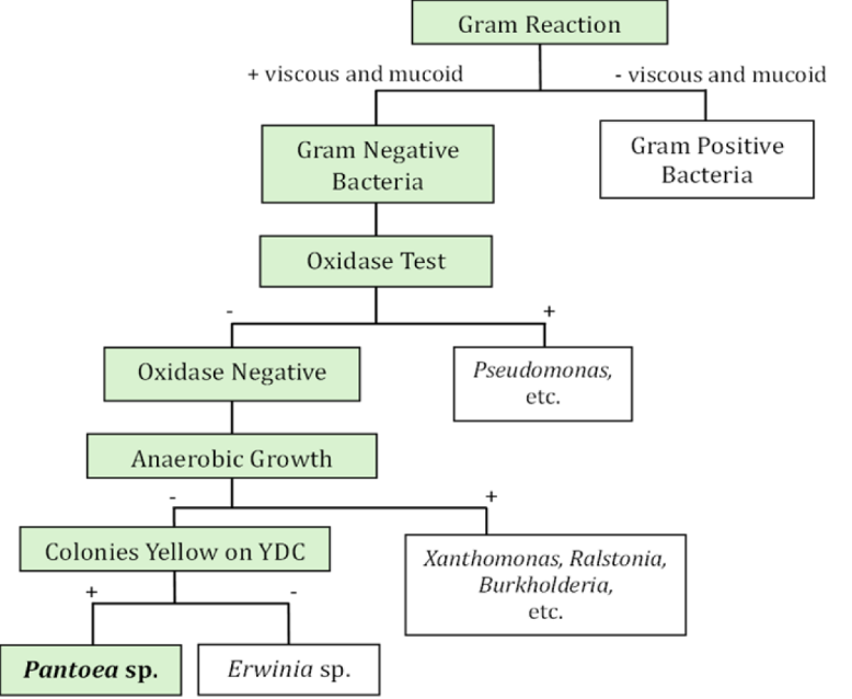
Flow chart for differentiation Pantoea sp. with other bacteria genera
Pathogenicity test
Pathogenicity tests were conducted using detached pulp of honey jackfruit and sweet corn cultivar Bonanza F1. Detached fruit-tissue assays facilitate simultaneous testing of multiple bacterial isolates under standardized environmental conditions and have previously been used for pathogenicity assessment of jackfruit bronzing-associated Pss ; ; . For jackfruit inoculation, healthy pulp was excised from surface-disinfected fruit, rinsed with sterile distilled water, dried on sterile tissue paper, and placed in Petri-dishes lined with sterile filter paper. Bacterial isolates were grown on NGA medium, suspended in sterile distilled water, and adjusted to 10⁸ CFU/mL. Each pulp sample was inoculated by injecting 1 mL of bacterial suspension using a sterile syringe, while sterile distilled water was used as a negative control. Each treatment consisted of three replicates. The inoculated samples were sealed and incubated at room temperature for two weeks. Symptom development was observed daily, and bronzing symptoms were evaluated based on pulp discoloration and the appearance of yellowish discoloration and bronzing specks.
For sweet corn inoculation, eight-day-old seedlings were injected in the seedling stem with 100 µL of bacterial suspension at approximately 10⁸ CFU/mL. Sterile distilled water was used as a negative control, and each treatment consisted of three replicates. The inoculated seedlings were maintained in clear polyethylene cages at 80% relative humidity and 25–27°C for 24–48 h and then observed for symptom development for up to 14 days post-inoculation. Symptoms recorded included water-soaking, necrosis parallel to the leaf veins, leaf blight, and wilting. After 14 days, bacteria were re-isolated from symptomatic jackfruit pulp and sweet corn seedlings on NGA medium as part of the assessment of Koch’s postulates and to determine whether bacteria with characteristics consistent with the inoculated isolates could be recovered from symptomatic tissues.
Disease symptom assessment
Disease severity was assessed 14 days after inoculation using a modified ordinal disease rating scale described by Abidin et al. (2021). Bronzing symptoms were evaluated based on changes in pulp color and the extent of rusty or bronzing specks with yellowish discoloration. Reddish discoloration was categorized following the color-based disease rating reference described by Abidin et al. (2021), where 0 indicated no symptom and higher scores represented increasing intensity of reddish-brown discoloration. Rusty or bronzing specks with yellowish discoloration were also scored from 0 to 4 based on the estimated percentage of symptomatic tissue area: 0 = no symptom, 1 = 1–24%, 2 = 25–49%, 3 = 50–74%, and 4 = >75% of the tissue area showing bronzing symptoms. Disease symptoms were photographed, and the negative control was used as a reference for asymptomatic tissue. Disease rating data were analyzed using the Kruskal–Wallis test because the severity scores were recorded on an ordinal scale (0–4). When a significant overall difference was detected, Dunn’s multiple-comparison test with Holm adjustment was used for pairwise comparisons among bacterial isolates. Statistical significance was determined at P < 0.05.
PCR test
Virulent bacterial isolates that produced symptoms on both jackfruit pulp and sweet corn seedlings were further subjected to PCR analysis for molecular confirmation. DNA was extracted using the Presto™ Mini gDNA Bacteria Kit (Geneaid Biotech Ltd., Taiwan). PCR amplification was performed using the Pantoea stewartii-specific primer pair ES16 (5′-GCGAACTTGGCAGAGAT-3′) and ESIG2c (5′-GCGCTTGCGTGTTATGAG-3′), targeting the 16S–23S rRNA/ITS region. The primer pair was selected based on previous reports for the detection of Pss ; . PCR reactions were prepared using MyTaq™ HS Red Mix, 2× (Bioline/Meridian Bioscience). Genomic DNA from a previously confirmed Pss strain BNL04 was included as the positive control. Nuclease-free water was substituted for the DNA template in the no-template control to verify the absence of contamination during amplification. The amplification program started with a 1-min heating step at 94°C. This was followed by 35 amplification cycles consisting of 15 s at 94°C, 15 s at 55–58°C, and 30 s at 72°C for denaturation, primer annealing, and DNA extension, respectively. The resulting amplicons were resolved in a 1% agarose gel by electrophoresis and subsequently examined using UV light. Samples showing a DNA fragment of approximately 920 bp, consistent with the expected amplicon size of the positive control, were interpreted as positive for putative P. stewartii subsp. stewartii. No amplification was expected from the negative control.
Results and Discussion
Field observation in south sulawesi
Field observations conducted in several jackfruit-growing areas in South Sulawesi revealed symptoms similar to those previously described as jackfruit bronzing disease ; . The affected fruits generally showed no obvious external symptoms; however, after the fruits were opened, discoloration was observed in the internal tissues, particularly on the pulp and rags. The symptoms included yellowish discoloration, reddish-brown discoloration, and rusty or bronzing specks on the edible pulp and surrounding tissues (Figure 2). These symptoms were not uniformly distributed across the fruit but appeared as localized reddish or rusty patches in several sections of the pulp.
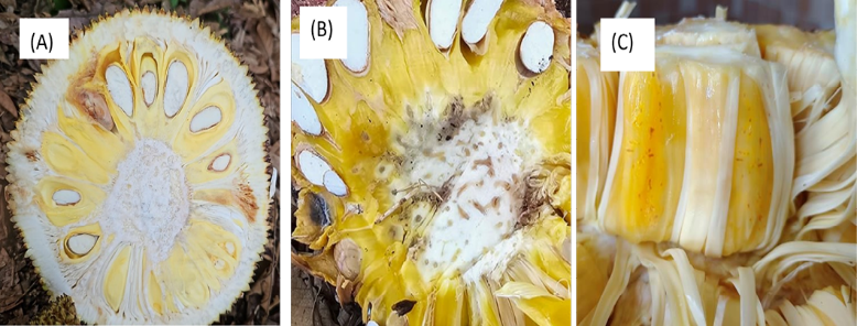
Field symptoms observed on jackfruits in South Sulawesi, Indonesia: (A) reddish-brown discoloration on jackfruit pulp and surrounding tissues; (B) yellowish to brown discoloration on the internal fruit tissues; and (C) rusty or bronzing specks with yellowish discoloration on jackfruit pulp. Black arrows indicate symptomatic areas.
The observed symptoms were consistent with previous descriptions of jackfruit bronzing disease ; ; . A characteristic feature of this disease is that affected fruits may show little or no obvious external symptoms, while discoloration develops predominantly in the internal fruit tissues ; ; . This symptom pattern is epidemiologically relevant because reliance on external visual inspection may result in affected fruits remaining undetected during harvesting and marketing. More generally, visual surveillance of emerging plant diseases can be limited when infection is asymptomatic or symptoms are not readily visible, emphasizing the importance of complementary diagnostic approaches ; . The detection of bronzing-like symptoms at all three surveyed locations in the present study indicates that the condition was not confined to a single sampling site. However, the incidence values reported here represent only the proportion of symptomatic fruits among those examined at the selected sites and should not be interpreted as an estimate of disease prevalence across South Sulawesi, because the survey was not designed as a randomized regional epidemiological survey. Notably, jackfruit bronzing has also been reported across multiple production areas in other countries; for example, recent reports documented its occurrence across several jackfruit-growing regions in Vietnam and multiple localities in Hainan, China ; . These observations highlight the need for broader, spatially representative and seasonally repeated surveys to determine the distribution, temporal dynamics, and potential sources of inoculum associated with jackfruit bronzing disease in Indonesia.
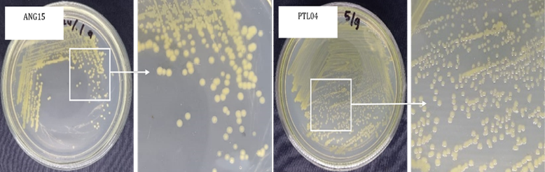
Representative colony morphology of bacterial isolates obtained from jackfruit showing bronzing-like symptoms in South Sulawesi. The isolates produced yellow-pigmented, circular, smooth, and convex colonies on Nutrient Glucose Agar.
Morphological and physiological characterization
Bacterial isolation from jackfruit with bronzing-like symptoms yielded 17 yellow-pigmented isolates with colony morphology resembling Pantoea spp. On Nutrient Glucose Agar, the colonies were pale yellow, cream-yellow, or lemon-yellow, circular, flat to convex, and had entire margins with approximately 1–2 mm in diameter (Figure 3; Table 2). These colony characteristics were similar to previous descriptions of Pantoea stewartii subsp. stewartii, which generally produces yellow-pigmented, circular, smooth, and flat to convex colonies ; ; . Physiological characterization showed that bacteria isolates were Gram-negative, facultatively anaerobic, yellow-pigmented on YDC medium, catalase-positive, oxidase-negative, and indole-negative (Table 3; Figure 4). These traits are consistent with the reported characteristics of Pantoea spp. and support their preliminary identification as putative Pantoea isolates ; ; .
Table 2: Colony morphology of bacterial isolates obtained from jackfruit showing bronzing-like symptoms in South Sulawesi
| No | Isolate code | Colony morphologya | |||
| Color | Form | Elevation | Margin | ||
| 1 | ANG04 | Pale Yellow | Circular | Convex | Entire |
| 2 | ANG07 | Lemon-yellow | Circular | Convex | Entire |
| 3 | ANG08 | Pale Yellow | Circular | Convex | Entire |
| 4 | ANG15 | Pale Yellow | Circular | Convex | Entire |
| 5 | ANG20 | Pale Yellow | Circular | Convex | Entire |
| 6 | ANG22 | Pale Yellow | Circular | Convex | Entire |
| 7 | ANG25 | Pale Yellow | Circular | Convex | Entire |
| 8 | MCL07 | Lemon-yellow | Circular | Flat | Entire |
| 9 | MCL10 | Cream-yellow | Circular | Convex | Entire |
| 10 | MCL12 | Pale Yellow | Circular | Flat | Entire |
| 11 | MCL16 | Lemon-yellow | Circular | Convex | Entire |
| 12 | MCL18 | Pale Yellow | Circular | Convex | Entire |
| 13 | PTL01 | Cream-yellow | Circular | Convex | Entire |
| 14 | PTL04 | Pale Yellow | Circular | Convex | Entire |
| 15 | PTL19 | Lemon-yellow | Circular | Convex | Entire |
| 16 | PTL21 | Pale Yellow | Circular | Flat | Entire |
| 17 | PTL22 | Cream-yellow | Circular | Convex | Entire |
aColony morphology characterization was done using protocol by Breakwell et al. (2016)
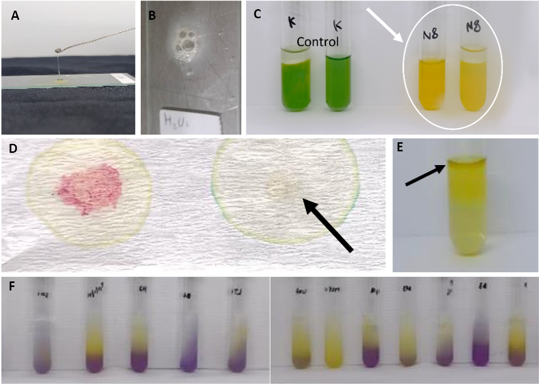
Physiological characteristics of bacterial isolates obtained from symptomatic jackfruit: (A) Gram-negative reaction indicated by mucus formation in the 3% KOH test; (B) catalase-positive reaction; (C) facultative anaerobic growth under aerobic and anaerobic conditions; (D) oxidase-negative reaction; (E) indole-negative reaction indicated by the absence of a red ring after Kovac’s reagent addition; and (F) acid production test, where a color change from purple to yellow indicates acid production.
However, the similarity in colony morphology and physiological traits among the 17 isolates did not necessarily correspond to pathogenicity or molecular identification. Only a subset of the phenotypically similar isolates subsequently reproduced bronzing symptoms and produced the expected Pss-associated PCR amplicon. This finding illustrates the limitation of relying solely on colony morphology and routine physiological tests for the identification of Pss. Closely related Pantoea species can share overlapping phenotypic characteristics, making additional pathogenicity and molecular tests important for more reliable identification ; .
Further differentiation based on acid production from lactose, maltose, arbutin, and glycerol showed variation among the isolates. Ten isolates, ANG04, ANG08, ANG15, ANG20, ANG22, ANG25, MCL10, PTL01, PTL04, and PTL19, did not produce acid from the tested carbon sources, which was consistent with the reported physiological profile of Pss ; ; . However, the observed variation among isolates further supports the use of physiological tests as preliminary screening rather than as definitive identification. Therefore, pathogenicity tests and PCR analysis were subsequently performed to evaluate the pathogenic potential and molecular identity of the selected isolates.
Pathogenicity test
Pathogenicity tests showed that five bacterial isolates, namely ANG15, ANG22, MCL10, PTL04, and PTL19, were able to reproduce bronzing-like symptoms on detached jackfruit pulp within 14 days after inoculation (Table 4). Disease severity differed significantly among the five pathogenic isolates at 14 dpi (Kruskal–Wallis test, H = 16.57, P = 0.0023). ANG15 consistently produced the highest disease rating (4.0 ± 0.00), whereas ANG22 produced the lowest rating (1.6 ± 0.55). Post-hoc Dunn’s test with Holm adjustment detected a significant difference between ANG15 and ANG22 (adjusted P = 0.0013), while the remaining pairwise comparisons were not statistically significant. The inoculated jackfruit pulp in
Table 3: Physiological characteristics of bacterial isolates associated with jackfruit bronzing-like symptoms in South Sulawesi
| No | Isolate Code | GRa | AGb | YCc | OTd | IPe | Acid Production fromf | |||
| Lactose | Maltose | Arbutin | Glycerol | |||||||
| 1 | ANG04 | + | + | + | - | - | - | - | - | - |
| 2 | ANG07 | + | + | + | - | - | + | + | - | - |
| 3 | ANG08 | + | + | + | - | - | - | - | - | - |
| 4 | ANG15 | + | + | + | - | - | - | - | - | - |
| 5 | ANG20 | + | + | + | - | - | - | - | - | - |
| 6 | ANG22 | + | + | + | - | - | - | - | - | - |
| 7 | ANG25 | + | + | + | - | - | - | - | - | - |
| 8 | MCL07 | + | + | + | - | - | + | - | - | - |
| 9 | MCL10 | + | + | + | - | - | - | - | - | - |
| 10 | MCL12 | + | + | + | - | - | + | + | - | - |
| 11 | MCL16 | + | + | + | - | - | + | + | + | - |
| 12 | MCL18 | + | + | + | - | - | + | - | - | - |
| 13 | PTL01 | + | + | + | - | - | - | - | - | - |
| 14 | PTL04 | + | + | + | - | - | - | - | - | - |
| 15 | PTL19 | + | + | + | - | - | - | - | - | - |
| 16 | PTL21 | + | + | + | - | - | + | + | + | - |
| 17 | PTL22 | + | + | + | - | - | + | + | + | + |
aGR, Gram Reaction (3% KOH Test): +, positive mucus formation (Gram-negative); −, negative mucus formation (Gram-positive) ; ;
bAG, Anaerobic Growth: +, positive can growth under anaerobic condition; −, can not growth under anaerobic condition
cYC, Yellow Colony in YDC Medium: +, positive; −, negative
dOT, Oxidase test: +, positive; −, negative
e IP, Indole production test: +, positive; −, negative
fAcid production from several carbon source: +, positive; −, negative
Table 4: Pathogenicity and disease severity of selected bacterial isolates on detached jackfruit pulp at 14 days post-inoculation
| Isolate | Symptom on jackfruit pulp | Disease rating at 14 dpia | Re-isolationb | Pathogenicity result |
| ANG15 | Bronzing specks with yellowish discoloration | 4.0 ± 0.00 | + | Positive |
| ANG22 | Reddish-brown discoloration | 1.6 ± 0.55 | + | Positive |
| MCL10 | Rusty specks with yellowish discoloration | 3.0 ± 0.00 | + | Positive |
| PTL04 | Severe bronzing specks | 2.4 ± 0.89 | + | Positive |
| PTL19 | Yellowish to reddish discoloration | 3.2 ± 0.84 | + | Positive |
| Negative control | No symptom | 0.0 ± 0.00 | − | Negative |
aValues are presented as mean ± standard deviation from five replicates. Differences among isolates were analyzed using the Kruskal–Wallis test followed by Dunn’s multiple-comparison test with Holm adjustment (P < 0.05). The overall Kruskal–Wallis test was significant (H = 16.57, P = 0.0023).
bRe-isolation: + indicates that bacteria with similar colony morphology were successfully re-isolated from symptomatic jackfruit pulp; − indicates that no similar bacteria were re-isolated.
in South Sulawesi and were also consistent with previous descriptions of jackfruit bronzing disease which is characterized by internal discoloration and rusty specks on the pulp and rags of infected fruit ; ; . The ability of these isolates to reproduce similar symptoms on healthy jackfruit pulp supports their association with the bronzing symptoms observed in the field. After symptom development, bacteria were re-isolated from symptomatic tissues, and the re-isolated cultures showed colony morphology and physiological characteristics similar to those of the original inoculated isolates.
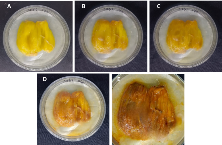
developed yellowish discoloration, reddish-brown discoloration, and rusty or bronzing specks, whereas the negative control inoculated with sterile distilled water remained asymptomatic. These symptoms were similar to the field symptoms observed
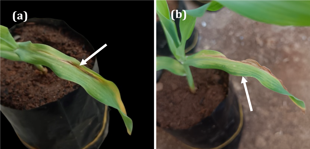
Symptoms observed on sweet corn cultivar Bonanza F1 after inoculation with pathogenic bacterial isolates obtained from symptomatic jackfruit in South Sulawesi. (a) Necrosis and water-soaked wilting symptoms; and (b) pale green to yellowish streaks developing parallel to leaf veins and progressing into necrosis.
Inoculation on sweet corn cultivar Bonanza F1 also showed that the five pathogenic isolates could induce Stewart’s wilt-like symptoms. Early symptoms appeared as pale green to yellowish streaks parallel to the leaf veins, followed by necrosis and water-soaked wilting symptoms within 14 days post-inoculation (Figure 6). These symptoms are consistent with typical Stewart’s disease symptoms on maize, including linear chlorotic streaks, necrotic lesions, and wilting ; . The development of symptoms on both jackfruit and sweet corn suggests that the isolates obtained from jackfruit were pathogenic not only to jackfruit but also to sweet corn seedlings, a known host of Pss ; . The ability of the same isolates to induce symptoms in both jackfruit pulp and sweet corn indicates cross-host pathogenic potential under experimental conditions, consistent with previous host-range studies of jackfruit-associated Pss isolates ; . This observation raises the possibility that jackfruit-associated Pss populations may retain pathogenic traits relevant to maize, the historically recognized primary host of the pathogen ; EFSA . However, the present inoculation assays do not demonstrate natural transmission between jackfruit and maize or establish either crop as an epidemiological reservoir for the other. The sweet corn assay involved artificial stem inoculation and therefore bypassed the natural infection and transmission processes occurring under field conditions, where Pss epidemiology in maize is associated with factors such as insect vectors and infected planting material (EFSA . Further studies using more naturally relevant inoculation routes, field surveys of adjacent jackfruit and maize crops, and molecular comparison of isolates from both hosts are needed to determine whether an epidemiological connection exists between the two crop systems.
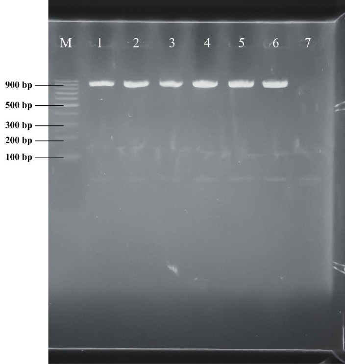
PCR detection using the ES16/ESIG2c specific primer pair for P. stewartii targeting the 16S–23S rRNA ITS region. Five isolates from jackfruit produced the expected 920 bp amplicon, supporting their identification as P. stewartii; M: DNA ladder; 1: ANG15, 2: ANG22, 3: MCL10, 4: PTL04, 5: PTL19, 6: Positive control-Pss strain BNL04, 7: no-template control.
PCR test
The PCR assay using the Pantoea stewartii–specific primer pair ES16/ESIG2c successfully amplified the expected 920 bp fragment in five bacterial isolates, namely ANG15, ANG22, MCL10, PTL04, and PTL19 (Figure 7). The presence of this specific amplicon indicates that these isolates are molecularly associated with Pss, whereas isolates without the expected band were considered negative under the PCR conditions used in this study. The ES16/ESIG2c primer pair targets the 16S–23S rRNA/ITS region, which has been reported as a useful molecular marker for bacterial identification because this non-coding region generally shows higher sequence variability than the conserved 16S rRNA gene, allowing better discrimination among closely related bacterial taxa .
The result is consistent with Coplin et al. (2002), who developed PCR primers and reported that ES16/ESIG2c amplified a 0.92 kb fragment from P. stewartii strains. In that study, all tested P. stewartii strains produced the expected amplicon, while most other related Pantoea and Erwinia species did not produce the same diagnostic product, supporting the specificity of this primer set for Pss detection . This primer pair has also been applied in previous jackfruit bronzing studies. Gapasin et al. (2014) reported amplification of the expected 0.92 kb ITS fragment from jackfruit bronzing bacterial isolates, while Abidin et al. (2020) used the same ES16/ESIG2c primer pair for molecular verification of Pss strains associated with jackfruit bronzing disease in Malaysia.
Therefore, the amplification of the 920 bp product in ANG15, ANG22, MCL10, PTL04, and PTL19 provides molecular evidence supporting the identification of these isolates as putative Pss and strengthens their association with jackfruit bronzing symptoms. Similar PCR-based confirmation has also been reported in China, where a 920 bp ES16/ESIG2c amplicon was obtained from jackfruit bronzing isolates and further supported by sequence similarity and pathogenicity test . However, PCR detection alone should be interpreted together with colony morphology, biochemical characteristics, pathogenicity tests, and, when possible, sequencing of the amplified fragment to provide stronger confirmation of the causal agent. This is particularly important because closely related yellow-pigmented Pantoea species may occur in plant tissues, and previous studies noted occasional weak or variable reactions in some non-target Pantoea strains .
Conclusions and Recommendations
This study provides preliminary evidence of the occurrence of jackfruit bronzing disease associated with Pss in South Sulawesi, Indonesia. Seventeen yellow-pigmented bacterial isolates were obtained from symptomatic jackfruit and showed morphological and physiological characteristics consistent with Pantoea spp. Among them, five isolates, ANG15, ANG22, MCL10, PTL04, and PTL19, reproduced bronzing-like symptoms on detached jackfruit pulp and induced Stewart’s wilt-like symptoms on sweet corn seedlings. The same isolates were successfully re-isolated from symptomatic tissues and produced the expected 920 bp amplicon using the ES16/ESIG2c primer pair, supporting their association with P. stewartii subsp. stewartii. These findings indicate that jackfruit bronzing disease may represent an emerging disease problem in Indonesia and highlight the need for further surveillance in jackfruit-producing areas. Further studies using DNA sequencing, multilocus sequence analysis, or whole-genome-based approaches are recommended to provides preliminary evidence the taxonomic identity, genetic diversity, host range, and epidemiological significance of the bacterial isolates associated with jackfruit bronzing disease in Indonesia.
Acknowledgements
The authors sincerely acknowledge the Ministry of Higher Education, Research, and Technology of the Republic of Indonesia for financial support through the PMDSU Scholarship (Contract No. 069/C3/DT.05.00/PL/2025). The authors also thank Prof. Dr. Tutik Kuswinanti for her valuable insights in data curation and manuscript review.
Novelty Statement
This study provides the first preliminary evidence of jackfruit bronzing disease associated with putative Pantoea stewartii subsp. stewartii isolates in South Sulawesi, Indonesia. The findings extend current knowledge of the occurrence of P. stewartii-like bacteria associated with jackfruit bronzing symptoms and their experimental pathogenic potential on jackfruit and sweet corn in Indonesia.
Authors’ Contribution
Fadyah Khamila Sahlan: Investigation, data collection, data analysis, visualization, and writing original draft.
Baharuddin Patandjengi: Conceptualization, supervision, validation, writing review and editing.
All authors read and approved the final manuscript.
Generative AI and AI-assisted technology statement
The authors declare that no generative AI and AI assisted technology was used in the creation of this manuscript.
Conflict of interest
The authors declare that there is no conflict of interest regarding the publication of this manuscript.
References
- Abidin, N., S.I. Ismail, G. Vadamalai, M.T. Yusof, M. Hakiman, D.S. Karam, N.W.I. Suhaimy, R. Ibrahim, D. Zulperi. 2020. Genetic diversity of Pantoea stewartii subspecies stewartii causing jackfruit-bronzing disease in Malaysia. PLoS ONE., 15(6): 1–18. https://doi.org/10.1371/journal.pone.0234350 [CrossRef]
- Abidin, N., S.I. Ismail, G. Vadamalai, M.T. Yusof, M. Hakiman, D.S. Karam, D. Zulperi. 2021. Pathogenic variability of the jackfruit-bronzing bacterium Pantoea stewartii subspecies stewartii infection to jackfruit varieties and its pivotal plant hosts in Malaysia. Agron., 11(11): 2113. https://doi.org/10.3390/agronomy11112113 [CrossRef]
- BPS. 2025a. Produksi tanaman buah-buahan dan sayuran tahunan menurut provinsi dan jenis tanaman, 2025. Badan Pusat Statistik, Jakarta, Indonesia. Retrieved on May 19, 2026.
- BPS. 2025b. Produksi tanaman buah-buahan dan sayuran tahunan menurut kabupaten/kota dan jenis tanaman di Provinsi Sulawesi Selatan, 2025. Badan Pusat Statistik Provinsi Sulawesi Selatan, Makassar, Indonesia. Retrieved on May 19, 2026.
- Breakwell, D., C. Woolverton, B. MacDonald, K. Smith, R. Robison. 2016. Colony morphology protocol. American Society for Microbiology. Available at: https://asm.org/protocols/colony-morphology-protocol
- Coplin, D.R. Majerczak, Y. Zhang, W.S. Kim, S. Jock and K. Geider. 2002. Identification of Pantoea stewartii subsp. stewartii by PCR and strain differentiation by PFGE. Plant Dis., 86(3): 304–311. https://doi.org/10.1094/PDIS.2002.86.3.304 [CrossRef]
- Cubero, J., P.J. Zarco-Tejada, S. Cuesta-Morrondo, A. Palacio-Bielsa, J.A. Navas-Cortés, P. Sabuquillo, T. Poblete, B.B. Landa, J. Garita-Cambronero. 2024. New approaches to plant pathogen detection and disease diagnosis. Phytopathol., 114(9): 1989–2006. https://doi.org/10.1094/PHYTO-10-23-0366-IA [CrossRef]
- EFSA Panel on Plant Health (PLH). 2018. Pest categorisation of Pantoea stewartii subsp. stewartii. EFSA Journal, 16(7), 5356. https://doi.org/10.2903/j.efsa.2018.5356 [CrossRef]
- EPPO. 2016. PM 7/60 (2) Pantoea stewartii subsp. stewartii. EPPO Bull. 46(2), 226–236. https://doi.org/10.1111/epp.12303 [CrossRef]
- Elevitch, C.R. and H.I. Manner. 2006. Artocarpus heterophyllus (jackfruit). In: Species Profiles for Pacific Island Agroforestry. Agroforestry Net, Inc., Hawaii.
- FAO. 2018. Minor tropical fruits: Mainstreaming a niche market. Food Outlook, July 2018: 67–75. Food and Agriculture Organization of the United Nations.
- Gapasin, R.M., R.P. Garcia, C.T. Advincula, C.S. Cruz, L.M. Borines. 2014. Fruit bronzing: a new disease affecting jackfruit caused by Pantoea stewartii (Smith) Mergaert et al. Ann. Trop. Res., 36(1): 17–31. https://doi.org/10.32945/atr3612.2014 [CrossRef]
- Gupta, A., A.R. Marquess, A.K. Pandey and A. Bishayee. 2023. Jackfruit (Artocarpus heterophyllus Lam.) in health and disease: A critical review. Crit. Rev. Food Sci. Nutr., 63(23): 6344–6378. https://doi.org/10.1080/10408398.2022.2031094. [CrossRef]
- Ha, V.T.N., L.K. Hoang, P.K. Huyen. 2024. Pantoea stewartii subsp. stewartii, the causative agent of Thai jackfruit’s bronzing disease and its possible host range in Vietnam. J. Plant Prot. Res., 64(2): 149–157. https://doi.org/10.24425/jppr.2024.150249 [CrossRef]
- Hanson, A. 2016. Oxidative-fermentative test protocol. American Society for Microbiology. Available at: https://asm.org/protocols/oxidative-fermentative-test-protocol
- Hernández-Morales, A., J.M. Pérez-Casillas, R.E. Soria-Guerra, J.B. Velázquez-Fernández, J.L. Arvizu-Gómez. 2017. First report of Pantoea stewartii subsp. stewartii causing jackfruit bronzing disease in Mexico. J. Plant Pathol., 99(3): 807
- Hernández-Morales, A., R.E. Soria-Guerra, N. Isiordia-Aquino, J. Campos-Guillén, J.R. Pacheco-Aguilar, A.B. Martínez-Rizo, J.L. Arvizu-Gómez. 2021. Association of Pantoea stewartii subsp. stewartii with ChrysSA genus flea beetles in jackfruit crops. J. Plant Pathol., 103: 539–547. https://doi.org/10.1007/s42161-021-00787-4 [CrossRef]
- Hu, S., X. Li, X. Yin, R. Li, R. Zhang, J. Zang and Y. Liu. 2022. Species-specific identification of Pseudomonas based on 16S–23S rRNA gene internal transcribed spacer (ITS) and its combined application with next-generation sequencing. BMC Microbiol., 22: 188. https://doi.org/10.1186/s12866-022-02607-w [CrossRef]
- Ibrahim, R., N.W. Ismail-Suhaimy, T. Shu-Qing, S.I. Ismail, N. Abidin, M. Hakiman, D.S. Karam, M.S. Ahmad-Hamdani, M.T. Yusof, D. Zulperi. 2020. Molecular characterization and phylogenetic analysis of Pantoea stewartii subspecies stewartii causing bronzing disease of jackfruit in Malaysia based on cps and hrp gene sequences. J. Plant Pathol., 102(1): 193–199. https://doi.org/10.1007/s42161-019-00383-7 [CrossRef]
- Ibrahim, R., S.I. Ismail, M.Y. Ina-Salwany, D. Zulperi. 2022. Biology, diagnostics, pathogenomics and mitigation strategies of jackfruit-bronzing bacterium Pantoea stewartii subspecies stewartii: What do we know so far about this culprit? Horticult., 8(8): 702. https://doi.org/10.3390/horticulturae8080702 [CrossRef]
- Kamil, R.Z., J. Marcelim, F.N. Aziz, N. Hasniah, H.B.P. Putra, R. Paramastuti and H. Rizqiati. 2026. Functional assessment of Lactiplantibacillus plantarum isolated from jackfruit. Int. J. Agric. Biol., 35: 350411. https://doi.org/10.17957/IJAB/15.2468 [CrossRef]
- Lin, X., C. Feng, T. Lin, A.J. Harris, Y. Li and M. Kang. 2022. Jackfruit genome and population genomics provide insights into fruit evolution and domestication history in China. Hortic. Res., 9: uhac173. https://doi.org/10.1093/hr/uhac173 [CrossRef]
- MacWilliams, M.P. 2016. Indole test protocol. American Society for Microbiology. Available at: https://asm.org/protocols/indole-test-protocol
- Mergaert J., L. Verdonck K. Kersters. 1993. Transfer of Erwinia ananas (synonym Erwinia uredovora) and Erwinia stewartii to the genus Pantoea emend. as Pantoea ananas (Serrano 1928) comb. nov. and Pantoea stewartii (Smith 1898) comb. nov., respectively, and description of Pantoea stewartii subsp. indologenes subsp. nov. Intern. J. Syst. Bacteriol., 43(1): 162–173. https://doi.org/10.1099/00207713-43-1-162 [CrossRef]
- Mulyono, D., S. Prabawati, Waryat, A. Sulistyaningrum, N.Q.H.M.P. Yufdy and Yusuf. 2026. Characterization and evaluation of kitubin jackfruit as a candidate for superior variety in Depok city. IOP Conf. Ser.: Earth Environ. Sci., 1584: 012013. https://doi.org/10.1088/1755-1315/1584/1/012013 [CrossRef]
- Najamuddin, E., E. Djaya, N. Djaenuddin, A. Muis, R. Fauriah and Salim. 2023. Status and recent developments of Pantoea stewartii subsp. stewartii causes the wilt disease in maize in Indonesia: A review. Caraka Tani: J. Sustain. Agric., 38(1): 26–39. https://doi.org/10.20961/carakatani.v38i1.60688. [CrossRef]
- Occhibove, F., D.S. Chapman, A.J. Mastin, S.S.R. Parnell, B. Agstner, R. Mato-Amboage, G. Jones, M. Dunn, C.R.J. Pollard, J.S. Robinson, M. Marzano, A.L. Davies, R.M. White, A. Fearne, S.M. White. 2020. Eco-epidemiological uncertainties of emerging plant diseases: The challenge of predicting Xylella fastidiosa dynamics in novel environments. Phytopathol., 110(11): 1740–1750. https://doi.org/10.1094/PHYTO-03-20-0098-RVW [CrossRef]
- Powers, E.M. 1995. Efficacy of the Ryu nonstaining KOH technique for rapidly determining Gram reactions of food-borne and waterborne bacteria and yeasts. Appl. Environ. Microbiol., 61(10): 3756–3758. https://doi.org/10.1128/aem.61.10.3756-3758.1995 [CrossRef]
- Rahma, H., M.S. Sinaga, M. Surahman and Giyanto. 2014. First report of Stewart’s wilt of maize caused by Pantoea stewartii subsp. stewartii in Bogor District, Indonesia. J. Int. Soc. Southeast Asian Agric. Sci., 20(2): 131–141.
- Roper, M.C. 2011. Pantoea stewartii subsp. stewartii: Lessons learned from a xylem-dwelling pathogen of sweet corn. Mol. Plant Pathol., 12(7): 628–637. https://doi.org/10.1111/j.1364-3703.2010.00698.x [CrossRef]
- Sahlan, F.K., A.M. Stevens, T. Kuswinanti, B. Patandjengi. 2026. Occurrence and identification of Stewart’s wilt pathogen (Pantoea stewartii subsp. stewartii) in maize from Sulawesi Island, Indonesia. Physiol. Molecul. Plant Pathol., 145: 103374. https://doi.org/10.1016/j.pmpp.2026.103374 [CrossRef]
- Scala, V., L. Faino, F. Costantini, V. Crosara, A. Albanese, N. Pucci, M. Reverberi, and S. Loreti. 2023. Analysis of Italian isolates of Pantoea stewartii subsp. stewartii and development of a real-time PCR-based diagnostic method. Front. Microbiol., 14: 1129229. https://doi.org/10.3389/fmicb.2023.1129229 [CrossRef]
- Schaad, N.W., J.B. Jones, W. Chun. 2001. Laboratory Guide for Identification of Plant Pathogenic Bacteria, Third Edition. St Paul (US): APS Press. ISBN 089054 263.
- Shin, G.Y., A. Smith, T.A. Coutinho, B. Dutta, & B.H. Kvitko. 2022. Validation of species-specific PCR assays for the detection of Pantoea ananatis, P. agglomerans, P. allii, and P. stewartii. Plant Dis., 106(10): 2563–2570. https://doi.org/10.1094/PDIS-08-21-1810-SC [CrossRef]
- Suryani, L., L.Q. Aini, A.N. Sugiharto and A.L. Abadi. 2012. Characterization of bacterial pathogen causing wilt and leaf blight on corn (Zea mays) by physiological, biochemical and molecular methods. Agrivita., 34(3): 286–295. https://doi.org/10.17503/Agrivita-2012-34-3-p286-295 [CrossRef]
- Zhao, Y., M. Xiao, C.B. Yan, F.C. Hu, S.Q. Zhang, X.H. Wang, H.Y. Fan. 2023. First report of bronzing disease caused by Pantoea stewartii on jackfruit in China. Plant Dis., 107(8): 2514. https://doi.org/10.1094/PDIS-11-22-2740 [CrossRef]
- Zulperi, D., N. Manaf, S.I. Ismail, D.S. Karam and M.T. Yusof. 2017. First report of Pantoea stewartii subsp. stewartii causing fruit bronzing of jackfruit (Artocarpus heterophyllus), a new emerging disease in Peninsular Malaysia. Plant Dis., 101(5): 831. https://doi.org/10.1094/PDIS-11-16-1689-PDN [CrossRef]

Services
Article usage statistics
Loading…
Related articles
- Foot and Mouth Disease Outbreak Public Awareness: A Study of Knowledge, Attitudes and Practices of Farmers in Baring Village, Pangkajene and Islands Regency, South Sulawesi, IndonesiaJournal of Animal Health and Production · 2025
- Multidimensional Assessment of Feed Corn Farming Sustainability: Ecological, Technological, Social and Economic Insights from South Sulawesi, IndonesiaSarhad Journal of Agriculture · 2025
- Re-Emergence of Foot and Mouth Disease Outbreak in Indonesia: A ReviewAdvances in Animal and Veterinary Sciences · 2023
- A Comparative Study of Serological Tests Used in The Diagnosis of Toxoplasma gondii Infection in Small Ruminants in South Sulawesi, IndonesiaAdvances in Animal and Veterinary Sciences · 2026
- Knowledge of Employee about Zoonoses-Associated with Reptiles in a Business Units that Imports and Sells Exotic Reptiles in IndonesiaAdvances in Animal and Veterinary Sciences · 2023
- Food Safety Ensuring by Street Food Vendors in Makassar City, South Sulawesi, IndonesiaSarhad Journal of Agriculture · 2026