
Alcoholic liver disease (ALD) involves both hepatic and intestinal alterations, including hepatic steatosis and changes in goblet cell abundance. Gabapentin is used in alcohol-related disorders and has demonstrated potential anti-inflammatory effects; however, its short-term effects on hepatic and intestinal histopathology remain insufficiently characterized. This study evaluated the effects of seven-day gabapentin treatment on hepatic steatosis and intestinal goblet cell proportion in a zebrafish model of ALD. Adult zebrafish were exposed to 0.5% ethanol for 28 days, followed by seven days of no pharmacological treatment, prednisone (5 mg/L), or gabapentin by immersion at 150, 200, or 300 mg/L. A normal unexposed group was included as a reference. Hepatic steatosis and intestinal goblet cell proportion were quantitatively assessed by histopathological examination. After seven days of treatment, the untreated ALD group showed marked hepatic steatosis (67.66±1.48%) and an increased intestinal goblet cell proportion (64.57±3.14%). Both parameters were significantly lower in the prednisone- and gabapentin-treated groups than in the untreated ALD group (p < 0.001), with progressively lower values observed across increasing gabapentin concentrations. Hepatic steatosis was strongly associated with intestinal goblet cell proportion (R²= 0.97, p < 0.001). Increasing gabapentin concentrations was inversely associated with hepatic steatosis (R²= 0.81, p < 0.001) and goblet cell proportion (R²= 0.84, p < 0.001). These findings indicate that seven-day gabapentin treatment is associated with dose-related reductions in hepatic steatosis and intestinal goblet cell proportion in zebrafish with ALD. Further molecular, biochemical, functional, and longer-term studies are recommended to clarify the mechanisms and durability of these effects.
Research Article · Volume 14, Issue 12, Pages 2763–2772, 5 November 2026 · Open Access
Effects of Seven-Day Gabapentin Treatment on Hepatic Steatosis and Intestinal Goblet Cell Proportion in a Zebrafish Model of Alcoholic Liver Disease
· · · ·
Affiliations & Notes ⌄
- 1Master Program in Basic Medical Sciences, Faculty of Medicine, Universitas Airlangga, Surabaya, Indonesia
- 2Department of Anatomy, Histology, and Pharmacology, Faculty of Medicine, Universitas Airlangga, Surabaya, Indonesia
- 3Research Group of Molecular and Biomedical Science,, Faculty of Medicine, Universitas Airlangga, Surabaya, Indonesia
- 4Research Center of Maternal Tropical Research Infection Research Excellence (MATRIX), Universitas Airlangga, Surabaya, Indonesia
- 5Department of Transfusion Medicine and Clinical Microbiology, Faculty of Allied Health Sciences, Chulalongkorn University, Bangkok, Thailand
- 6Immunomodulation of Natural Products Research Unit, Chulalongkorn University, Bangkok, Thailand
- *Correspondence | Annette d’Arqom; Email: annette-d-a@fk.unair.ac.id
Article Info ⌄
- Journal:
- Advances in Animal and Veterinary Sciences
- Submitted Date:
- 28 August 2026
- Accepted Date:
- 20 October 2026
- Published Date:
- 5 November 2026
Abstract
Introductıon
Alcohol consumption remains a major global public health problem and contributes substantially to morbidity and mortality worldwide (WHO, 2024). Alcohol use disorder is characterized by a problematic pattern of alcohol consumption associated with impaired control and continued use despite adverse consequences. Persistent excessive alcohol intake is an important risk factor for the development and progression of alcoholic liver disease (ALD) . ALD encompasses a spectrum of hepatic abnormalities ranging from steatosis and alcoholic hepatitis to fibrosis, alcohol-associated cirrhosis, and hepatocellular carcinoma ; . Hepatic steatosis is one of the earliest pathological manifestations of chronic alcohol exposure and is closely related to disturbances in hepatic lipid metabolism. Ethanol metabolism generates acetaldehyde and reactive oxygen species, while alterations in the ratio of reduced to oxidized nicotinamide adenine dinucleotide (NADH/NAD⁺), fatty acid oxidation, and lipogenic pathways further promote lipid accumulation in hepatocytes ; .
In addition to its direct effects on the liver, chronic alcohol exposure can alter intestinal homeostasis and contribute to gut-liver axis dysfunction. Increased intestinal permeability facilitates the translocation of bacterial products, including lipopolysaccharide, into the portal circulation, thereby promoting hepatic inflammatory responses . Alcohol exposure may also affect intestinal epithelial cell populations, including goblet cells, which produce mucins that constitute an important component of the intestinal mucus barrier. Alterations in goblet cell number and mucin production may occur in response to epithelial injury and disruption of the gut microenvironment, potentially influencing mucus barrier integrity and gut–liver interactions ; . Therefore, assessment of hepatic steatosis and intestinal goblet cell proportion may provide complementary histopathological information on alcohol-associated hepatic and intestinal alterations.
Current therapeutic options for ALD remain limited and depend on disease severity and clinical presentation . Corticosteroids are used in selected patients with severe alcoholic hepatitis, although their clinical application may be limited by adverse effects and contraindications . Gabapentin is commonly used in alcohol-related disorders, particularly for the management of alcohol withdrawal symptoms . In addition to its neurological effects, experimental evidence has suggested that gabapentin may exert anti-inflammatory activity (d’Arqom et al., 2026). In an experimental model of intestinal inflammation, gabapentin reduced histological injury and inflammatory responses through mechanisms involving peroxisome proliferator-activated receptor gamma (PPAR-γ) signaling . However, the short-term histopathological effects of gabapentin on both hepatic and intestinal abnormalities after alcohol exposure remain insufficiently characterized.
Zebrafish (Danio rerio) are increasingly used as experimental models in biomedical and liver research because of their conserved metabolic and inflammatory pathways, practical advantages, and suitability for pharmacological studies ; . Alcohol exposure in zebrafish can reproduce hepatic abnormalities relevant to ALD, while intestinal alterations also make this model useful for studying liver-intestinal interactions ; . The present study therefore aimed to evaluate the effects of seven-day gabapentin treatment on hepatic steatosis and intestinal goblet cell proportion at a single endpoint in an alcohol-exposed zebrafish model. In addition, differences across increasing gabapentin concentrations were examined to determine whether the hepatic and intestinal histopathological outcomes showed a dose-related pattern.
Materials and Methods
Study design
This study was a randomized controlled laboratory experiment evaluating the effects of seven-day gabapentin treatment on hepatic and intestinal histopathology in a zebrafish model of alcoholic liver disease. The experiment was conducted at Department of Anatomy, Histology, and Pharmacology, Faculty of Medicine, Universitas Airlangga, Surabaya, Indonesia. All experimental procedures were approved by the Ethical Committee, Faculty of Medicine, Universitas Airlangga (No. 212/EC/KEPK/FKUA/2025, 4 August 2025). Wild-type zebrafish (Danio rerio) were obtained from a local commercial distributor and species identification was verified by the Faculty of Fisheries and Marine, Universitas Airlangga (No. 008/UP-FISH/UA.FPK/08/2025). The present analysis focused on histopathological outcomes, hepatic steatosis and goblet cell proportion, assessed seven days after completion of the ethanol exposure period.
Animal model and treatment
Adult male and female zebrafish aged 3–4 months were maintained at a density of eight fish/L under a 14:10-h light–dark cycle at 26–28°C . Fish were fed Tetrabits Complete (Tetra, Melle, Germany) twice daily . A total of 48 adult zebrafish were randomly allocated into six experimental groups. One group was maintained without ethanol exposure or pharmacological treatment and served as the normal control. The remaining five groups were exposed to 0.5% ethanol (Merck, Darmstadt, Germany) for 28 days to induce alcoholic liver disease . The 0.5% ethanol exposure solution was renewed daily throughout the 28-day induction period. The actual ethanol concentration in the exposure water was not analytically verified during the induction period. Following 28 days of ethanol exposure, ethanol administration was discontinued and the fish underwent a seven-day treatment period. One group received no pharmacological treatment and served as the untreated ALD group, while one group received prednisone (Sigma-Aldrich, MO, USA) at 5 mg/L. The remaining three groups received gabapentin (Xi’an Faithfull Biological Technology, Shaanxi, China) by immersion at concentrations of 150, 200, and 300 mg/L, respectively. The gabapentin immersion concentrations of 150, 200, and 300 mg/L were selected using an exploratory conversion approach based on human daily dose levels of 900, 1,200, and 1,800 mg/day, respectively . The corresponding experimental groups were designated G900, G1200, and G1800. These concentrations were used to establish exploratory treatment levels and should not be interpreted as pharmacokinetically equivalent to human oral doses. Treatment solutions were renewed daily throughout the seven-day intervention period. The minimum sample size was determined using the Federer approach, resulting in four zebrafish per group. Eight fish were initially allocated to each group to account for potential mortality. At the day-35 endpoint, four surviving zebrafish per group were randomly selected for histopathological analysis (n= 24 total). Histopathological processing was limited to the predefined minimum sample size because of available research resources.
Histopathological analysis
Four fish were randomly selected from each experimental group on day 35 and euthanized using an ice bath. The liver and gastrointestinal tract were dissected and fixed in 10% neutral-buffered formalin for 24–48 h. Fixed tissues were processed using standard paraffin histology procedures, including dehydration through graded ethanol, clearing in xylene, paraffin infiltration and embedding, and sectioning at 5 µm using a rotary microtome. Sections were mounted on glass slides, deparaffinized, rehydrated, and stained with hematoxylin and eosin (H and E) for histopathological analysis, following established histological procedures . Histopathological analysis was independently performed by two observers who were blinded to the experimental group allocation. For each tissue sample, five microscopic fields were evaluated using a 40× objective lens and a 10× eyepiece, corresponding to a total magnification of 400×. Measurements from the five microscopic fields were averaged for each observer. Measurements were independently recorded by two blinded observers. Any discrepancies were reviewed and resolved by consensus, and the values from both observers were averaged to obtain a single final value for each fish. Hepatic steatosis was assessed by counting steatotic and normal hepatocytes, and the proportion of steatotic hepatocytes was expressed as a percentage of the total hepatocytes evaluated ; . Steatotic changes were identified based on the presence of microvesicular or macrovesicular cytoplasmic vacuolation, with hepatocellular ballooning and peripheral displacement of the nucleus in affected hepatocytes . Both patterns were included in the overall quantification of hepatic steatosis and were not analyzed separately as distinct subtypes. As the zebrafish intestinal mucosa is organized into irregular folds rather than true mammalian-type villi and lacks intestinal crypts, conventional measurements such as villus height and crypt depth were not applicable. Therefore, intestinal analysis focused on the proportion of goblet cells within the epithelial lining . Goblet cells and absorptive epithelial cells were counted in five microscopic fields. Goblet cells were identified by their abundant mucin-containing cytoplasm and peripherally displaced nuclei. The goblet cell proportion was calculated as the number of goblet cells divided by the total number of epithelial cells evaluated and expressed as a percentage. Increased goblet cell abundance has been described as a histopathological alteration in the zebrafish intestine .
Statistical analysis
Data are presented as mean ± standard deviation (SD). Differences among the six experimental groups seven days after completion of ethanol exposure were analyzed using one-way analysis of variance (ANOVA), followed by Bonferroni post hoc testing for multiple comparisons. Simple linear regression using individual fish data was used to assess the relationship between hepatic steatosis and intestinal goblet cell proportion across all experimental groups. Separate linear regression analyses were performed using individual fish data from the untreated ALD control group and the three gabapentin-treated groups (150, 200, and 300 mg/L; n = 16) to evaluate the association between gabapentin concentration and hepatic steatosis or intestinal goblet cell proportion. The coefficient of determination (R²) and p-values were reported. A p value < 0.05 was considered statistically significant. Statistical analyses were performed using GraphPad Prism version 5.0.
Results
Survival rate and gross examination
Survival during the 28-day exposure period varied across groups, with no additional mortality during the subsequent seven-day treatment period. Four surviving zebrafish per group were randomly selected for histopathological analysis (n= 4/group). Animal numbers at each experimental stage are summarized in Table 1. The specific causes of mortality were not determined, and no consistent external macroscopic abnormalities were observed among the experimental groups. Representative gross appearances of zebrafish from each group are shown in Figure 1.
Table 1: Number of zebrafish at each experimental stage and final histopathological analysis.
| Group | D-0 N (%) |
D-28 N (%) |
D-35 N(%) |
Histopathology (n) |
| Normal control | 8 (100%) | 7 (87.5%) | 7 (87.5%) | 4 |
| Untreated ALD | 8 (100%) | 6 (75%) | 6 (75%) | 4 |
| Prednisone | 8 (100%) | 5 (62.5%) | 5 (62.5%) | 4 |
| G900 | 8 (100%) | 7 (87.5%) | 7 (87.5%) | 4 |
| G1200 | 8 (100%) | 6 (75%) | 6 (75%) | 4 |
| G1800 | 8 (100%) | 6 (75%) | 6 (75%) | 4 |
Note: Survival percentages at D28 and D35 were calculated based on the initial number of eight zebrafish per group (n = 8). Four surviving zebrafish from each group were randomly selected for histopathological analysis at D35.
Effects of seven-day gabapentin treatment on hepatic steatosis
Seven days after completion of ethanol exposure, marked hepatic steatosis was observed in the untreated ALD group (67.66 ± 1.48%), whereas the normal control group showed minimal steatotic changes (6.25 ± 0.85%). Prednisone treatment significantly reduced hepatic steatosis to 30.24 ± 4.67% compared with the untreated ALD group (p < 0.001). Similarly, all gabapentin-treated groups showed significantly lower hepatic steatosis, with values of 35.63 ± 1.50%, 33.19 ± 2.98%, and 31.26 ± 3.66% in the G900, G1200, and G1800 groups, respectively (p < 0.001 vs. untreated ALD group). A graded reduction in hepatic steatosis was observed across increasing gabapentin concentrations. No statistically significant differences in hepatic steatosis were observed between the gabapentin-treated groups and the prednisone-treated group. Representative histopathological findings are shown in Figure 2 and Table 2.
Table 2: Hepatic steatosis and intestinal goblet cell proportion after seven days of treatment.
| Group | Hepatic steatosis (%) | Intestinal goblet cells proportion (%) |
| Normal control | 6.25 ± 0.85 | 5.49 ± 0.30 |
| ALD control | 67.66 ± 1.48 | 64.57 ± 3.14 |
| Prednisone | 30.24 ± 4.67ᵃ | 27.25 ± 5.49ᵃ |
| G900 | 35.63 ± 1.50ᵃ | 35.54 ± 1.99ᵃᵇ |
| G1200 | 33.19 ± 2.98ᵃ | 32.55 ± 3.06ᵃ |
| G1800 | 31.26 ± 3.66ᵃ | 29.94 ± 2.25ᵃ |
Data are presented as mean ± SD (n = 4 zebrafish per group). ᵃ p < 0.001 vs. ALD Control. ᵇ p < 0.05 vs. Prednisone.
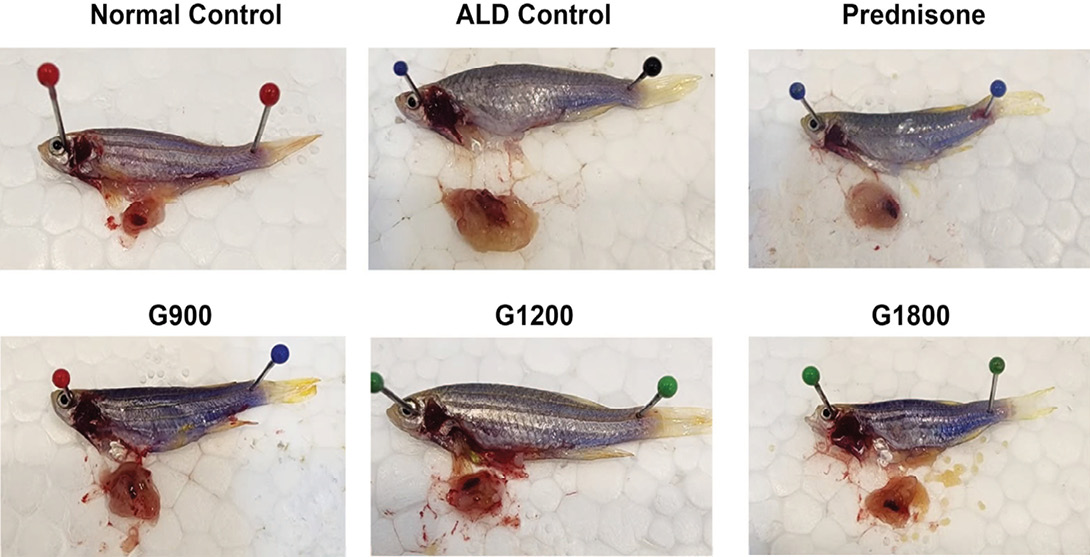
Representative gross appearance of zebrafish after seven days of treatment following chronic ethanol exposure. Normal Control: normal group without ethanol exposure or pharmacological treatment; ALD Control: ethanol-exposed group without pharmacological treatment; Prednisone: ethanol-exposed group treated with prednisone; G900, G1200, and G1800: ethanol-exposed groups treated with gabapentin at immersion concentrations of 150, 200, and 300 mg/L, respectively. No consistent or clearly distinguishable external macroscopic abnormalities were observed among the experimental groups.
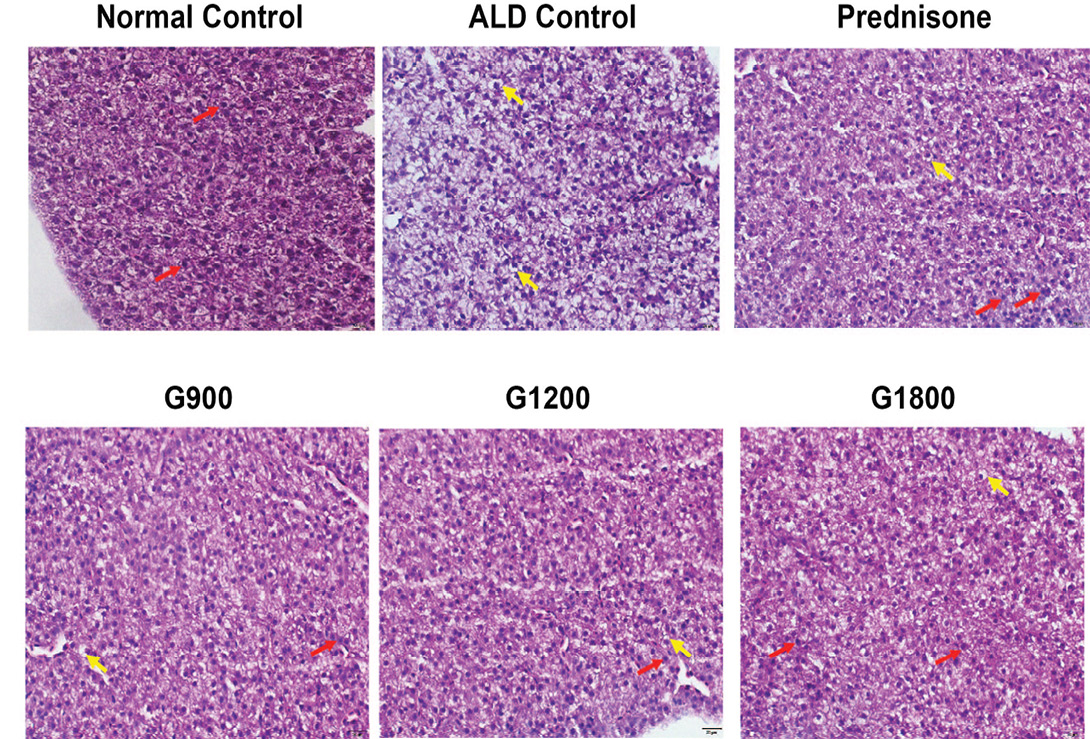
Representative liver histopathology after seven days of treatment following chronic ethanol exposure. Hematoxylin and eosin (H&E) staining, 400× magnification. Normal Control: normal group without ethanol exposure or pharmacological treatment; ALD Control: ethanol-exposed group without pharmacological treatment; Prednisone: ethanol-exposed group treated with prednisone; G900, G1200, and G1800: ethanol-exposed groups treated with gabapentin at immersion concentrations of 150, 200, and 300 mg/L, respectively. Yellow arrows indicate hepatocytes with steatotic changes, whereas red arrows indicate normal hepatocytes. Data are representative of four zebrafish per group.
Effects of seven-day gabapentin treatment on intestinal goblet cell proportion
Seven days after completion of ethanol exposure, the untreated ALD group showed a markedly increased intestinal goblet cell proportion (64.57 ± 3.14%) compared with the normal control group (5.49 ± 0.30%). Prednisone treatment significantly reduced the goblet cell proportion to 27.25 ± 5.49% (p < 0.001 vs. untreated ALD group). Gabapentin treatment was also associated with significantly lower goblet cell proportions, with values of 35.54 ± 1.99%, 32.55 ± 3.06%, and 29.94 ± 2.25% in the G900, G1200, and G1800 groups, respectively (p < 0.001 vs. untreated ALD group). A graded reduction in goblet cell proportion was observed across increasing gabapentin concentrations. The G900 group showed a significantly higher goblet cell proportion than the prednisone-treated group (p < 0.05), whereas no significant differences were observed between the G1200 and G1800 groups and the prednisone-treated group. Representative intestinal histopathology is shown in Figure 3 and Table 2.
Relationship between hepatic steatosis, intestinal goblet cell proportion, and gabapentin dose
At the day-35 endpoint, simple linear regression using individual fish data from all six experimental groups (n = 24) demonstrated a strong positive association between hepatic steatosis and intestinal goblet cell proportion (R²= 0.97, p < 0.001; Figure 4A). Using individual fish data from the untreated ALD control group and the three gabapentin-treated groups (n = 16), increasing gabapentin concentration was associated with a significant inverse linear trend in hepatic steatosis (R² = 0.81, p < 0.001; Figure 4B) and intestinal goblet cell proportion (R² = 0.84, p < 0.001; Figure 4C). These findings indicate that higher gabapentin immersion concentrations were associated with lower hepatic steatosis and intestinal goblet cell proportions at the seven-day treatment endpoint.
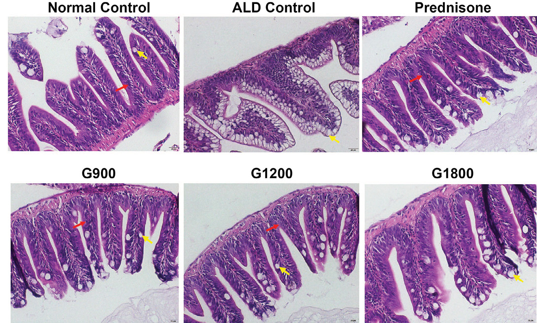
Representative intestinal histopathology after seven days of treatment following chronic ethanol exposure. Hematoxylin and eosin (H&E) staining, 400× magnification. Normal Control: normal group without ethanol exposure or pharmacological treatment; ALD Control: ethanol-exposed group without pharmacological treatment; Prednisone: ethanol-exposed group treated with prednisone; G900, G1200, and G1800: ethanol-exposed groups treated with gabapentin at immersion concentrations of 150, 200, and 300 mg/L, respectively. Yellow arrows indicate goblet cells, whereas red arrows indicate absorptive epithelial cells. Data are representative of four zebrafish per group.
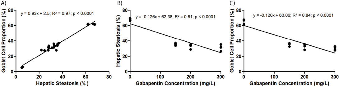
Linear relationships among gabapentin immersion concentration, hepatic steatosis, and intestinal goblet cell proportion at the day-35 endpoint. (A) Linear relationship between hepatic steatosis and intestinal goblet cell proportion. (B) Linear relationship between gabapentin immersion concentration and hepatic steatosis. (C) Linear relationship between gabapentin immersion concentration and intestinal goblet cell proportion. Gabapentin groups were designated G900, G1200, and G1800 based on human clinical dose categories of 900, 1,200, and 1,800 mg/day, corresponding to experimental immersion concentrations of 150, 200, and 300 mg/L, respectively. Higher gabapentin immersion concentrations were associated with lower hepatic steatosis and intestinal goblet cell proportions.
Discussion
The present study demonstrated that seven-day gabapentin treatment was associated with lower hepatic steatosis and intestinal goblet cell proportion following chronic ethanol exposure. Both outcomes showed a graded pattern across increasing gabapentin concentrations, while hepatic steatosis was positively associated with intestinal goblet cell proportion. These findings suggest that the short-term gabapentin treatment is associated with concurrent hepatic and intestinal histopathological changes in this experimental model.
Hepatic steatosis is an early and characteristic feature of alcohol-associated liver injury. Previous studies in adult zebrafish have shown that chronic ethanol exposure induces hepatic lipid accumulation and liver injury through disturbances in lipid metabolism, including increased lipogenesis and impaired fatty acid oxidation ; . Consistent with these findings, marked steatosis remained evident in untreated ALD zebrafish after the seven-day post-ethanol period, whereas substantially lower steatosis values were observed in all gabapentin-treated groups. This difference suggests that the histopathological state of the liver after seven days was more favorable in fish receiving gabapentin than in untreated ALD controls.
An additional finding was the graded reduction in hepatic steatosis across increasing gabapentin concentrations. Steatosis decreased from 35.63 ± 1.50% in G900 to 33.19 ± 2.98% in G1200 and 31.26 ± 3.66% in G1800, whereas the untreated ALD group showed substantially higher steatosis. The inverse association between gabapentin treatment level and hepatic steatosis further supports a dose-related pattern. However, this relationship should be interpreted cautiously. The present study assessed histopathological outcomes rather than direct molecular or biochemical markers of lipid metabolism, and therefore the observed decrease in steatosis cannot be attributed to a specific metabolic pathway. Although modulation of PPAR-related signaling and inflammatory pathways has been proposed as biologically relevant in alcohol-associated liver injury, these mechanisms were not directly measured in the present experiment. Thus, the current findings support a dose-related histopathological association rather than a definitive mechanism of action.
The intestinal findings were also consistent with previous reports showing that chronic alcohol exposure can disrupt intestinal epithelial homeostasis and alter mucus-associated responses ; . In the present study, the intestinal goblet cell proportion remained marked in untreated ALD zebrafish, whereas the prednisone- and gabapentin-treated groups showed substantially lower goblet cell proportion after seven days of treatment. This pattern suggests that the histopathological differences associated with short-term treatment were not confined to hepatic tissue but were also observed in the intestinal epithelium.
Similar to hepatic steatosis, intestinal goblet cell proportion showed a graded pattern across increasing gabapentin treatment levels. Values declined from 35.54 ± 1.99% in G900 to 32.55 ± 3.06% in G1200 and 29.94 ± 2.25% in G1800. The inverse association between gabapentin treatment level and goblet cell proportion further supports a dose-related intestinal response. Nevertheless, lower goblet cell proportion should not automatically be interpreted as direct evidence of restored intestinal barrier function. Intestinal permeability, mucus thickness, mucin expression, bacterial translocation, and microbiome composition were not evaluated in this study. Therefore, the present findings are best interpreted as histopathological evidence of differences in intestinal epithelial status rather than functional proof of barrier restoration.
The strong positive association between hepatic steatosis and intestinal goblet cell proportion represents an important finding of this study. Hepatic steatosis was closely associated with intestinal goblet cell proportion (R²= 0.97, p < 0.001), indicating that higher hepatic steatosis values tended to occur alongside higher intestinal goblet cell proportions. This parallel pattern is consistent with the concept that alcohol-associated injury involves interconnected hepatic and intestinal responses. The gut-liver axis provides a biologically plausible framework for this association because intestinal disturbances can influence hepatic inflammation through portal delivery of microbial products, while liver-derived inflammatory signals may also influence intestinal homeostasis ; . However, the present association is cross-sectional and does not establish the direction of interaction or prove a causal gut-to-liver mechanism. The observed relationship should therefore be interpreted as evidence of coordinated histopathological involvement rather than mechanistic confirmation of gut–liver signaling.
Gabapentin is primarily recognized for its neurological indications, including its use in alcohol-related disorders, but experimental evidence also suggests potential anti-inflammatory effects. In an experimental model of intestinal inflammation, gabapentin reduced histological injury and inflammatory responses through mechanisms involving PPAR-γ signaling . Such findings provide a possible biological context for the lower hepatic and intestinal histopathological abnormalities observed in the present study. Nevertheless, the current experiment did not measure PPAR signaling, inflammatory cytokines, oxidative stress markers, or calcium-dependent inflammatory pathways. Consequently, the present data cannot determine whether the observed histopathological differences resulted from anti-inflammatory activity, altered lipid metabolism, effects on epithelial homeostasis, or a combination of these mechanisms. Further mechanistic studies are required to establish how gabapentin influences hepatic and intestinal responses following chronic ethanol exposure.
Comparison with prednisone provides an additional experimental reference for interpreting the magnitude of the observed effects. At the seven-day endpoint, hepatic steatosis and goblet cell proportions in the highest gabapentin concentration approached the values observed in the prednisone-treated group. The similarity in histopathological values is noteworthy because corticosteroids remain an established treatment option for selected patients with severe alcohol-associated hepatitis . However, this study was not designed to establish therapeutic equivalence, non-inferiority, or clinical interchangeability between gabapentin and prednisone. The sample size was limited, the intervention period was short, and the study was conducted in zebrafish rather than patients with clinically defined alcoholic hepatitis. Therefore, the comparison with prednisone should be regarded as an experimental benchmark rather than evidence that gabapentin can replace corticosteroid therapy.
The seven-day treatment period also provides a distinct perspective on the early histopathological effects of gabapentin. Rather than evaluating long-term structural recovery, this study focused on whether measurable differences in hepatic and intestinal pathology could be detected after a relatively brief intervention. The persistence of marked steatosis and goblet cell proportion in untreated ALD fish at the same endpoint suggests that spontaneous improvement during this period was limited relative to that observed in the treated groups. At the same time, because only one post-treatment endpoint was analyzed, the exact onset and trajectory of these differences cannot be determined. The present findings therefore characterize the histopathological status after seven days of treatment but do not establish when treatment-related changes first emerged or whether further improvement would occur with longer administration.
This study has several limitations. First, histopathological assessment was restricted to a single endpoint following seven days of treatment and therefore does not establish the temporal trajectory of hepatic or intestinal responses. Second, the sample size was limited to four zebrafish per group, which may restrict the precision and generalizability of the observed effects. The survival rate of 87.5% in the normal control group resulted from the death of a single fish among eight initially allocated animals. Given the small group size, this single mortality event substantially influenced the calculated survival percentage. The cause of death was not determined. Third, hepatic and intestinal outcomes were evaluated primarily by H and E histopathology without molecular, biochemical, or functional confirmation. Measures of hepatic lipid metabolism, inflammatory mediators, oxidative stress, intestinal permeability, mucus production, and epithelial barrier function were not included. Fourth, although the correlation between hepatic steatosis and goblet cell proportion was strong, the cross-sectional nature of the analysis prevents causal interpretation. In addition, sex distribution was not systematically recorded across experimental groups and was not included as a factor in the analysis. Therefore, potential sex-related differences in the hepatic and intestinal responses could not be evaluated. Although histopathological assessments were performed independently by two blinded observers, inter-observer reliability was not formally quantified using a statistical reliability measure. Finally, the gabapentin immersion concentrations used in zebrafish cannot be directly extrapolated to clinically relevant human dosing. Further studies incorporating larger sample sizes, additional treatment durations, and molecular, biochemical, and functional assessments are required to clarify the mechanisms, dose-response characteristics, and durability of the observed effects.
Taken together, seven-day gabapentin treatment was associated with significantly lower hepatic steatosis and intestinal goblet cell proportion in zebrafish with alcoholic liver disease compared with untreated controls. Both histopathological outcomes showed a graded pattern across increasing gabapentin doses, with the highest dose producing values approaching those observed in the prednisone-treated group. These findings suggest that short-term gabapentin treatment may beneficially affect both hepatic and intestinal histopathological alterations in this experimental model. Further studies incorporating molecular, biochemical, and functional assessments are required to clarify the underlying mechanisms and determine whether these effects are sustained over longer treatment periods.
Conclusions
Seven-day gabapentin treatment was associated with significantly lower hepatic steatosis and intestinal goblet cell proportion in zebrafish following chronic ethanol exposure. Both histopathological outcomes showed a graded reduction across increasing gabapentin concentrations. The parallel changes in hepatic steatosis and intestinal goblet cell proportion suggest that short-term gabapentin treatment is associated with histopathological changes in both hepatic and intestinal tissues. However, these findings are limited to histopathological outcomes at a single post-treatment endpoint. Further studies incorporating molecular, biochemical, functional, and longer-term assessments are required to clarify the underlying mechanisms and durability of the observed effects.
Acknowledgement
The authors thank AT for assistance with animal handling and S for assistance with histopathological processing.
Novelty Statement
The novelty of this study lies in evaluating the dose-related effects of gabapentin on both hepatic and intestinal histopathological changes in an ethanol-induced zebrafish model of alcoholic liver disease. This integrated assessment provides preliminary evidence of concurrent hepatic and intestinal responses to short-term gabapentin treatment.
Author’s Contribution
HSZ: Data collection, analysis, interpretation, statistics, and manuscript drafting. AdA: Concept and design, data collection, analysis, funding acquisition, manuscript drafting, and critical revision. R: Data analysis, interpretation, and critical revision. MAE: Concept and design, administrative and technical support. KA: Critical revision. All authors have read and approved the final manuscript.
Data availability
The data supporting the findings of this study are available from the corresponding author upon reasonable request.
Funding
This study was funded by Direktorat Penelitian dan Pengabdian Kepada Masyarakat, Direktorat Jenderal Riset dan Pengembangan, and Kementerian Pendidikan Tinggi, Sains dan Teknologi (SK No. 0419/C3/DT.05.00/2025; Contract No. 059/C3/DT.05.00/PL/2025; No. 2421/B/UN3.LPPM/PT.01.03/2025).
Ethical clearance
All procedures were approved by Ethical Committee, Faculty of Medicine, Universitas Airlangga (No. 212/EC/KEPK/FKUA/2025, 4 August 2025).
Generative AI and AI assisted technology statement
The authors declare that no generative AI and AI assisted technology was used in the creation of this manuscript.
Conflict of interest
The authors have declared no conflict of interest.
References
- Aleström P, D’Angelo L, Midtlyng PJ, Schorderet DF, Schulte-Merker S, Sohm F (2020). Zebrafish: Housing and husbandry recommendations. Lab. Anim., 54(3): 213-224. https://doi.org/10.1177/0023677219869037 [CrossRef]
- Birchenough GM, Johansson ME, Gustafsson JK, Bergström JH, Hansson GC (2015). New developments in goblet cell mucus secretion and function. Mucosal Immunol., 8(4): 712-719. https://doi.org/10.1038/mi.2015.32 [CrossRef]
- Borges RS, Pereira ACM, Souza GCD, Carvalho JCT (2019). Histopathology of Zebrafish (Danio rerio) in nonclinical toxicological studies of new drugs. Zebrafish in biomedical research. Y. Bozkurt. London, IntechOpen.
- d’Arqom A, Rizky KA, Aqilah NM, Huda F, Lee MT, Tansy BA (2026). Immunomodulatory and anti-inflammatory effects of gabapentin: A systematic review and risk of bias analysis of preclinical studies. Immunol., 6(2): 30. https://doi.org/10.3390/immuno6020030 [CrossRef]
- Dai M, Guo S, Xu Y, Zhu X, Li C (2024). Conversion method for converting zebra fish into human dosage for efficacy evaluation. Chinese Patent CN113496071B. China National Intellectual Property Administration.
- Darniwa AV, Cahyanto T, Rahayu SN (2024). Effect of Light Intensity on Zebra Fish (Danio rerio) Cell Regeneration. J. Pijar Mipa, 19(2): 260-264. https://doi.org/10.29303/jpm.v19i2.4364 [CrossRef]
- de Brito TV, Júnior GJD, da Cruz Júnior JS, Silva RO, da Silva Monteiro CE, Franco AX (2020). Gabapentin attenuates intestinal inflammation: Role of PPAR-gamma receptor. Eur. J. Pharmacol., 873: 172974. https://doi.org/10.1016/j.ejphar.2020.172974 [CrossRef]
- Flores EM, Nguyen AT, Odem MA, Eisenhoffer GT, Krachler AM (2020). The zebrafish as a model for gastrointestinal tract-microbe interactions. Cell Microbiol., 22(3): e13152. https://doi.org/10.1111/cmi.13152 [CrossRef]
- Guan H, Zhang X, Kuang M, Yu J (2022). The gut-liver axis in immune remodeling of hepatic cirrhosis. Front. Immunol., 13: 946628. https://doi.org/10.3389/fimmu.2022.946628 [CrossRef]
- Hammer AM, Morris NL, Earley ZM, Choudhry MA (2015). The first line of defense: The effects of alcohol on post-burn intestinal barrier, immune cells, and microbiome. Alcohol Res., 37(2): 209-222. https://doi.org/10.35946/arcr.v37.2.06 [CrossRef]
- Hartmann P, Seebauer CT, Schnabl B (2015). Alcoholic liver disease: The gut microbiome and liver cross talk. Alcohol. Clin. Exp. Res., 39(5): 763-775. https://doi.org/10.1111/acer.12704 [CrossRef]
- Hyun J, Han J, Lee C, Yoon M, Jung Y (2021). Pathophysiological aspects of alcohol metabolism in the liver. Int. J. Mol. Sci., 22(11): 5717. https://doi.org/10.3390/ijms22115717 [CrossRef]
- Jeon S, Carr R (2020). Alcohol effects on hepatic lipid metabolism. J. Lipid Res., 61(4): 470-479. https://doi.org/10.1194/jlr.R119000547 [CrossRef]
- Jophlin LL, Singal AK, Bataller R, Wong RJ, Sauer BG, Terrault NA (2024). ACG clinical guideline: Alcohol-associated liver disease. Am. J. Gastroenterol., 119(1): 30-54. https://doi.org/10.14309/ajg.0000000000002572 [CrossRef]
- Levine AR, Carrasquillo L, Mueller J, Nounou MI, Naut ER, Ibrahim D (2019). High-dose gabapentin for the treatment of severe alcohol withdrawal syndrome: A retrospective cohort analysis. Pharmacotherapy, 39(9): 881-888. https://doi.org/10.1002/phar.2309 [CrossRef]
- Li H, Li N, Lu Q, Yang J, Zhao J, Zhu Q (2022). Chronic alcohol-induced dysbiosis of the gut microbiota and gut metabolites impairs sperm quality in mice. Front. Microbiol., 13: 1042923. https://doi.org/10.3389/fmicb.2022.1042923 [CrossRef]
- Lin JN, Chang LL, Lai CH, Lin KJ, Lin MF, Yang CH (2015). Development of an animal model for alcoholic liver disease in zebrafish. Zebrafish, 12(4): 271-280. https://doi.org/10.1089/zeb.2014.1054 [CrossRef]
- Park K-H, Kim S-H (2019). Low dose of chronic ethanol exposure in adult zebrafish induces hepatic steatosis and injury. Biomed. Pharmacother., 117: 109179. https://doi.org/10.1016/j.biopha.2019.109179 [CrossRef]
- Ramkissoon R, Shah VH (2022). Alcohol use disorder and alcohol-associated liver disease. Alcohol Res., 42(1): 13. https://doi.org/10.35946/arcr.v42.1.13 [CrossRef]
- Schneider AC, Gregório C, Uribe-Cruz C, Guizzo R, Malysz T, Faccioni-Heuser MC (2017). Chronic exposure to ethanol causes steatosis and inflammation in zebrafish liver. World J. Hepatol., 9(8): 418-426. https://doi.org/10.4254/wjh.v9.i8.418 [CrossRef]
- Tadokoro T, Morishita A, Himoto T, Masaki T (2023). Nutritional support for alcoholic liver disease. Nutrients, 15(6): 1360. https://doi.org/10.3390/nu15061360 [CrossRef]
- Teame T, Zhang Z, Ran C, Zhang H, Yang Y, Ding Q (2019). The use of zebrafish (Danio rerio) as biomedical models. Anim. Front., 9(3): 68-77. https://doi.org/10.1093/af/vfz020 [CrossRef]
- Thakur PC, Stuckenholz C, Rivera MR, Davison JM, Yao JK, Amsterdam A (2011). Lack of de novo phosphatidylinositol synthesis leads to endoplasmic reticulum stress and hepatic steatosis in cdipt-deficient zebrafish. Hepatology, 54(2): 452-462. https://doi.org/10.1002/hep.24349 [CrossRef]
- Ventura-Cots V, Rachakonda V, Bataller R (2019). Alcoholic liver disease, management of in encyclopedia of gastroenterology (Second Edition): 56-63. Academic Press. https://doi.org/10.1016/B978-0-12-801238-3.65788-4 [CrossRef]
- World Health Organization (WHO) (2024). Alcohol. WHO Fact Sheet, 28 June 2024. Available at: https://www.who.int/news-room/fact-sheets/detail/alcohol (accessed 8 September 2026).
- You M, Arteel GE (2019). Effect of ethanol on lipid metabolism. J. Hepatol., 70(2): 237-248. https://doi.org/10.1016/j.jhep.2018.10.037 [CrossRef]
- Zanandrea R, Bonan CD, Campos MM (2020). Zebrafish as a model for inflammation and drug discovery. Drug Discov. Today, 25(12): 2201-2211. https://doi.org/10.1016/j.drudis.2020.09.036 [CrossRef]
- Zheng J, Li Z, Xu H (2023). Intestinal microbiotas and alcoholic hepatitis: Pathogenesis and therapeutic value. Int. J. Mol. Sci., 24(19): 14809. https://doi.org/10.3390/ijms241914809 [CrossRef]
- Zulfatim HS, Afrina V, d’Arqom A, Sutantyo QE, Amornsupak K, Nualkaew T (2025). Anti-inflammatory drugs for alcoholic liver disease: A systematic review on survival and adverse events. Int. J. Hepatol., 2025: 8535952. https://doi.org/10.1155/ijh/8535952 [CrossRef]

Services
Article usage statistics
Loading…
Related articles
- Protective Response of Zinc Oxide Nanoparticle with Chitosan Oligosaccharide on Intestinal Integrity, Goblet Cell Count and Meat Quality of Broiler Chicken under Heat StressJournal of Animal Health and Production · 2024
- Biochemical Evaluation of Bone Marrow Mesenchymal Stem Cells and GLP-1 Therapy as a New Approach for Type II Diabetes Treatment in Rat ModelAdvances in Animal and Veterinary Sciences · 2024
- Protective Effects of Intestinal Trefoil Factor against Endotoxin-Induced Injury of Intestinal Mucosal Epithelial Cells in PigletsPakistan Journal of Zoology · 2017
- Effects of Calcitonin on Porcine Intestinal Epithelial Cells Proliferation, Phosphorus Absorption, and NaPi-IIb ExpressionPakistan Journal of Zoology · 2019
- Anti-angiogenic Effect of Ginsenoside Rh2 by Downregulation of VEGF in a Zebrafish ModelPakistan Journal of Zoology · 2020
- Histochemical Characterization of Convict Cichlid (Amatitlania nigrofasciata) Intestinal Goblet CellsVeterinary Sciences: Research and Reviews · 2017