
Avian Mycoplasmosis, primarily caused by Mycoplasma gallisepticum (M. gallisepticum) and Mycoplasma synoviae (MS), is a major threat to the poultry industry in Pakistan. This review synthesizes current epidemiological, pathogenic, diagnostic, and economic insights on avian Mycoplasmosis due to Mycoplasma gallisepricum (M. gallisepticum). The disease leads to chronic respiratory disease (CRD), infectious synovitis, and systemic infections, causing significant economic losses through decreased egg production, reduced growth rates, poor feed conversion, and increased mortality. Pakistan ranks among the top poultry-producing countries globally, with over 1.5 million workers engaged in the sector. However, rising seroprevalence rates as high as 76.6% in young broiler breeders in Lahore and 92% in field samples nationwide indicate increasing mycoplasmosis due to M. gallisepticum. Spatial analysis reveals hotspots in Punjab and Khyber Pakhtunkhwa, while temporal trends highlight higher prevalence during winter, particularly in older and female birds. Risk factors such as inadequate biosecurity, vertical transmission, and antibiotic overuse further complicate control efforts. Pathogenesis involves host immune evasion, cytokine dysregulation, and colonization of the respiratory tract, leading to systemic spread. Current diagnostic methods include SPA, ELISA, PCR, and culture, with PCR considered the most confirmatory. Control strategies emphasize biosecurity, monitoring, and vaccination, though eradication remains elusive due to poor farm-level practices. This review underscores the urgent need for spatial-temporal mapping, national surveillance, and integrated management strategies to mitigate the impact of avian mycoplasmosis in Pakistan and secure the sustainability of its poultry sector.
Research Article · Volume 39, Issue 4, Pages 1–13, 26 October 2026 · Open Access
Emerging Trends of Mycoplasma gallisepticum (M. gallisepticum) in Pakistan’s Poultry Industry
· · · · ·
Affiliations & Notes ⌄
- 1Animal Health Program, Animal Sciences Institute, National Agricultural Research Centre Islamabad, Pakistan
- 2Faculty of Veterinary & Animal Sciences, PMAS Arid Agriculture University Rawalpindi, Pakistan
- 3University Laboratory of Food-borne Parasitic Zoonoses, Department of Translational Physiology, Infectiology, and Public Health, Faculty of Veterinary Medicine, Ghent University, 133 Salisburylaan, B-9820 Merelbeke, Belgium
- *Correspondence | Bilal Javid; Email: bilaljavid824@gmail.com
Article Info ⌄
- Journal:
- Pakistan Journal of Agricultural Research
- Submitted Date:
- 21 July 2026
- Accepted Date:
- 11 September 2026
- Published Date:
- 26 October 2026
From the article
- Mycoplasma gallisepticum (M. gallisepticum) is the principal cause of avian Mycoplasmosis, which continues to pose a serious danger to Pakistan’s quickly expanding poultry sector.
- The data presented in this study underscore a disturbingly high and variable seroprevalence of M. gallisepticum across Pakistan, with reports ranging from 5.5% to as high as 76.6%, depending on bird type, season, and region.
- Spatial analysis points to disease hotspots in Rawalpindi, Faisalabad, and Khyber Pakhtunkhwa, while temporal trends suggest a continuous rise in M. gallisepticum prevalence which is driven by increased poultry density, poor vaccination practices, and potential vertical transmission from breeders to offspring.
- A unified, evidence-based approach involving veterinarians, researchers, policymakers, and poultry producers is essential to mitigate the threat posed by Mycoplasma gallisepticum and secure the future of Pakistan’s poultry sector.
Selected from the article's own text.
Abstract
Introduction
Pakistan poultry industry is at great risk due to occurrence of infectious and zoonotic diseases because it cause huge economic loss . Pakistan has a high incidence of avian mycoplasmosis, with recent reports of seroprevalence rates as high as 43.33% . The Punjab, Pakistan being the largest province in terms of both the number of people and poultry, commercial poultry flourished due to the agricultural foundation and access to inexpensive labor. This province raised 709.5 million chickens in fiscal year 2010–11, comprising 631.97 million broilers, 27.75 million layers, 12.55 million breeders, and 37.23 million backyard chickens . In Pakistan, the domestic and commercial poultry populations were estimated at 95.50 million and 1,968.71 million birds, respectively. The poultry industry provides employment to more than 1.5 million people and represents a vibrant component of the livestock sector. With an investment of more than Rs. 1,056 billion, the industry has experienced an average annual growth rate of 7.3% over the past decade. This expansion has made Pakistan the eleventh-largest poultry producer in the world, with considerable potential for further growth and development. Furthermore, the poultry sector contributes approximately 40.7% of the country’s gross meat production (Ministry of . Mycoplgallisepticum was initially identified in turkeys because infectious sinusitis caused prominent clinical signs that facilitated early recognition. Later studies confirmed that the pathogen infects both turkeys and chickens, with variations in disease expression reflecting differences in host susceptibility, clinical manifestations, and production systems . mycoplasmosis are tiny, self-replicating bacteria with minimal genetic material and have low G+C content about 23-40 % . There are four main, extremely prevalent, infectious species that cause mycoplasmosis: Mycoplasma gallisepticum (M. gallisepticum), Mycoplasma synoviae (MS), Mycoplasma meleagridis (MM), and Mycoplasma iowae (MI). Avian mycoplasmosis is economically important and highly contagious disease of poultry globally. It is mostly caused by Mycoplasma gallisepticum (M. gallisepticum) and Mycoplasma synoviae (MS), which cause infectious synovitis and chronic respiratory disease (CRD) in chickens, respectively . M. gallisepticum bacteria mainly infect upper respiratory tract of birds, especially poultry, due to its aerobic nature . Gram-negative Mycoplasma pathogens are members of the Mollicutes order-I class. There are more than 100 species of Mycoplasmatales, Mycoplasmataceae, and Mycoplasma in various birds. Different Mycoplasma species have genomic sizes ranging from 600 -1350 kbp. The growth of the Mycoplasma pathogen requires a variety of particular nutrients. mycoplasmosis are small, pleomorphic bacteria that are surrounded by a triple-layered plasma membrane and lack a cell wall Nasal discharge, coughing, rales, decreased egg production, lowered feed conversion efficiency, and high mortalities are the symptoms of mycoplasmosis (Ferguson et al., 2020). This causes severe economic burdens on commercial poultry farming, particularly in countries such as Pakistan where poultry sector is an important component of food security and the agricultural economic stability . Poor biosecurity, inadequate vaccination protocols, and the existence of live bird markets that serve as reservoirs and vectors have all contributed to the disease’s recent decades-long increase . Pakistan faces significant obstacles in its fight against avian mycoplasmosis, including the absence of thorough epidemiological data that accounts for both temporal and spatial factors. Even while many laboratory studies and regional surveys have established the occurrence of Mycoplasma spp. in isolated areas, there are still relatively few comprehensive national-level assessments that map the spread of diseases and their variations over time . These types of spatial and temporal analysis are essential for identifying high-risk areas, understanding seasonal trends, tracking outbreak dynamics, and developing targeted treatments. Because of differences in climate, poultry population, and management techniques, for instance, studies have shown that the prevalence of M. gallisepticum can vary greatly across Pakistan’s northern and southern regions . Geographic patterns are important because they can reveal high illness hotspots, regional clustering, and transmission channels.
Due to their highly concentrated commercial chicken farms, some Pakistani provinces, like Khyber Pakhtunkhwa and Punjab, have an excessive population . The use of geographic mapping of instances can help prioritize monitoring operations and allocate veterinary resources to disease-burdened areas. Furthermore, geographical research helps clarify the importance of factors such farm size, the proximity of live bird markets, and the existence of backyard poultry by finding associations between agricultural practices and disease frequency . A number of studies have shown that the prevalence of mycoplasmosis in Pakistan has been rising, particularly since the 1990s (Shaoib, 2019) and by looking at short-term patterns like seasonal peaks or multi-year epidemic cycles, farmers and veterinarians can better prepare for periods of higher risk . The study of avian mycoplasmosis in Pakistan is significantly influenced by the economic factor. Recent estimates indicate that the disease costs Pakistan’s chicken industry millions of dollars per year since it causes greater veterinary expenses, carcass condemnation, mortality, and poor production efficiency . In broiler production, infection results in decreased growth rates, elevated feed conversion ratios, and declining meat quality . As a result of reduced egg production, poor eggshell quality, and unnecessary culling, the disease in layers has a direct influence on profitability . These economic ramifications have broader effects on rural lives and national food security in addition to endangering the sustainability of commercial chicken businesses. The poultry business is one of the busiest segments of Pakistan’s animal industry. Approximately 1.5 million people in Pakistan rely on the chicken industry for their livelihoods . More than 30% of Pakistan’s total meat production comes from the poultry sector (Qadir and Irum. 2023). The emergence of zoonotic and infectious illnesses puts Pakistan’s poultry sector at serious risk since they result in significant financial losses . Punjab Province is the biggest province in terms of poultry production and commercial poultry sector flourished due to inexpensive labor. The Punjab province raised 709.5 million chickens in fiscal year 2010–11, comprising 631.97 million broilers, 27.75 million layers, 12.55 million breeders, and 37.23 million backyard chickens . Avian mycoplasmosis was initially discovered in Turkey in 1926 and then in 1936 in chicken . Pakistan has a high prevalence of avian mycoplasmosis, with seroprevalence rates as high as 43.33% reported recently . There are four main, extremely prevalent, infectious species that cause mycoplasmosis: M. gallisepticum (M. gallisepticum), M. synoviae (MS), M. meleagridis (MM), and M. iowae (MI). Turkeys are susceptible to mycoplasmosis diseases caused by MM and MI, while chickens are primarily affected by M. gallisepticum and MS. Gram-negative Mycoplasma pathogens are members of the Mollicutes order-I class. There are more than 100 species of Mycoplasmatales, Mycoplasmataceae, and Mycoplasma in various birds. Different Mycoplasma species have genomic sizes ranging from 600 -1350 kbp. The growth of the Mycoplasma pathogen requires a variety of particular nutrients. Mycoplasmas are small, pleomorphic bacteria that are surrounded by a triple-layered plasma membrane and lack a cell wall . In Northern areas of Pakistan, the seroprevalence and role of M. gallisepticum (M. gallisepticum) in chronic respiratory disease (CRD) cases in poultry is not well documented. A thorough and efficient surveillance program will help in light of the high seropositivity and M. gallisepticum isolation from CRD suspected birds examined in Pakistan’s northern region .
Sign and symptoms
Coughing, sneezing, and dyspnea are among the respiratory symptoms of M. gallisepticum-induced CRD . The birds exhibit respiratory distress, including coughing, wheezing, sneezing, ocular discharge, enlarged eyelids, frothy exudate from the eye, trouble breathing, and gargling sounds during breathing . MS causes synovitis and airsacculitis in birds, while M. gallisepticum causes infectious sinusitis in turkeys and chronic respiratory disease in chickens. Both horizontal and vertical transmission are possible for the infection. The three primary components of avian Mycoplasmosis prevention and control strategies are immunization, treatment, and biosecurity. Commercially available vaccinations against M. gallisepticum and MS infections for birds include live attenuated, recombinant live poxvirus, and inactivated bacterins . Certain eggs from infected breeders can spread M. gallisepticum to offspring both horizontally and vertically through contaminated water, feed, and environmental conditions. It can also be passed on to others by farmers . The upper respiratory tract becomes colonized during M. gallisepticum-caused respiratory tract infections, which can then result in air sacculitis and tracheal irritation. Prior to research on conjunctivitis, arthritis, salpingitis, and meningo-encephalopathy in chickens and turkeys revealed that the organism is not limited to the respiratory system alone. Pathogenic M. gallisepticum R strain experimental infection demonstrated systemic spread to the heart, brain, liver, spleen, and kidneys, revealing M. gallisepticum’s capacity to penetrate the respiratory tract’s mucosal barrier and spread to internal body organs . The control of avian mycoplasma infections is fairly easy and straightforward, especially since the pathogenic avian mycoplasmas are egg transmitted and lack a cell wall, making them vulnerable to environmental influences. The first step in effective control is placing mycoplasma-free replacement stock on a single-age farm with all-in, all-out type management, good biosecurity, and a regular monitoring program. Multi-age commercial egg producers are primarily positive for M. gallisepticum and MS, and both infections are common in commercial broiler production in some regions of the world. M. gallisepticum vaccines are increasingly used in situations where control is impractical, such as large populations of poultry in small geographic areas and multiple-age farms that never depopulate . To prevent Mycoplasma infections, the National Poultry Improvement Plan (NPIP) suggested three fundamental control methods: strict security measures, serological surveillance, and prompt removal of diseased birds . For the purpose of planning an antibiotic therapy to limit the disease, serological identification is helpful in the early diagnosis of CRD caused by M. gallisepticum. The history and symptoms of the CRD, a positive serological test leads to the supposition that first diagnosis. The organism should thereafter be isolated and identified . Several serological methods, including the serum plate agglutination (SPA) test, the haemagglutination inhibition (HI) test, and the enzyme linked immunosorbant assay (ELISA), are used to identify M. gallisepticum in its early stages. Conventional diagnostic procedures can be effectively replaced by molecular techniques. Certain primers target particular M. gallisepticum genes . Efficient immunoprophylaxis using both killed and live M. gallisepticum vaccines is the only way to control the disease, as chronic forms are not cost-efficient or effective to treat . Since MS infections are frequently associated with other infections (mixed infections), PCR is regarded as a highly accurate diagnostic technique for identifying that particular pathogen. Because serological testing can produce false-positive findings, PCR is considered a confirmatory method to ensure satisfactory results and the true state of the illness in birds .
Pathogenecity of mycoplasmosis
The flask-shaped terminal organelle of Mycoplasma gallisepticum contains major cytadhesion-associated proteins, including GapA and CrmA, which are essential for attachment to host respiratory epithelial cells and contribute to virulence . Recent studies have further highlighted that surface lipoproteins, membrane-associated proteins, and antigenic variation contribute to host adaptation and immune evasion during M. gallisepticum infection . Initially, M. gallisepticum adhesion and attachment to the host cell surface are facilitated by the presence of cyto-adherence molecules. M. gallisepticum (M. gallisepticum) begins infection by first attaching to host cells, which stimulates an inflammatory reaction that leads to increased mucus production at epithelial surfaces. To avoid detection and elimination by the immune system, M. galliseptium penetrates into cells that are normally non-phagocytic, allowing it to spread more effectively throughout the host. The infection also prompts the release of various cytokines, molecules that play a key role in disease development by activating white blood cells and driving the inflammatory process . M. gallisepticum moves to the lower respiratory tract after colonizing the upper respiratory tract, where it causes pneumonia, bronchitis, air-sacculitis and other respiratory infections . The respiratory tract is the main site of infection for M. gallisepticum which first colonizes the airway and bronchial epithelial cells before securely adhering to their surfaces. Once established, this bacterium interferes with DNA and protein synthesis, disrupting vital cellular functions, hence lowering mucus output and compromising normal cell function. M. gallisepticum penetrates deeper tissues as the infection worsens, resulting in a systemic infection that spreads widely. This leads to major output losses, especially in poultry, in addition to serious tissue damage and immunosuppression. By combining these pathogenic pathways, M. gallisepticum is able to evade host defenses and create persistent infections . The pathogenesis of M. gallisepticum often starts when the bacteria penetrate the respiratory system of the host, adhere to, and invade healthy cells that line the airways. Immunological response When M. gallisepticum colonizes, it interferes with cellular processes, causing structural harm and weakens mucosal defenses. As it suppresses host defenses and avoids immune detection, the infection subsequently spreads throughout the body. Chronic inflammation brought on by this ongoing invasion weakens the host and exacerbates tissue damage. Immunosuppression, cellular damage, and metabolic disturbance all work together to cause serious health decline and decreased output over time, especially in poultry . The cumulative effect of M. gallisepticum infection highlights the disease’s status as a serious pathogen in avian populations, resulting in both financial losses and concerns for animal welfare. The innate immune response provides the first line of defense against M. gallisepticum infection. Although innate immunity initially limits pathogen invasion, M. gallisepticum employs immune evasion strategies that frequently allow persistent infection despite these early host defenses. As a result, the pathogen gradually establishes itself within the respiratory tract, facilitating chronic infection. Adaptive immune responses involving both humoral and cellular immunity subsequently become essential for controlling disease progression (Guanson et al., 2000). Chickens show a strong humoral immune response after infection by direct contact or aerosol transmission, as evidenced by a sharp increase in antibody titers directed against pathogenic M. gallisepticum proteins. Although M. gallisepticum’s immune evasion tactics frequently permit chronic infection in spite of this defense mechanism, the host’s strong antibody production represents their attempt to fight the infection. Systemic IgM antibodies were found during the first week of infection, before any appreciable cellular infiltration was noticed. Around three weeks after exposure, the infection worsened and the tracheal mucosa showed a noticeable increase in B-lymphocytes. The development of localized adaptive immunity in impacted respiratory tissues follows the host’s initial fast humoral defense through IgM synthesis, as seen by this sequential immunological response. The delayed tracheal B-cell response indicates that M. gallisepticum gradually establishes itself on mucosal surfaces as the immune system gradually concentrates its defenses at the main infection site. The development of localized mucosal immunity was indicated by the tracheal mucosa exhibiting significantly elevated IgA antibody levels around two weeks after M. gallisepticum infection by 95% . A progressive immune activation, wherein specialized mucosal defenses subsequently enhance the initial systemic IgM synthesis, is indicated by this delayed IgA response. By targeting the pathogen at its primary site of colonization, the host’s adaptive immune system may be able to inhibit bacterial adherence and transmission through the respiratory epithelium, as evidenced by the development of secretory IgA at this stage. Following the first systemic reaction but before the complete formation of a chronic infection, this mucosal antibody response is a crucial stage in the host–pathogen interaction ; . The significant accumulation of B lymphocytes observed in the tracheal mucosa following M. gallisepticum infection underscores their vital role in the host’s immune defense. The adaptive immune system’s targeted response at the main site of infection is demonstrated by this marked B-cell infiltration, where these cells most likely aid in local antibody synthesis and antigen presentation. By producing pathogen-specific immunoglobulins that may restrict bacterial adhesion and spread across respiratory epithelial surfaces, B cells—rather than other lymphocyte subsets—suggest a particularly significant humoral defense mechanism against M. gallisepticum in avian hosts . Both T and B cells are essential for establishing a strong immune response to M. gallisepticum (M. gallisepticum) infection. Experiments reveal that compared to immunocompetent controls, surgically thymectomized and bursectomized birds—which lack functional T and B cell populations, respectively—developed much more severe M. gallisepticum infections. The significantly poorer illness outcomes in these birds with reduced lymphocytes demonstrate how humoral (B cell-mediated) and cellular (T cell-mediated) immunity cooperate to regulate M. gallisepticum development. Adaptive immune responses including both lymphocyte populations are crucial for limiting disease development and severity in avian hosts, even while innate immunity pathways offer initial protection (Guanson et al., 2000). As early as four days after infection, CD4+ helper T cells and CD8+ cytotoxic T cells can be seen in the tracheal lamina propria, indicating the quick development of the immune response to M. gallisepticum infection. The onset of adaptive immunological responses at the primary site of infection is suggested by this early T-cell infiltration ; Balanchard and Browning, 2005). At the same time, a variety of cytokines and chemokines orchestrate the recruitment and activation of distinct immune cell populations, making significant contributions to immunological dynamics. Both the initial inflammatory response and the subsequent development of specific immunity against M. gallisepticum are influenced by these signaling molecules. The course and result of M. gallisepticum infection in avian hosts are determined by the complex immunological environment created by the coordinated action of soluble factors (cytokines/chemokines) and cellular immunity (mediated by T-cell subsets) ; Balanchard and Browning, 2005). B and T lymphocytes are non-specifically activated by pro-inflammatory cytokines and chemokines during M. gallisepticum infection. M. gallisepticum lipoproteins trigger TLR-2 signaling, disrupting expression of immune-related genes (CCL-20, IL-8, IL-12, IFN-γ, IL-1β, MIP-1β, RANTES, CXCL-13, CXCL-14) via both TLR-dependent and NF-κB-mediated pathways. The increased inflammatory response that characterizes M. gallisepticum pathogenesis is a result of this dysregulated immune gene expression . M. gallisepticum causes a lymphoproliferative response in the respiratory tract that is mediated by cytokines and chemokines. Through phagocytic activity, macrophages play a major role in the pathogen’s efficient stimulation of both innate and adaptive immune pathways. Monitoring B and T cell populations during vaccine development and evaluation is crucial because, although they are necessary for immunological defense against M. gallisepticum, their responses may also affect the course of the disease . Figure 1 shows the overall general flow of mycoplasmosis pathogenesis in chicken.
Prevalence of mycoplasmosis
The occurrence of Mycoplasma gallisepticum (M. gallisepticum) in poultry varies significantly depending on location, type of bird, diagnostic methods used, and the year of study, as indicated by surveillance data from across Pakistan. For instance, in Faisalabad M. gallisepticum in 20.65% and 15.22% of samples using SPAT and PCR methods, respectively, among layer chickens . Meanwhile, in Quetta, researchers reported 10 positive PCR samples out of 22 healthy chickens. This finding suggests that asymptomatic carrier birds may act as reservoirs of infection, facilitating the silent spread of the pathogen within and between flocks and complicating disease surveillance and control efforts. . Researchers noted a notably high seroprevalence of M. gallisepticum in Lahore, with rates of 76.60% in young broilers and 33.17% in adults, detected through indirect ELISA as shown in Table 1. In Quetta, Pishin, and Kuchlak M. gallisepticum prevalence was obderved in broilers at 7.86% using SPAT and

The pathway of Mycoplasma gallisepticum from respiratory colonization and immune evasion to inflammation, systemic dissemination, and tissue destruction is shown in above figure.
11.19 via ELISA, while in layers, the figures were 18.6% and 15.33%, respectively . Seasonal incidence rates of 5.68% for broilers and 5.52% for layers based on clinical signs and other testing methods in the Khushab district . In Rawalpindi, [] the researchers provided a detailed breakdown showing M. gallisepticum prevalence at 7.14% for broilers, 44.9% for layers, and 59.6% for broiler breeders . In Khyber Pakhtunkhwa, researchers reported an overall prevalence of 35.03% using SPAT, with marked district-specific variations: D.I. Khan (43.33%), Peshawar (36.67%), Abbottabad (31.46%), Mansehra (20.86%), and Tank (12.67%) . The considerable variation in prevalence among districts may be attributed to differences in farm biosecurity measures, climatic conditions, poultry management practices, flock density, and movement of birds, all of which can influence the transmission and persistence of M. gallisepticum. These findings emphasize the importance of implementing region-specific surveillance and disease control strategies. The considerable variation in prevalence among districts may be attributed to differences in farm biosecurity measures, climatic conditions, poultry management practices, flock density, and movement of birds, all of which can influence the transmission and persistence of M. gallisepticum. These findings emphasize the importance of implementing region-specific surveillance and disease control strategies. The research conducted in three districts of Balochistan, provided the finding of M. gallisepticum prevalence rates of 10.47% and 19.76% in broilers and 19.44% and 31.66% in layers when using SPAT and ELISA . Seroprevalence among broiler breeder farms in Punjab via ELISA was identified as 53.40% . In the Rawalpindi division 43.32% positivity rate for M. gallisepticum based on PCR results from commercial chickens . Research conducted in Northern Pakistan (specifically Haripur, Abbottabad, and Mansehra) reported a prevalence of 46.56% in poultry and 27.2% in pheasants by ELISA. Culture positivity was 17.1%, whereas PCR detected markedly higher positivity rates of 82.4% in Haripur, 82.3% in Abbottabad, and 60.5% in Mansehra . The substantially higher detection rate by PCR compared with culture suggests the greater sensitivity of molecular techniques for identifying M. gallisepticum. However, PCR may also detect DNA from non-viable organisms, whereas culture confirms only viable bacteria. Therefore, the observed discrepancy between the two methods should be considered when interpreting prevalence estimates and assessing active infection as illustrated in Figure 2. Conclusively, samples from Abbottabad and Rawalpindi, reporting M. gallisepticum seroprevalence rates of 75% in layers, 70% in breeders, and 50% in broilers. Culture positivity rates were 26.5% for breeders, 21% for layers, and 9% for broilers, with PCR indicating an overall M. gallisepticum positivity of 37.1% .
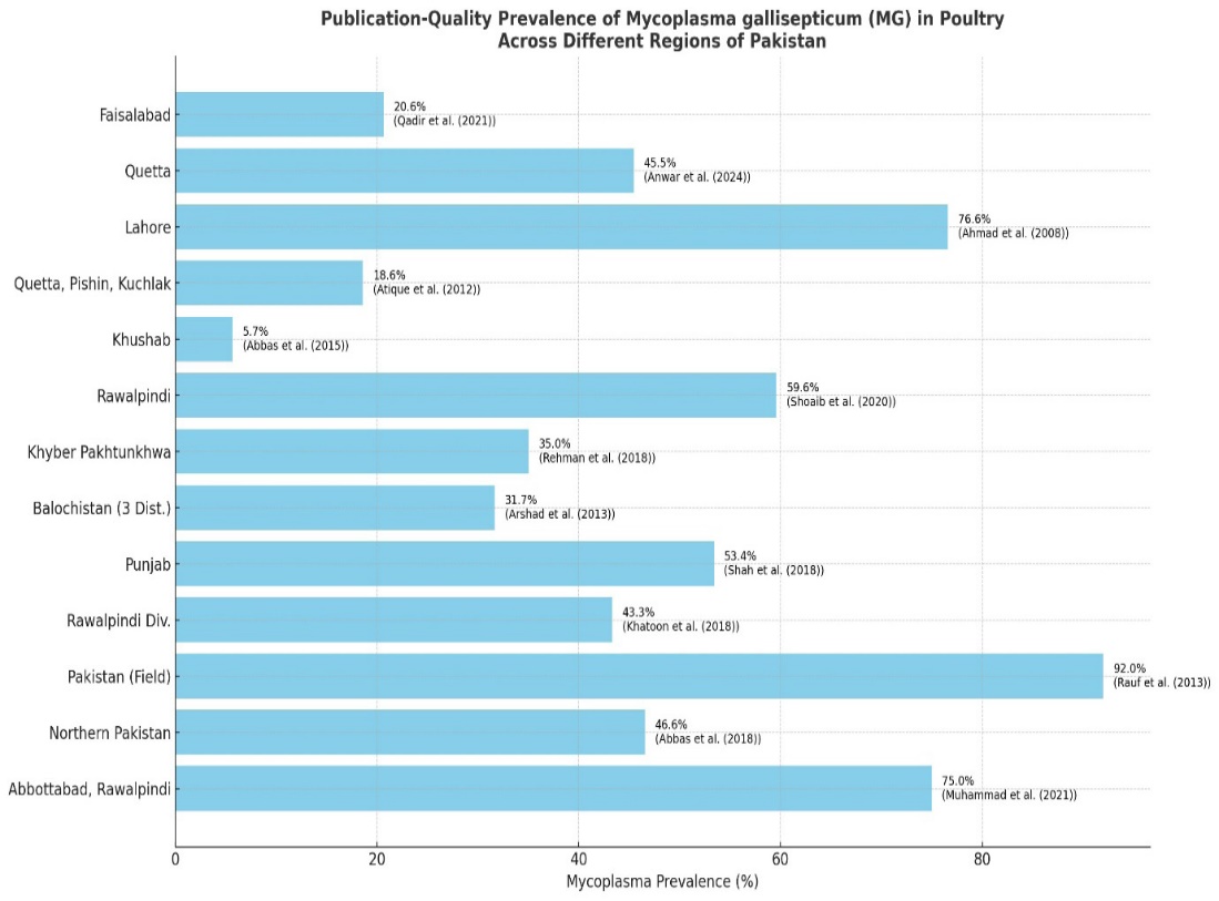
Shows the prevalence of Mycoplasmosis in poultry in different regions of Pakistan.
Table 1: Provides a detailed overview of Mycoplasmosis prevalence in poultry across various regions of Pakistan.
| City | Poultry type | No. of samples | Diagnostic test | M. gallisepticum Prevalence | Reference |
| Faisalabad | Layer chicken | 92 | SPAT and PCR | 20.65 and 15.22% positive samples respectively by SPAT and PCR | |
| Quetta | Healthy chickens | 22 | PCR | 10 positive and 12 negative | |
| Lahore distric | Broiler breeding stock | 2777 serum sample | Indirect ELISA | 76.60%I in young and 33.17% in adult birds | |
| Quetta, Pishin and Kuchlak districts of Balochistan, Pakistan | Broiler and layer flocks | 600 | SPAT,ELISA | 7.86 and 11.19% in broilers while 18.6 and 15.33% in layers by SPAT and ELISA respectively. | |
| Khushab District, Pakistan | Broilers and commercial layers | (360 poultry forms) | Biochemical test, culturing on McConkey agar, clinical sign and post _mortem | Seasonal incidence 5.68% in broilers on quarterly basis and 5.52% in layers | |
| Rawalpindi, Pakistan | Breeder, broiler and layer birds | 1647 | SPAT | M. gallisepticum, MS and SPG in case of broilers was 7.14, 10 and 5.35%, in the layer birds was 44.9, 42.6 and 51.32% and in broiler breeders was 59.6, 50.13 and 44.1% respectively. | (Shaoib et al., 2020) |
| Dera Ismail khan, Peshawar, Abbottabad, Tank, Mansehra | Broilers and backyard poultry | 648 | SPAT | 35.03% overall Dera Ismail khan 43.33%,Peshawar 36.67%,abbottabad 31.46%,tank 12.67% and Mansehra 20.86% | |
| Three district of Baluchistan | Broiler and layer flocks | 600 | SPAT and ELISA | In broilers SPAT and ELISA are 10.47 and 19.76 while in layers 19.44 and 31.66 by SPAT and ELISA respectively. | |
| Punjab | Broiler breeder farms | 103 | ELISA | 53.40% | |
| Rawalpindi division | Commercial chickens | 124 | PCR | 43.32% | |
| Lahore (Pakistan Field samples) | White Leghorn commercial layers | Trachea (n=154), Lungs (n=113), Air sacs (n=113) | Culture Test, Growth Inhibition Test (GIT), PCR | 21.84% (Overall confirmed M. gallisepticum by culture + GIT) 27.6% (M. gallisepticum suspected from culture) 80.51% (Trachea), 72.2% (Lungs), 74.19% (Air sacs) | |
| Northern Pakistan (Haripur, Abbottabad, Mansehra) | Multiple Broilers breeders, layers+ pheasantry | 2341 poultry and 24 pheasantry birds 1609 swab culture and 2341 sera for ELISA | PCR, Culture, ELISA | 46.56% and 27.2% among poultry and pheasantry birds, Culture positively;17.1% PCR isolates from Haripur 82.4%, Abbottabad 82.3% and Mansehra 60.5%. | |
| Abbottabad and Rawalpindi | Broilers, Layers, Breeders | 600 serum and 600 swab samples | ELISA, PCR, Culture | Seroprevalence Layers 75%,breeders 70%,broilers 50% Culture positivity 26.5%, 21%, 9% in breeders, layers and broilers respectively. PCR:37.1% | |
| Layer flock | 358 Serum samples | Indirect ELISA and RPA | Indirect ELISA 29.9%, RPA 20.7% Peak in younger birds in winter |
Risk factors
A variety of host-related, environmental, genetic, and management factors have been associated with the prevalence and transmission dynamics of Mycoplasma gallisepticum and Mycoplasma synoviae in poultry across Pakistan. Age has been found to be a significant determinant, a data reporting that birds older than 20 weeks had markedly higher infection rates—M. gallisepticum prevalence reaching 73.94% in breeders and MS at 54.09% in layers, indicating increased susceptibility with advancing age. Gender also influences prevalence; the same study documented higher infection rates in female birds (M. gallisepticum: 52%; SPG: 24%) than in males (M. gallisepticum: 46%; SPG: 15%) . Seasonal variations further contribute to disease outbreaks, particularly during colder months. Data showed that M. gallisepticum prevalence rose from 47.74% in summer to 61.8% in winter (p<0.01), emphasizing the role of cold-induced stress in respiratory disease susceptibility. Moreover, regional factors play a role, with Faisalabad showing significant M. gallisepticum clustering during winter . The type of poultry also matters in determining the M. gallisepticum prevalence, as M. gallisepticum was significantly higher in layers (44.9%) than broilers (7.14%), likely due to the longer production cycles and more prolonged exposure in layers . In line with this, broiler breeder flocks have shown the highest seroprevalence across both pathogens as M. gallisepticum at 59.6% and MS at 50.14%, highlighting their vulnerability and their potential as reservoirs for vertical transmission . Indeed, vertical transmission is a critical genetic-related risk . M. gallisaptium infection in progeny from positive breeder flocks as high as 60 to 73%, reinforcing the need for rigorous screening of breeding stock. Management practices such as biosecurity levels have a profound effect on M. gallisepticum/MS prevalence. Breeder farms with inadequate disinfection and hygiene reported MS levels up to 50.14%, indicating horizontal transmission thrives under poor farm sanitation .
Antibiotic use also plays a complex role in disease detection. Mycoplasma Synoviae (MS) frequency may seem lower or concealed in broiler flocks where antibiotics are regularly administered. A 10% MS prevalence in such highly medicated broiler flocks was observed Indicating that excessive antibiotic usage may impede proper surveillance by suppressing clinical symptoms (Figure 3). All together, these results show that Mycoplasmosis in poultry is influenced by a variety of factors, including age, sex, breed, environment, and husbandry techniques. Designing successful preventative and control measures suited to Pakistan’s varied chicken production systems requires a thorough understanding of these risk variables.
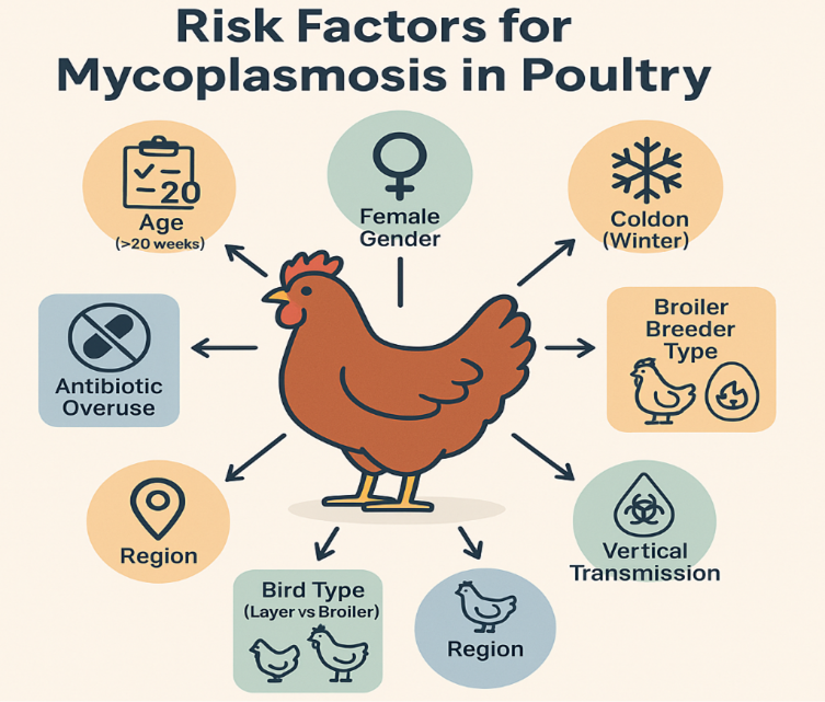
shows the different types of risk factors which are responsible for the occurrence of Mycoplasmosis.
Conclusions and Recommendations
Mycoplasma gallisepticum (M. gallisepticum) is the principal cause of avian Mycoplasmosis, which continues to pose a serious danger to Pakistan’s quickly expanding poultry sector. The widespread distribution of M. gallisepticum across diverse geographic regions—from the densely populated poultry zones of Punjab to the scattered flocks in Balochistan and Khyber Pakhtunkhwa—reflects both the endemic nature of the pathogen and the systemic weaknesses in biosecurity, surveillance, and disease control infrastructure.
The data presented in this study underscore a disturbingly high and variable seroprevalence of M. gallisepticum across Pakistan, with reports ranging from 5.5% to as high as 76.6%, depending on bird type, season, and region. Broiler breeders and layer flocks were particularly susceptible, with infection peaks notably occurring during the winter months. Spatial analysis points to disease hotspots in Rawalpindi, Faisalabad, and Khyber Pakhtunkhwa, while temporal trends suggest a continuous rise in M. gallisepticum prevalence which is driven by increased poultry density, poor vaccination practices, and potential vertical transmission from breeders to offspring. The pathogen’s ability to cause chronic respiratory disease, synovitis, and systemic infection is compounded by its immune evasion mechanisms, leading to persistent infections and economic losses. Despite existing diagnostic tools (e.g., ELISA, PCR, culture) and vaccine options, control remains challenging due to inconsistent monitoring, inadequate farmer awareness, and variable field biosecurity. There is an urgent need for comprehensive control strategies that include coordinated national surveillance, regulated antibiotic usage, enhanced farm-level biosecurity, prompt diagnosis, and vaccination. Building a spatial-temporal illness database is equally crucial since it would enable early outbreak detection and focused intervention. In conclusion, Avian Mycoplasmosis is not merely a veterinary concern but a national challenge with implications for food security, rural livelihoods, and public health. A unified, evidence-based approach involving veterinarians, researchers, policymakers, and poultry producers is essential to mitigate the threat posed by Mycoplasma gallisepticum and secure the future of Pakistan’s poultry sector.
Acknowledgements
We acknowledge the guidance provided by academic mentors and appreciate the collective efforts of fellow researchers, faculty members, and technical staff whose constructive feedback and encouragement played a significant role in shaping the direction of this review
Novelty Statement
This review provides a comprehensive Pakistan-specific assessment of Mycoplasma gallisepticum infection in poultry, integrating epidemiological patterns, diagnostic advances, risk factors, economic impacts, and control challenges. It highlights regional and seasonal trends and identifies key gaps in surveillance and disease management to guide integrated control strategies in Pakistan.
Author’s Contribution
Bilal Javid contributed to the conceptualization of the study, major writing, and preparation of the original manuscript. Muhammad Usman Mukhtar contributed to data collection, data organization, and methodology. Syed Musa Raza Naqvi contributed to the literature search and interpretation of the findings. Aayesha Riaz provided methodological guidance and academic oversight throughout the study. Sami Tahir Qureshi contributed to the critical revision of the manuscript. Muhammad Athar Abbas contributed to research oversight and validation of the study. All authors reviewed and approved the final version of the manuscript.
Generative AI and AI-assisted technology statement
The authors declare that no generative AI and AI assisted technology was used in the creation of this manuscript.
Conflict of interest
The authors declare that there is no conflict of interest associated with this manuscript.
Source of funding
This study received no specific grant from any public, commercial, or not-for-profit funding agency.
Animal Ethics
This article is a review study and does not involve any experimental work with live animals. Therefore, ethical approval for animal use was not required.
References
- Aamir Islam, A.I., A.A. Asim Aslam, Z.I. Chaudhry, M.U.D.A. Mansoor-ud-Din Ahmed, H.U.R. Habib-ur-Rehman, K.S. Khalid Saeed, and I.A. Ishtiaq Ahmed. 2011. Pathology of Mycoplasma gallisepticum in naturally infected broilers and its diagnosis through PCR. Prevalen., 48(22): 46.
- Abbas, G., S.H. Khan, M. Hassan, S. Mahmood, S. Naz, and S.S. Gilani. 2015. Incidence of poultry diseases in different seasons in Khushab district, Pakistan. J. Advan. Veterin. Anim. Res., 2(2): 141–145. https://doi.org/10.5455/javar.2015.b65 [CrossRef]
- Abbas, N., M. Suleman, N.A. Khan, I. Ali, M. Rauf, and S. Rahman. 2018. Prevalence of Mycoplasma gallisepticum in poultry and wild life birds suspected of chronic respiratory disease in Northern Pakistan. Pak. J. Zool., 50(3): 1071–1077. https://doi.org/10.17582/journal.pjz/2018.50.3.1071.1077 [Full text] [CrossRef]
- Ahmad, A., M. Rabbani, T. Yaqoob, A. Ahmad, M.Z. Shabbir, and F. Akhtar. 2008. Status of IgG antibodies against Mycoplasma gallisepticum in non-vaccinated commercial poultry breeder flocks. Int. J. Poult. Sci., 18(2-3): 61–63.
- Anwar, S., J. Khan, A. Abid, A. Wahid, A. Wajid, and A. Raheem. 2024. Surveillance of Mycoplasma gallisepticum in bird’s retail stalls in Quetta district. Nature’s Symph., 2(1): 01–08. https://doi.org/10.69547/NS.21.01 [CrossRef]
- Arshad, A. et al. 2013. Development of duplex PCR assay for the detection of Mycoplasma gallisepticum and Mycoplasma synoviae prevalence in Pakistan. Int. J. Anim. Veterin. Advan., 5(3): 114–119. https://doi.org/10.19026/ijava.5.5586 [CrossRef]
- Atique, M.A., F. Abbas, M.A. Awan, M.M. Tariq, Z. Ahmed, I. Ali, and M.A. Alam. 2012. Identification of avian Mycoplasma species in commercial broilers and layers with respiratory symptoms in Balochistan. Afric. J. Biotechnol., 11(100): 16557–16559.
- Blanchard, A. and G. Browning, (Eds.). 2005. Mycoplasmas: molecular biology pathogenicity and strategies for control. CRC Press. [CrossRef]
- Economic Survey of Pakistan. 2025. Government of Pakistan finance Division. https://www.finance.gov.pk/survey_2025.html.
- Farooq, S., A.A. Shah, N. Siddique, S. Rafique, A. Sharif, M.A. Abbas, and K. Naeem, 2020. Biological characterization of locally circulating Mycoplasma gallisepticum in poultry. Pak. Veterin. J. https://doi.org/10.29261/pakvetj. [CrossRef]
- Ferguson-Noel, N., N.K. Armour, A.H. Noormohammadi, M. El-Gazzar, and J.M. Bradbury. 2020. Mycoplasmosis. Diseases of poultry, 907-965. https://doi.org/10.1002/9781119371199.ch21 [CrossRef]
- Gaunson, J.E., C.J. Philip, K.G. Whithear, & G.F. Browning. 2006. The cellular immune response in the tracheal mucosa to Mycoplasma gallisepticum in vaccinated and unvaccinated chickens in the acute and chronic stages of disease. Vacc., 24(14): 2627–2633. https://doi.org/10.1016/j.vaccine.2005.12.008 [CrossRef]
- Gaunson, J.E., C.J. Philip, K.G. Whithear, and G.F. Browning. 2000. Lymphocytic infiltration in the chicken trachea in response to Mycoplasma gallisepticum infection. Microbiol., 146(5): 1223–1229. https://doi.org/10.1099/00221287-146-5-1223 [CrossRef]
- Haider, S., A. Maqbool, T. Pervez, S. Parveen, A. Ahmad, Z. Iqbal, and S. Umar. 2019. Seroprevalence of Mycoplasma gallisepticum in commercial poultry birds showing respiratory clinical signs in Chakwal District, Pakistan. Pak. J. Zool., 51(5): 1–4. https://doi.org/10.17582/journal.pjz/2019.51.5.sc7 [Full text] [CrossRef]
- Hasni, M.S., M. Chaudhry, M.H. Mushtaq, A.Z. Durrani, H. Bin Rashid, S.S. Gill, ... & H. Sattar. 2021. Prevalence and associated risk factors of avian influenza H9 in backyard poultry populations of two agroecological zones of Pakistan. Pak. Veterin. J., 41(1). https://doi.org/10.29261/pakvetj/2020.085 [CrossRef]
- Islam, A., A. Aslam, Z.I. Chaudhry, M.U.D. Ahmed, H.U. Rehman, K. Saeed, & I. Ahmed. 2011. Pathology of Mycoplasma gallisepticum in naturally infected broilers and its diagnosis through PCR. Prevalen., 48(22): 46.
- Javed, M.A., S. Frasca Jr, D. Rood, K. Cecchini, M. Gladd, S.J. Geary, & L.K. Silbart. 2005. Correlates of immune protection in chickens vaccinated with Mycoplasma gallisepticum strain GT5 following challenge with pathogenic M. gallisepticum strain Rlow. Infect. Immun., 73(9): 5410–5419. https://doi.org/10.1128/IAI.73.9.5410-5419.2005 [CrossRef]
- Jelani, G., M.H.R. Farhan, R. Asrar, M. Ahmad, C. Naseem, Z.A. Khoso, and M.H. Soomro. 2023. Mycoplasma gallisepticum infection, a perpetual problem. Res. J. Veterin. Practition., 11(1): 1–6. https://doi.org/10.17582/journal.rjvp/2023/11.1.1.6 [Full text] [CrossRef]
- Khatoon, H. 2018. Prevalence of Mycoplasmosis and antibiotic susceptibility of Mycoplasma gallisepticum in commercial chicken flocks of Rawalpindi Division, Pakistan. Pak. Veterin. J., 38(4): 446–448. https://doi.org/10.29261/pakvetj/2018.081 [CrossRef]
- Majumder, S., F. Zappulla, and L.K. Silbart. 2014. Mycoplasma gallisepticum lipid associated membrane proteins up-regulate inflammatory genes in chicken tracheal epithelial cells via TLR-2 ligation through an NF-κB dependent pathway. PLoS One., 9(11): e112796. https://doi.org/10.1371/journal.pone.0112796 [CrossRef]
- Masukagami, Y., B. Nijagal, C.W. Tseng, S. Dayalan, K.A. Tivendale, P.F. Markham, and F.M. Sansom. 2018. Metabolite profiling of Mycoplasma gallisepticum mutants, combined with bioinformatic analysis, can reveal the likely functions of virulence-associated genes. Veterin. Microbiol., 223: 160–167. https://doi.org/10.1016/j.vetmic.2018.08.001 [CrossRef]
- Michiels, T., S. Welby, M. Vanrobaeys, C. Quinet, L. Rouffaer, L. Lens, A. Martel, and P. Butaye. 2016. Prevalence of Mycoplasma gallisepticum and Mycoplasma synoviae in commercial poultry, racing pigeons and wild birds in Belgium. Avian Pathol., 45(2): 244–252. https://doi.org/10.1080/03079457.2016.1145354 [CrossRef]
- Ministry of Finance, Government of Pakistan. (2024). Pakistan economic survey 2023–24. https://www.finance.gov.pk/survey/chapter_24/2_agriculture.pdf
- Mugunthan, S.P., G. Kannan, H.M. Chandra, and B. Paital. 2023. Infection, transmission, pathogenesis and vaccine development against Mycoplasma gallisepticum. Vaccin., 11(2): 469. https://doi.org/10.3390/vaccines11020469 [CrossRef]
- Muhammad, J., M. Rabbani, A.A. Sheikh, A.A. Rabaan, A. Khan, I.U. Haq ... & A. Akbar. 2021. Molecular detection of Mycoplasma gallisepticum in different poultry breeds of Abbottabad and Rawalpindi, Pakistan. Brazil. J. Biol., 83: e246514. https://doi.org/10.1590/1519-6984.246514 [CrossRef]
- Mukhtar, M., M.M. Awais, M.I. Anwar, Z. Hussain, N. Bhatti, & S. Ali, 2012. Seroprevalence of Mycoplasma gallisepticum among commercial layers in Faisalabad, Pakistan. J. Basic Appl. Sci., 8(1): 183-186.
- Mukhtar, M., M.M. Awais, M.I. Anwar, Z. Hussain, N. Bhatti, and S. Ali. 2012. Seroprevalence of Mycoplasma gallisepticum among commercial layers in Faisalabad, Pakistan. J. Basic Appl. Sci., 8(1): 183–186.
- Nunoya, T., K. Kanai, T. Yagihashi, S. Hoshi, K. Shibuya, and M. Tajima. 1997. Natural case of salpingitis apparently caused by Mycoplasma gallisepticum in chickens. Avian Pathol., 26(2): 391–398. https://doi.org/10.1080/03079459708419221 [CrossRef]
- Papazisi, L., S. Frasca Jr, M. Gladd, X. Liao, D. Yogev, and S.J. Geary. 2002. GapA and CrmA coexpression is essential for Mycoplasma gallisepticum cytadherence and virulence. Infect. Immun., 70(12): 6839–6845. https://doi.org/10.1128/IAI.70.12.6839-6845.2002 [CrossRef]
- Qadir, M.F. 2020. Epidemiological and diagnosis status of Mycoplasma gallisepticum in chickens around the globe. EC Veterin. Sci., 5: 01–07.
- Qadir, M.F. and A. Irum. 2023. Epidemiological and diagnostic status of Mycoplasma synoviae in Pakistan and India: A review. https://doi.org/10.47278/journal.abr/2023.006 [CrossRef]
- Qadir, M.F., A. Khan, M.K. Saleemi, S.T. Gul, A. Khan, and Q. Mujahid. 2021. Epidemiological and pathological status of Mycoplasma gallisepticum in layer chicks at Faisalabad, Pakistan. Pak. J. Agric. Sci., 58(1).
- Qadir, M.F., A. Khan, M.K. Saleemi, S.T. Gul, A. Khan, and Q. Mujahid. 2021. Epidemiological and pathological status of Mycoplasma gallisepticum in layer chicks at Faisalabad, Pakistan. Pak. J. Agric. Sci., 58(1).
- Rauf, M., Z.I. Chaudhary, M. Younus, A.A. Anjum, M.A. Ali, A.N. Ahmad, and M.U.R. Khan. 2013. Identification of Mycoplasma gallisepticum by polymerase chain reaction and conventional diagnostics from white leghorn layer flocks.
- Rehman, A., A. Shah, and S. Rahman. 2018. Seroprevalence of Mycoplasma gallisepticum and Mycoplasma synoviae in commercial broilers and backyard poultry in five districts of Khyber Pakhtunkhwa-Pakistan. Pak. Veterin. J., 38(2): 149–152. https://doi.org/10.29261/pakvetj/2018.047 [CrossRef]
- Rehman, H., N. Fawad, G. Abbas, G. Naheed, B. Siddique, F. Afzal, and M. Javed. 2013. Surveillance of poultry diseases in Punjab province, Pakistan; special reference to Newcastle disease. Res. J. Vet. Pract., 1: 1-4.
- Sarfaraz, S., K. Muhammad, T. Yaqub, A. Aslam, M. Rabbani, M. Khalil, & R. Riaz. 2017. Antibody response of broilers to oil-based combined avian influenza (H9N2) and Mycoplasma gallisepticum vaccine. J. Anim. Plant Sci., 27(4).
- Seifi, S. and M.R. Shirzad. 2012. Incidence and risk factors of Mycoplasma synoviae infection in broiler breeder farms of Iran. Veterin. World., 5(5). https://doi.org/10.5455/vetworld.2012.265-268 [CrossRef]
- Shah, A.H. 2018. Seroprevalence of Mycoplasma gallisepticum and Mycoplasma synoviae in commercial broilers and backyard poultry in five districts of Khyber Pakhtunkhwa-Pakistan. Pak. Veterin. J., 38(2). https://doi.org/10.29261/pakvetj/2018.047 [CrossRef]
- Shoaib, M. 2019. Mycoplasmosis in poultry, a perpetual problem. J, Microbiol., Biotechnol. Food Sci., 8(6): 1271. https://doi.org/10.15414/jmbfs.2019.8.6.1271-1275 [CrossRef]
- Shoaib, M., A. Riaz, A. Yousaf, M.A. Zafar, M. Kamran, R.M. Amir, and A.M. Malik. 2020. Sero-prevalence and associated risk factors of Mycoplasma gallisepticum, Mycoplasma synoviae and Salmonella pullorum/gallinarium in poultry. https://doi.org/10.29261/pakvetj/2019.097 [CrossRef]
- Umar, S., M.T. Munir, Z. Ur-Rehman, S. Subhan, T. Azam, and M.A.A. Shah. 2017. Mycoplasmosis in poultry: Update on diagnosis and preventive measures. World’s Poult. Sci. J., 73(1): 17–28. https://doi.org/10.1017/S0043933916000830 [CrossRef]
- Whithear, K.G., K.E. Harrigan, and S.H. Kleven. 1996. Standardized method of aerosol challenge for testing the efficacy of Mycoplasma gallisepticum vaccines. Avian Diseas., 654-660 https://doi.org/10.2307/1592277. [CrossRef]
- Yadav, J.P., P. Tomar, Y. Singh, and S.K. Khurana. 2022. Insights on Mycoplasma gallisepticum and Mycoplasma synoviae infection in poultry: a systematic review. Anim. Biotechnol., 33(7): 1711-1720. https://doi.org/10.1080/10495398.2021.1908316 [CrossRef]

Services
Article usage statistics
Loading…
Related articles
- Seroprevalence of Mycoplasma gallisepticum in Commercial Poultry Birds Showing Respiratory Clinical Signs in Chakwal District, PakistanPakistan Journal of Zoology · 2019
- Prevalence of Mycoplasma gallisepticum in Poultry and Wild Life Birds Suspected of Chronic Respiratory Disease in Northern PakistanPakistan Journal of Zoology · 2018
- Potential Economic Impact of Newcastle Disease Virus Isolated from Wild Birds on Commercial Poultry Industry of Pakistan: A ReviewHosts and Viruses · 2019
- Detection of <i>Mycoplasma</i> <i>gallisepticum</i> from field samples of Poultry using conventional PCRAdvances in Animal and Veterinary Sciences · 2013
- A Review: Fruit Production Industry of Pakistan Trends, Issues and Way ForwardPakistan Journal of Agricultural Research · 2022
- Detection of Sub Clinical Infection of Mycoplasma gallisepticum in Commercial Chicken by Indirect ELISAAdvances in Animal and Veterinary Sciences · 2016