
Porcine epidemic diarrhea (PED), caused by porcine epidemic diarrhea virus (PEDV), is a highly contagious enteric disease that causes substantial economic losses to pig production in Vietnam and other endemic countries. This study aimed to determine the seroprevalence of PEDV and identify risk factors associated with infection in non-vaccinated sows in the Mekong Delta, Vietnam. A total of 1,605 serum samples were collected from non-vaccinated sows on 431 commercial sow farms, together with 1–5 fecal samples per farm for PEDV detection. Anti-PEDV antibodies were detected using a commercial indirect enzyme-linked immunosorbent assay (ELISA). PEDV RNA was detected in 101 of the 431 herds (23.4%). The overall seroprevalence was 25.4% (408/1,605) at the individual level and 47.8% (206/431) at the farm level. By herd size, the highest PEDV infection prevalence was found in herds of 50–100 sows (27.04%), followed by herds of over 100 sows (26.83%) and 20 to less than 50 sows (26.76%); the lowest was in herds of 10 to less than 20 sows and less than 10 sows (25.21% and 19.75%, respectively). The mean sample-to-positive (S/P) ratio was significantly higher in farms with a history of PED outbreaks (1.340–1.797) than in farms without previous outbreaks (0.740–0.885). These findings demonstrate that PEDV is widely circulating among pig populations in the Mekong Delta and highlight the importance of vaccination and strict biosecurity measures for effective disease prevention and control.
Research Article · Volume 14, Issue 12, 5 November 2026 · Open Access
Seroprevalence of Porcine Epidemic Diarrhea Virus Infection in Vietnam
·
Affiliations & Notes ⌄
- 1Vemedim Research and Development Center, No. 322A, 30/4 street, Ninh Kieu ward, Can Tho city, Vietnam, Vietnam
- 2Faculty of Veterinary Medicine, College of Agriculture, Can Tho University, Campus II, 3/2 street, Ninh Kieu ward, Can Tho city, Vietnam
- *Correspondence | Nguyen Thu Tam; Email: nttamty@ctu.edu.vn
Article Info ⌄
- Journal:
- Advances in Animal and Veterinary Sciences
- Submitted Date:
- 3 August 2026
- Accepted Date:
- 4 October 2026
- Published Date:
- 5 November 2026
Abstract
INTRODUCTION
Porcine epidemic diarrhea virus (PEDV) causes a rapidly spreading enteric disease in pigs, leading to severe economic losses due to high morbidity and mortality. The disease affects pigs of all ages and is clinically characterized by vomiting and diarrhea . PEDV was first reported in the United Kingdom in 1971, later spreading to Europe in the 1970s and Asia in the 1980s . In Vietnam, PEDV was officially documented in the southeastern provinces in 2008 and has since spread nationwide. Genetic studies show that PEDV strains circulating in Vietnam belong to both G1 and G2 genotypes, closely related to strains in neighboring countries ; Than et al., 2020).
Although PEDV typically does not cause mortality in adult pigs, it can impair reproductive performance. Sows infected during their first parity may experience long-term reproductive issues ; . Following infection, anti-PEDV IgA antibody levels rise in blood, milk, and the ileum but gradually decline over time. Neutralizing antibodies remain elevated for up to six months post-outbreak . Recovered sows retain PEDV-specific antibodies, which are transferred via colostrum and milk to protect piglets. Antibodies have also been detected in finisher pigs more than 100 days post-infection, including in subclinically infected animals ; .
Diagnosing PED on-farm during an outbreak can be inaccurate without appropriate diagnostic tools, such as rapid PEDV test kits. After recovery, pigs develop PEDV-specific antibodies in their blood; these antibodies can be transferred to piglets via colostrum and milk, thereby providing protection against PED during the suckling period. Furthermore, these specific antibodies can persist for years in the blood of recovered sows. ELISA-based detection of specific antibodies is commonly used in exploratory or retrospective studies to assess exposure to various animal pathogens (e.g., classical swine fever, PRRS, and avian influenza). However, research assessing PEDV exposure in sows under field conditions by IgG antibody testing has received limited attention. Previous serological surveys in northern Vietnam found no significant differences in PEDV prevalence by locality, age, breed, or farm size . However, in the Mekong Delta - one of Vietnam’s key pig-producing regions - comprehensive serological investigations remain limited. Therefore, this study aimed to determine the seroprevalence of PEDV in sows across seven provinces in the Mekong Delta, providing new insights into the endemic status of PEDV in Vietnam.
Materials and methods
Ethical approval
Sample collection and transportation were conducted in accordance with the guidelines of the Vietnam Department of Animal Health, with permission obtained from the respective Provincial Sub-Departments of Animal Health. The study involved routine field sampling of serum and fecal specimens by qualified veterinarians as part of animal health surveillance and did not involve any experimental intervention; formal approval by an institutional animal ethics committee was therefore not required. Verbal informed consent was obtained from each herd owner before sampling.
Sample collection and location
A total of 1,605 serum samples from non-vaccinated sows were collected across 431 pig farms in seven provinces of the Mekong Delta: Ben Tre, Can Tho, Dong Thap, Hau Giang, Soc Trang, Tien Giang, and Vinh Long. Anti-PEDV antibodies were detected using a commercial indirect ELISA kit (Swinecheck® PED Indirect, Biovet, Canada; Cat# TRM 559). According to the manufacturer, the relative sensitivity of the assay is 100.0% (95% CI: 96.70–100.0%) and the relative specificity is 95.73% (95% CI: 93.60–97.30%), with a kappa coefficient of 0.874 relative to immunofluorescence assay and virus neutralization test results; according to testing conducted by the manufacturer, this kit exhibits no cross-reactivity with other swine viruses.
Blood was collected from the anterior vena cava after the sampling site had been disinfected with 70% alcohol. A volume of 2–5 mL of blood was drawn using a 23-gauge needle. The needle was then removed, and the blood was gently dispensed so that it flowed down the wall of a sterile tube. Each tube was labeled with identifying information (ear-tag number and sampling date). The blood was allowed to clot, and the samples were transported to the laboratory at 4–8°C within 24 hours. Samples were centrifuged at 3,000 rpm for 10 minutes to separate the serum, which was transferred into sterile tubes and stored at −20°C until analysis.
Identification of both PEDV-exposed and non-exposed farms
To determine farm infection status, 1–5 fecal samples were collected per farm and tested for PEDV. RNA was extracted using the GeneJET Viral DNA and RNA Purification Kit (Thermo Fisher Scientific, USA), followed by cDNA synthesis with the RevertAid First Strand cDNA Synthesis Kit (Thermo Fisher Scientific, USA). PEDV was amplified using primers PED-F (GGG CGC CTG TAT AGA GTT TA) and PED-R (AGA CCA CCA AGA ATG TGT CC), targeting a 412 bp fragment (Li et al., 2016). PCR products were visualized on 0.8% agarose gels.
A total of 1,605 serum samples were collected, including 412 from PEDV-outbreak farms and 1,193 from non-exposed farms. Three to five samples were obtained from each farm. Additional factors recorded included farm location, herd size, and sow parity.
Antibody detection using ELISA
The indirect ELISA was performed according to the manufacturer’s instructions. Serum samples were diluted 1:200 by sequential dilution (10 µL serum + 190 µL diluent, followed by 20 µL of the first dilution + 180 µL diluent). Positive and negative controls were used without dilution.
Diluted sera (50 µL/well) were added to 96-well plates coated with PEDV antigens. Plates were incubated at 23°C for 1 hour, washed three times, and sequentially incubated with conjugate A and substrate B under the same conditions. After a final incubation with substrate for 10 minutes, reactions were stopped with stop solution, and optical density (OD) was measured at 450 nm.
Antibody titers were expressed as sample-to-positive (S/P) ratios, calculated from OD values. Validity criteria had to be met to validate the results. In particular, the negative control’s mean OD (MOD) had to be less than 0.40, and the positive control’s corrected OD had to be greater than 0.70. Interpretation: samples with an S/P ratio < 0.4 were considered negative, and those with an S/P ratio ≥ 0.4 were considered positive. The percentage of positive sera was determined as: % positive sera = (Total seropositive samples / Total serum samples) × 100
Data execution and statistical analysis
Raw data were processed in Microsoft Excel, and aggregated data were analyzed using Minitab 16.0 (Minitab Inc., 2010). The chi-square test was used for categorical comparisons, and one-way ANOVA followed by Tukey’s test was used for multiple-group comparisons of continuous variables. A significance level of p < 0.05 was applied.
Results
The PEDV-seroprevalence based on herd size
The results presented in Table 1 show that the proportion of individual sows with anti-PEDV antibodies was highest in herds of 50–100 sows (27.04%), followed by herds of over 100 sows (26.83%), herds of 20 to <50 sows (26.76%), and lowest in herds of <10 sows (19.75%) (p < 0.05).
For each province, differences in PEDV prevalence by herd size were not statistically significant (p > 0.05). However, Tien Giang and Ben Tre had no farms with fewer than 10 sows, so samples in these two provinces were collected only from pig farms with more than 10 sows. These provinces are key livestock production areas in the Mekong Delta and showed high PEDV infection rates, particularly in herds of 50–100 sows (36.54% and 30.91%, respectively), compared with other provinces.
Although no significant differences were observed in the S/P ratios of seropositive sows among the five herd sizes in the study (0.90–1.29) (Figure 1B), the levels of anti-PEDV antibodies were relatively high.
The PEDV-seroprevalence based on parity
The PEDV infection rates in this study were analyzed by parity and are presented in Table 2. The results indicated a tendency for sows in the 4th parity to have higher levels of anti-PEDV antibodies compared to other parities, particularly when compared to unproductive gilts, which exhibited lower PEDV antibody levels. However, across the six litter groups, there were no significant differences in the percentage of seropositivity or S/P ratios (24.68%–27.59% and 1.01–1.28, respectively) (Figure 2B).
To assess PEDV infection rates by parity across seven provinces, a comparison was conducted. The results showed that only in Tien Giang was there a significant difference in PEDV prevalence among sows by parity, with the highest prevalence in the 4th parity (52%) and the lowest in the 2nd parity (26.23%) (p < 0.05, Table 2).
The PEDV-seroprevalence based on locations
Using an indirect ELISA method to assess the presence of PEDV in sows across farms in seven provinces of the Mekong Delta, this study conducted randomized testing for IgG antibodies against PEDV in serum samples from unvaccinated sows. The results are presented in Table 3.
Of the 431 farms surveyed in this study, 206 (47.80%) were PEDV-seropositive. Ben Tre had the highest PEDV seroprevalence at 59.62% (31/52), significantly higher than Dong Thap’s 37.80% (p < 0.05, Table 3). The PEDV seropositive rate was detected in 35/66 farms (53.03%) in Vinh Long, 25/48 farms (52.08%) in Tien Giang, 28/59 farms (47.46%) in Soc Trang, 32/68 farms (47.06%) in Can Tho, and 24/56 farms (42.86%) in Hau Giang. Additionally, 25.42% (408/1,605) of sow sera collected from the Mekong Delta tested positive for anti-PEDV antibodies. Notably, the percentages of PEDV-seropositive sows in Tien Giang, Ben Tre, and Vinh Long (29.39%–33.72%) were significantly higher than those in Hau Giang, Soc Trang, and Dong Thap (17.06%–20.48%) (p < 0.05, Table 3). Additionally, the mean serum S/P ratio of positive sows on outbreak farms in Tien Giang was 1.797 ± 1.648, significantly higher than those in Dong Thap, Vinh Long, and Can Tho (1.367 ± 0.543, 1.304 ± 0.629, and 1.258 ± 0.445, respectively) (p < 0.05, Figure 3). Among non-outbreak farms, the mean S/P ratio in Hau Giang was 0.885 ± 0.374, significantly higher than that in Can Tho (0.740 ± 0.715) (Table 4).
This study found that 25.42% of sows and 47.80% of farms surveyed had anti-PEDV antibodies, despite none of the 431 pig farms or 1,605 sow serum samples in the Mekong Delta having received PEDV vaccination. These findings indicate that the sows on these farms were naturally infected with PEDV.
The PEDV-seroprevalence based on exposure status
Testing for PEDV across 431 farms revealed that 101
Table 1: The PEDV-seroprevalence based on herd size.
Location
|
Less 10 sows | 10 – less 20 sows | 20 – less 50 sows | 50 – 100 sows | > 100 sows | Total | ||||||||||||||
| No sample | No positive | Positive rate (%) | No sample | No positive | Positive rate (%) | No sample | No positive | Positive rate (%) | No sample | No positive | Positive rate (%) | No sample | No positive | Positive rate (%) | No sample | No positive | Positive rate (%) | |||
| Ben Tre | - | - | - | 53 | 16 | 30.19 | 85 | 26 | 30.59 | 67 | 21 | 31.34 | 55 | 17 | 30.91 | 260 | 80 | 30.77 | ||
| Can Tho | 54 | 13 | 24.07 | 37 | 9 | 24.32 | 57 | 14 | 24.56 | 38 | 9 | 23.68 | - | - | - | 186 | 45 | 24.19 | ||
| Dong Thap | 86 | 14 | 16.28 | 69 | 12 | 17.39 | 68 | 12 | 17.65 | 29 | 5 | 17.24 | - | - | - | 252 | 43 | 17.06 | ||
| Hau Giang | 42 | 8 | 19.05 | 19 | 4 | 21.05 | 46 | 10 | 21.74 | 32 | 7 | 21.88 | 27 | 5 | 18.52 | 166 | 34 | 20.48 | ||
| Soc Trang | 43 | 8 | 18.60 | 58 | 12 | 20.69 | 48 | 10 | 20.83 | 46 | 9 | 19.57 | 40 | 7 | 17.50 | 235 | 46 | 19.57 | ||
| Tien Giang | - | - | - | 57 | 18 | 31.58 | 101 | 34 | 33.66 | 52 | 19 | 36.54 | 51 | 17 | 33.33 | 261 | 88 | 33.72 | ||
| Vinh Long | 18 | 5 | 27.78 | 64 | 19 | 29.69 | 77 | 23 | 29.87 | 54 | 16 | 29.63 | 32 | 9 | 28.13 | 245 | 72 | 29.39 | ||
| Total | 243 | 48 | 19.75b | 357 | 90 | 25.21ab | 482 | 129 | 26.76a | 318 | 86 | 27.04a | 205 | 55 | 26.83a | 1,605 | 408 | 25.42 | ||
Values with different letters a, b in the same row indicated that the difference is statistically significant at p <0.05;A dash (–) indicates that no surveyed herd in that province fell into the given size category: no herd in Ben Tre or Tien Giang kept fewer than 10 sows, and no herd in Can Tho or Dong Thap kept more than 100 sows.
Table 2: The PEDV-seroprevalence based on parity .
| Location | Gilt | 1st parity | 2nd parity | 3rd parity | 4th parity | ≥ 5th parity | ||||||||||||
| No sample | No positive | Positive rate (%) | No sample | No positive | Positive rate (%) | No sample | No positive | Positive rate (%) | No sample | No positive | Positive rate (%) | No sample | No positive | Positive rate (%) | No sample | No positive | Positive rate (%) | |
| Ben Tre | 30 | 9 | 30.00 | 49 | 15 | 30.61 | 64 | 20 | 31.25 | 45 | 14 | 31.11 | 29 | 9 | 31.03 | 43 | 13 | 30.23 |
| Can Tho | 17 | 4 | 23.53 | 33 | 8 | 24.24 | 37 | 9 | 24.32 | 49 | 12 | 24.49 | 24 | 6 | 25.00 | 26 | 6 | 23.08 |
| Dong Thap | 18 | 3 | 16.67 | 29 | 5 | 17.24 | 51 | 9 | 17.65 | 56 | 10 | 17.86 | 46 | 8 | 17.39 | 52 | 8 | 15.38 |
| Hau Giang | 31 | 6 | 19.35 | 32 | 6 | 18.75 | 37 | 8 | 21.62 | 33 | 7 | 21.21 | 18 | 4 | 22.22 | 15 | 3 | 20.00 |
| Soc Trang | 49 | 9 | 18.37 | 41 | 8 | 19.51 | 45 | 9 | 20.00 | 49 | 10 | 20.41 | 29 | 6 | 20.69 | 22 | 4 | 18.18 |
| Tien Giang | 37 | 12 | 32.43ab | 56 | 19 | 33.93ab | 61 | 16 | 26.23b | 59 | 17 | 28.81b | 25 | 13 | 52.00a | 23 | 11 | 47.83ab |
| Vinh Long | 53 | 15 | 28.30 | 50 | 14 | 28.00 | 44 | 13 | 29.55 | 38 | 12 | 31.58 | 32 | 10 | 31.25 | 28 | 8 | 28.57 |
| Total | 235 | 58 | 24.68 | 290 | 75 | 25.86 | 339 | 84 | 24.78 | 329 | 82 | 24.92 | 203 | 56 | 27.59 | 209 | 53 | 25.36 |
Values with different letters a, b in the same row indicated that the difference is statistically significant at p <0.05.
farms tested positive for PEDV, accounting for 23.43% of the surveyed farms. Consequently, the farms were dividedinto two groups: PEDV-exposed farms and non-PEDV-exposed farms. Of the samples from exposed farms, 59.22% (244/412) were seropositive for PEDV, significantly higher than the 13.75% (164/1,193) from non-exposed farms (p < 0.05, Table 4). Furthermore, the S/P ratio of PEDV-positive samples from exposed farms was significantly higher than that from non-exposed farms (1.448 ± 0.846 vs. 0.794 ± 0.261, respectively) (p < 0.001, Table 4). These findings suggest that the presence of PEDV on sow farms significantly influences both the PEDV seropositivity rate and the levels of anti-PEDV antibodies in sows.
Discussion
The circulation of endemic and epidemic viruses in livestock populations at the farm, regional, and national levels significantly impacts animal welfare and production. In this study, the occurrence of PEDV in sow farms was influenced by provincial prevalence, herd size, parity, and farm exposure status. A PEDV-specific indirect ELISA was used to measure IgG antibodies in serum samples from unvaccinated sows across seven Mekong Delta provinces.
Table 3: The PEDV-seroprevalence based on locations /provinces.
| Location | No of pig farm | No of positive farm | Positive rate (%) | No sample | No positive (+) | Rate of positive sows (%) |
| Ben Tre | 52 | 31 | 59.62a | 260 | 80 | 30.77ab |
| Can Tho | 68 | 32 | 47.06ab | 186 | 45 | 24.19bc |
| Dong Thap | 82 | 31 | 37.80b | 252 | 43 | 17.06c |
| Hau Giang | 56 | 24 | 42.86ab | 166 | 34 | 20.48c |
| Soc Trang | 59 | 28 | 47.46ab | 235 | 46 | 19.57c |
| Tien Giang | 48 | 25 | 52.08ab | 261 | 88 | 33.72a |
| Vinh Long | 66 | 35 | 53.03ab | 245 | 72 | 29.39ab |
| Total | 431 | 206 | 47.80 | 1,605 | 408 | 25.42 |
Values with different letters a, b in the same column indicated that the difference is statistically significant at p <0.05.
Table 4: The positive rate and response level of anti-PEDV antibodies in sows in PED farms and non-PED farms.
| Location | PED exprosed farms | PEDV non-exposed farms | ||||||
| No sample | No positive | Rate of positive (%) | S/P ± SD | No sample | No positive | Rate of positive (%) | S/P ± SD | |
| Ben Tre | 97 | 61 | 62.89a | 1.506a ± 0.967 | 163 | 19 | 11.66b | 0.753b ± 0.233 |
| Can Tho | 47 | 30 | 63.83a | 1.258a ± 0.445 | 139 | 15 | 10.79b | 0.740b ± 0.175 |
| Dong Thap | 60 | 26 | 43.33a | 1.376a ± 0.543 | 192 | 17 | 8.85b | 0.774b ± 0.243 |
| Tien Giang | 113 | 71 | 62.83a | 1.797a ± 1.648 | 148 | 17 | 11.49b | 0.812b ± 0.233 |
| Vinh Long | 95 | 56 | 58.95a | 1.304a ± 0.629 | 150 | 16 | 10.67b | 0.804b ± 0.264 |
| Hau Giang | - | - | - | - | 166 | 34 | 20.48b | 0.885 ± 0.374 |
| Soc Trang | - | - | - | - | 235 | 46 | 19.57b | 0.787 ± 0.304 |
| Total | 412 | 244 | 59.22a | 1.448a ± 0.846 | 1193 | 164 | 13.75b | 0.794b ± 0.261 |
Values with different letters a, b in the same row indicated that the difference is statistically significant at p <0.05.
The prevalence of PEDV in sows showed statistically significant differences among the seven provinces, such as between Tien Giang and Can Tho, Hau Giang, Soc Trang, and Dong Thap, or between Ben Tre and Vinh Long versus Hau Giang, Soc Trang, and Dong Thap. This variation may be attributed to the fact that Tien Giang, Ben Tre, and Vinh Long are among the three provinces with the largest livestock populations in the Mekong Delta region, leading to a higher likelihood of disease spread during outbreaks. During the sampling period, PEDV outbreaks were reported in these provinces, whereas in Dong Thap and Can Tho, PEDV appeared only sporadically on some farms. Notably, no PEDV cases were detected in Hau Giang and Soc Trang during the sampling period. The percentage of seropositive farms in the three northern provinces was even higher than that in the seven Mekong Delta provinces, with PEDV detected in 9/10 (90%) commercial farms in Hai Duong and in all farms tested in Hung Yen (10/10) and Thai Binh (10/10). The overall farm-level seropositivity rate in these three provinces was 96.7% . Additionally, PEDV prevalence has been reported in northern Vietnam, where 30.9% of farms were PEDV-positive. The highest farm-level positivity was recorded in nucleus production farms (70%; 7/10). Higher proportions of positive farms were found in the Northeast and Red River Delta, the major pig-producing regions . A farm-level prevalence of 16.96% was also reported in the southeastern provinces of Vietnam . These findings indicate that PEDV is widespread in pig farms across Vietnam.
Following infection, sows maintain stable IgG and IgA antibody levels for up to six months . In this study, serum samples from unvaccinated PEDV sows tested positive for antibodies against PEDV, suggesting the presence of PEDV-infected animals among these sows. Additionally, although the seven provinces in the Mekong Delta region are geographically contiguous, they exhibit different patterns of PEDV circulation. According to Goede et al. (2013), PEDV can be detected in air samples at distances of up to 16 km (10 miles) from infected farms, putting farms in closer proximity at greater risk of infection. Due to the geographical spread of the seven Mekong Delta provinces over many kilometers, combined with the dense network of rivers and canals, there is a natural separation among these provinces, which may localize the disease to specific areas. This finding is consistent with Pensaert and Yeo (2006), who described the progression of PED in Asia, indicating a highly contagious disease circulating within immune sow populations. These results also confirm that PEDV is currently circulating in some Mekong Delta provinces,
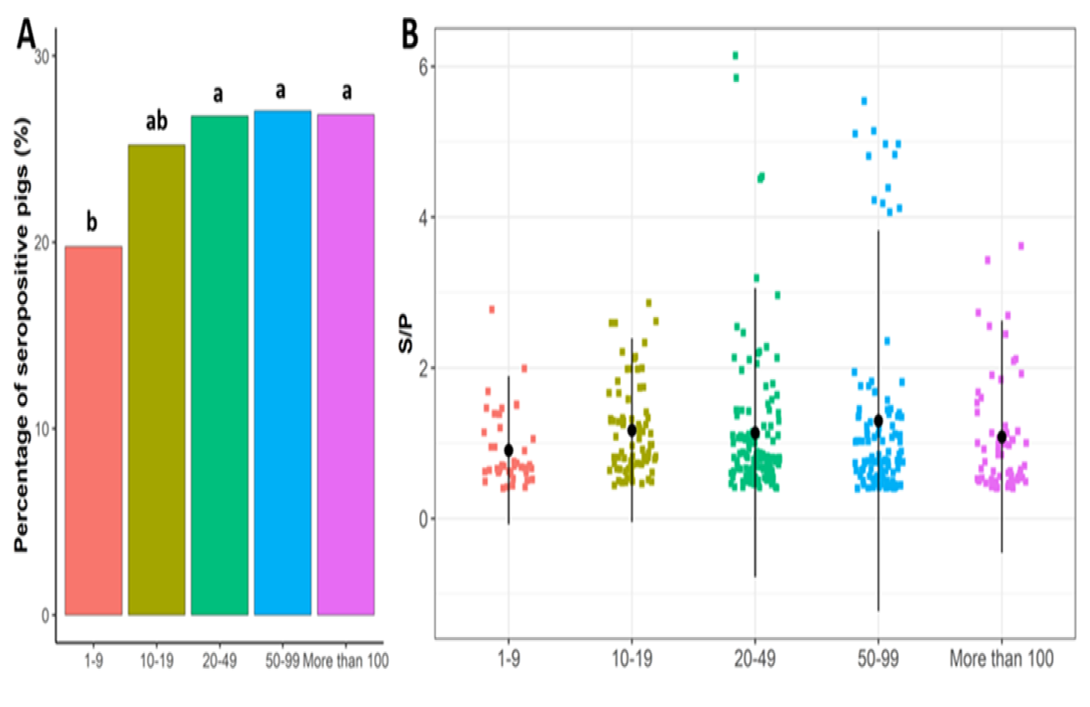
PEDV seropositive rates at sample levels (A) and the S/P ratio of PEDV-positive sows (B) based on herd size. a, b, and c indicate that the difference is statistically significant at p < 0.05).
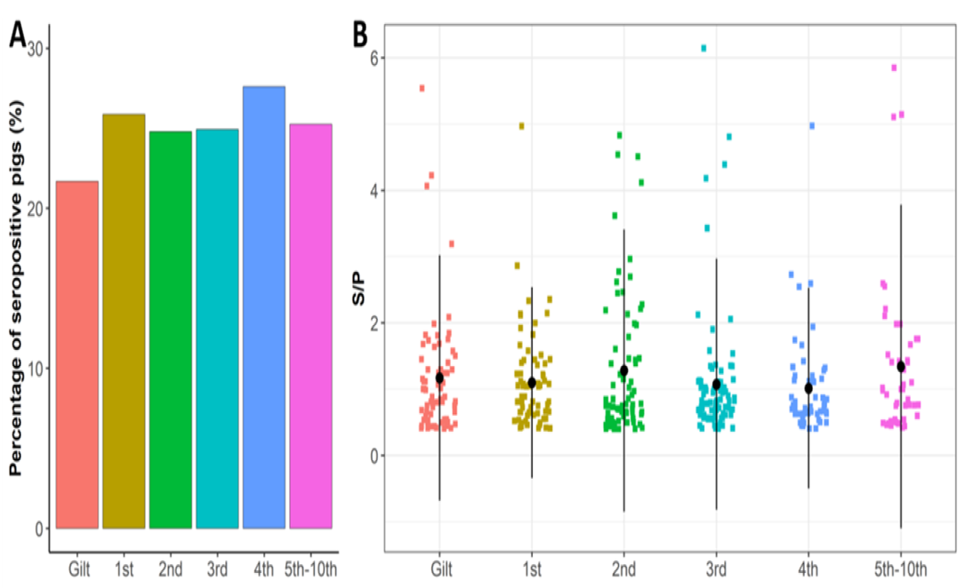
PEDV seropositive rates at sample levels (A) and the S/P ratio of PEDV-positive sows (B) based on parities.
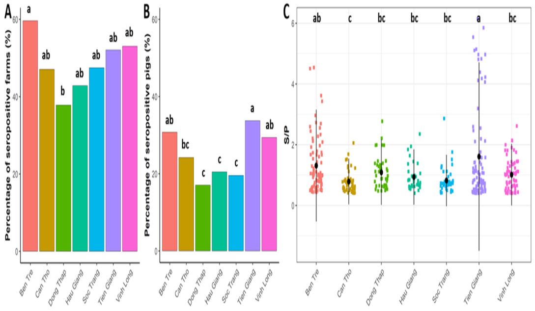
PEDV seropositive rates at farm (A) and sample (B) levels; the S/P ratio of PEDV-positive sows (C) from seven provinces of the Mekong Delta (a, b, c indicate that the difference is statistically significant at p < 0.05).
providing an important foundation for further studies on the epidemiological characteristics of PED in the Mekong Delta.
Herd size was a major factor influencing infection rates. Farms with 50–100 sows and those with >100 sows had the highest antibody prevalence, consistent with reports that large, high-density farms are more vulnerable to PEDV spread ; ; . Sasaki et al. (2016) reported a strong association between PEDV prevalence and farm size, as well as the number of feed mills and pig slaughterhouses in a given area. Other studies have attributed widespread PEDV dissemination to the movement of pigs, agricultural vehicles, farmers, farm visitors, commercial feed, and other materials ; . PED is rapidly transmitted and difficult to prevent. Infected pigs readily shed the virus in feces, and it can also be spread by intermediate carriers such as people, objects, and vehicles. Transmission occurs readily among herds on both smallholder and commercial farms. Infected sows can harbor and shed the virus into the environment, exposing other herds. Contact between carrier and susceptible pigs therefore increases infection rates, particularly on farms with poor biosecurity and hygiene . Pospischil et al. (2002) reported that regular antiseptic hygiene practices effectively limited PEDV spread. However, Myint et al. (2021) found no significant differences in PEDV seropositivity among large commercial farms ranging from 500 to 2,386 pigs. This finding suggests that, once farms exceed a certain commercial scale, herd size alone may not further differentiate seropositivity.
Parity was another factor associated with PEDV spread. Serum samples were obtained from breeding herds of different parities, including gilts and sows from the first to the fifth parity. Fourth-parity sows had higher anti-PEDV antibody levels, although exposure rates did not differ significantly among parity groups. These findings indicate that PEDV-infected sows mount an immune response and that antiviral antibodies can be detected in the serum of infected pigs. According to Bjustrom-Kraft et al. (2016), PEDV-specific antibodies persist in infected pigs for several months, and immunity in sows of second or higher parity typically lasts at least 7 months. Jung et al. (2014) reported that infected pigs shed large amounts of PEDV in feces for at least 56 days, continually contaminating floors, wastewater, and other environmental surfaces. Breeding facilities usually house sows of mixed parities; therefore, during a PED outbreak, the risk of infection is similar across parity groups. For this reason, several studies have suggested that industry-recommended biosecurity practices are important for limiting PEDV spread.
In infected herds, only 59.22% of sows tested antibody-positive. Thus, even in herds in which PEDV was detected by PCR, approximately 40% of sows remained antibody-negative. PEDV-specific serum IgG typically appears 7–10 days after infection, peaks at 2–4 weeks, and can persist for several months. During the early stage of infection, therefore, antibody levels may be undetectable by ELISA because they have not yet developed or remain low. In addition, housing areas for gestating sows, lactating sows, and replacement gilts are usually separate, and samples were collected randomly across these groups. As a result, some sows in affected herds may not yet have been infected and would therefore lack anti-PEDV antibodies.
Following PEDV infection, the levels of anti-PEDV IgA antibodies increase in blood, milk, and the ileum, but subsequently decline over time, with lower anti-PEDV IgA levels corresponding to lower S/P ratios. To date, the duration of anti-PEDV antibody persistence in infected pigs has not been fully characterized. Bjustrom-Kraft et al. (2016) observed a gradual decline in serum IgG in finishing pigs on a commercial farm after PEDV infection; by 111 days post-infection, only 60% of pigs still had antibodies detectable by a PEDV-specific ELISA. PEDV exposure is commonly assessed by detecting PEDV-specific IgG in serum. IgG antibodies appear in the blood 7–10 days after infection, peak at 2–4 weeks, and persist for several months. PEDV-specific IgG and IgA have also been detected in previously infected sows for up to five months after farrowing . According to Bjustrom-Kraft et al. (2016), in herds that have experienced an acute PEDV outbreak, immunity in sows of second or higher parity typically persists for at least seven months. Therefore, detection of PEDV-specific antibodies by indirect ELISA may be useful for determining whether a farm has previously been infected with PEDV. Based on our observations, an S/P ratio greater than 1 indicates a recent outbreak, and sera from sows on farms with active PEDV infections typically show S/P ratios in the range of 2.5–4.
Conclusions
This study confirmed that PEDV was widespread in pig farms in the Mekong Delta, Vietnam, with 47.80% positive farms and 25.42% positive serum samples. The seropositivity rate and the S/P value significantly differed among the seven provinces. In addition, large-scale or PEDV-positive farms had higher seropositive rates than small-scale or PEDV-negative farms. No differences in seroprevalence were found among different sow parities.
These findings demonstrate that PEDV is widely circulating among pig populations in the Mekong Delta and highlight the importance of vaccination and strict biosecurity measures for effective disease prevention and control
Acknowledgment
This research was supported with facilities and equipment by R&D Center – Vemedim.
Novelty statement
This study presents the first comprehensive serological assessment of PEDV in unvaccinated sows across seven provinces of the Mekong Delta, Vietnam, covering 431 farms and 1,605 sows. Unlike previous studies in Vietnam that focused mainly on northern provinces or isolated outbreaks, this study fills a critical gap by quantifying PEDV exposure at both the farm and individual animal levels in one of the country’s most important pig-producing regions. The results reveal significant provincial variations in seroprevalence and identify herd size and farm exposure status as key risk factors, thereby establishing a valuable baseline for future surveillance and control programs.
Author’s Contribution
This is the first serological survey of PEDV in unvaccinated sow herds across multiple provinces of the Mekong Delta, using ELISA to detect PEDV-specific antibodies. The findings provide a baseline for future surveillance and control programs.
Generative AI and AI-assisted technology statement
This work forms part of the empirical research for a doctoral thesis. All data were generated through field sampling and laboratory analysis and were not produced by AI. Generative AI was used only to improve the English language of the manuscript. The authors reviewed and take full responsibility for the content.
Conflict of interest
We have no conflict of interest.
References
- Bjustrom-Kraft J, Woodard K, Giménez-Lirola L, Rotolo M, Wang C, Sun Y, Lasley P, Zhang J, Baum D, Gauger P (2016). Porcine epidemic diarrhea virus (PEDV) detection and antibody response in commercial growing pigs. BMC vet. Res., 12, 1-8. https://doi.org/10.1186/s12917-016-0725-5 [CrossRef]
- Diep N, Sueyoshi M, Izzati U, Fuke N, Teh A, Lan N, Yamaguchi R (2018). Appearance of US‐like porcine epidemic diarrhoea virus (PEDV) strains before US outbreaks and genetic heterogeneity of PEDV s collected in Northern Vietnam during 2012-2015. Transboundary Emerg. Dis., 65, e83-e93. https://doi.org/10.1111/tbed.12681 [CrossRef]
- Do DT, Nguyen TT, Suphasawatt P, Roongroje T (2011). Genetic characterization of porcine epidemic diarrhea virus (PEDV) isolates from southern Vietnam during 2009-2010 outbreaks. Thai. J. Vet. Med., 41, 55. https://doi.org/10.56808/2985-1130.2280 [CrossRef]
- Goede D, Robbins R, Dufresne L, Engle M, Morrison RB (2013), Detection of porcine epidemic diarrhea virus in air samples at varying distances to epidemic farms in Oklahoma. In: Allen D. Leman, 2013. Swine Conference. English, 40: pp 212-216.
- Horie M, Kabemura M, Masatani T, Matsuu A, Ozawa M (2016). Isolation and molecular characterization of porcine epidemic diarrhea viruses collected in Japan in 2014. Arch. Virol., 161, 2189-2195. https://doi.org/10.1007/s00705-016-2900-1 [CrossRef]
- Jung K, Wang Q, Scheuer KA, Lu Z, Zhang Y, Saif LJ (2014). Pathology of US porcine epidemic diarrhea virus strain PC21A in gnotobitic pig. Emerging infect. Dis., 20: 662 – 665. https://doi.org/10.3201/eid2004.131685 [CrossRef]
- Koike N, Mai TN, Shirai M, Kubo M, Hata K, Marumoto N, Watanabe S, Sasaki Y, Mitoma S, Notsu K (2018). Detection of neutralizing antibody against porcine epidemic diarrhea virus in subclinically infected finishing pigs. J. Vet. Med. Sci., 80, 1782-1786. https://doi.org/10.1292/jvms.18-0132 [CrossRef]
- Lee C (2015). Porcine epidemic diarrhea virus: an emerging and re-emerging epizootic swine virus. Virol. J., 12, 1-16. https://doi.org/10.1186/s12985-015-0421-2 [CrossRef]
- Li R-F, Tian X-Q, Liu Y, Xu J, Liu D-Y (2016). Isolation and genetic analysis of a variant porcine epidemic diarrhea virus in China. Pol. J. Vet. Sci., 19, 65-73. https://doi.org/10.1515/pjvs-2016-0009 [CrossRef]
- Mai TN, Yamazaki W, Bui TP, Nguyen VG, Le Huynh TM, Mitoma S, Daous HE, Kabali E, Norimine J, Sekiguchi S (2020). A descriptive survey of porcine epidemic diarrhea in pig populations in northern Vietnam. Trop. Anim. health Prod., 52, 3781-3788. https://doi.org/10.1007/s11250-020-02416-1 [CrossRef]
- Minitab Inc. (2010). Minitab Statistical Software, Release 16 for Windows. State College, Pennsylvania, USA.
- Myint O, Hoa NT, Fuke N, Pornthummawat A, Lan NT, Hirai T, Yoshida A, Yamaguchi R (2021). A persistent epidemic of porcine epidemic diarrhea virus infection by serological survey of commercial pig farms in northern Vietnam. BMC Vet. Res., 17, 1-6. https://doi.org/10.1186/s12917-021-02941-7 [CrossRef]
- Nguyen TT, Nguyen ĐQ, Trinh TTH, Do TD, Tran TD, Nguyen TPN, Nguyen TTN (2012). Detection and identification of Porcine Epidemic Diarrhea Virus (PEDV) isolates from Southern-Eastern provinces of Vietnam. J. Vet. Sci. Tech. XIX, 26-30.
- Olanratmanee E-O, Kunavongkrit A, Tummaruk P (2010). Impact of porcine epidemic diarrhea virus infection at different periods of pregnancy on subsequent reproductive performance in gilts and sows. Anim. Reprod. Sci., 122, 42-51. https://doi.org/10.1016/j.anireprosci.2010.07.004 [CrossRef]
- Ouyang K, Shyu D-L, Dhakal S, Hiremath J, Binjawadagi B, Lakshmanappa YS, Guo R, Ransburgh R, Bondra KM, Gauger P (2015). Evaluation of humoral immune status in porcine epidemic diarrhea virus (PEDV) infected sows under field conditions. Vet. Res., 46, 1-11. https://doi.org/10.1186/s13567-015-0285-x [CrossRef]
- Pensaert MB, Yeo SG (2006). Porcine epidemic diarrhea: Diseases of swine, 9th ed. (Straw, JJ. Zimmerman SD and DJ Taylor eds.), Oxford: Wiley-Blackwell, pp 367 - 372.
- Pospischil A, Struedli A, Kiupel M (2002). Update on porcine epidemic diarrhoea. Journal Swine Health and Production 10, 81-85. https://doi.org/10.54846/jshap/348 [CrossRef]
- Sasaki Y, Alvarez J, Sekiguchi S, Sueyoshi M, Otake S, Perez A (2016). Epidemiological factors associated to spread of porcine epidemic diarrhea in Japan. Prev. Vet. Med. 123, 161-167. https://doi.org/10.1016/j.prevetmed.2015.11.002 [CrossRef]
- Schelkopf, A. C., Magstadt, D. R., Arruda, B. L., Arruda, P. H. E., Zimmerman, J. J., Wetzell, T., Dee, S. A., and Madson, D. M., (2015). Lactogenic protection against porcine epidemic diarrhea virus in piglets following homologous challenge. In: Allen D. Leman, 2015. Swine Conference. English 42: 28.
- Song D, Oh J, Kang B, Yang J, Song J, Moon H, Kim T, Yoo H, Jang Y, Park B (2005). Fecal shedding of a highly cell-culture-adapted porcine epidemic diarrhea virus after oral inoculation in pigs. J. Swine Health Prod., 13, 269-272. https://doi.org/10.54846/jshap/452 [CrossRef]
- Sung M-H, Lin C-N, Chiou M-T, Cheng I-J, Thanh Q-H, Chao D-Y, Lan Y-C (2019). Phylogeographic investigation of 2014 porcine epidemic diarrhea virus (PEDV) transmission in Taiwan. PloS one 14, e0213153. https://doi.org/10.1371/journal.pone.0213153 [CrossRef]
- Suzuki T, Murakami S, Takahashi O, Kodera A, Masuda T, Itoh S, Miyazaki A, Ohashi S, Tsutsui T (2015). Molecular characterization of pig epidemic diarrhoea viruses isolated in Japan from 2013 to 2014. Infection, Genetics and Evolution 36, 363-368. https://doi.org/10.1016/j.meegid.2015.10.017 [CrossRef]
- Weng L, Weersink A, Poljak Z, de Lange K, von Massow M (2016). An economic evaluation of intervention strategies for Porcine Epidemic Diarrhea (PED). Prev. Vet. Med., 134, 58-68. https://doi.org/10.1016/j.prevetmed.2016.09.018 [CrossRef]
- Yin M, Chen X-W, Xie Z-J, Wang C, Song Z-C, Chen J-Q (2024). Identification and functional analysis of miRNAs in PEDV-Infected PK-15 Cells. Pak. J. Zool., 56: 3605-3608. https://doi.org/10.17582/journal.pjz/20230526010532 [CrossRef]

Services
Article usage statistics
Loading…
Related articles
- RNA-Seq Reveals Differentially Expressed Genes of Pig Vaccinated with Modified Live Attenuated Porcine Epidemic DiarrheaPakistan Journal of Zoology · 2017
- Prevalence of Feline Panleukopenia Virus, Feline Herpesvirus and Feline Calicivirus Infection in Cats at the Clinic, Ho Chi Minh City, VietnamAdvances in Animal and Veterinary Sciences · 2025
- Seroprevalence of Rift Valley Fever Virus Infection among Slaughtered Ruminants in Jos, North-Central, NigeriaHosts and Viruses · 2020
- Alterations of Immunologic Responses of Calves Experimentally Infected with Virulent Either Type I or 2 Non-Cytopathogenic Strains of Bovine Viral Diarrhea VirusJournal of Virological Sciences · 2024
- Establishment of Multiplex PCR for Detection of Calf Diarrhea Associated Virus and Analysis of its Clinical Infection StatusPakistan Journal of Zoology · 2023
- The Seroprevalence of Zika Virus Infection among HIV Positive and HIV Negative Pregnant Women in Jos, NigeriaHosts and Viruses · 2020