
Digital flexural deformity (DFD) is a common congenital condition seen in neonatal calves, often causing knuckling, poor limb extension, and delayed growth. Severe cases usually need surgical correction, but standardized field protocols are limited. This report aimed to describe surgical management of bilateral DFD in a neonatal Holstein Frisian crossbred calf under remote field conditions. A three-day old, 35 kg female Holstein Frisian crossbred calf was examined at the owner’s premises in Feni, Bangladesh, with abnormal forelimb posture. Examination revealed fetlock flexion, dorsal weight bearing on the pasterns and restricted passive extension. Diagnosis was based on the physical orthopedic examination as imaging facilities were unavailable in the field area. Considering the bilateral tendon shortening, Z-tenoplasty of the superficial and deep digital flexor tendons was performed under sedation and local anesthesia, followed by plaster of Paris immobilization. The cast was removed on day 14, and the calf regained normal standing and walking ability by day 28 without any complications. This case report demonstrate that Z-tenoplasty combined with postoperative casting is an effective, low-resource approach for correcting Digital flexural deformity (DFD) in calves under field conditions.
Case Report · Issue 1, Pages 25–30, 19 November 2026 · Open Access
Z-tenoplasty for Correction of Bilateral Digital Flexural Deformity in a Neonatal Calf under Field Conditions: A Case Report
, , , ,
Affiliations & Notes ⌄
- 1Sigma Vet Centre, Feni, Bangladesh
- 2Chattogram Veterinary and Animal Sciences University, Bangladesh
- *Correspondence | Md Thanim Islam Ifty; Email: thanim3537@my.cvasu.ac.bd
Article Info ⌄
- Journal:
- Veterinary Sciences: Research and Reviews
- Submitted Date:
- 1 September 2026
- Accepted Date:
- 4 November 2026
- Published Date:
- 19 November 2026
Abstract
Introduction
Digital flexural deformity (DFD) in neonatal calves is a congenital orthopedic condition caused by contracture of the flexor tendons, typically resulting in persistent knuckling of the fetlock joint and less commonly carpal joint . This condition demonstrates restricted limb extension and abnormal gait leading to postural instability. Secondary complications like skin ulceration, septic arthritis, delayed colostrum intake and impaired growth have also been reported in several cases . DFD is usually observed in forelimbs, rarely hindlimbs and very often in all four limbs. Epidemiological studies have documented bilateral flexural deformity especially in the fetlock joint in the most of case . In addition, it is one of the most commonly reported congenital musculoskeletal abnormalities in calves and foals . However, the condition has been more extensively documented in calves, whereas in foals it is generally described as flexural limb deformities which includes both congenital and acquired. In calves, DFD has been reported across several breeds . A retrospective study of 93 affected calves in Austria found 83.9% were Simmental, whereas an earlier study of neonatal dairy calves in India reported a predominance of crossbred Jersey calves, along with cases in Holstein-Frisian and local non-descriptive breeds ; .
Moreover, It is primarily caused by disproportionate shortening and increased tension of the superficial and deep digital flexor tendons relative to bone growth . In addition, hereditary factors, intrauterine mispositioning, neuromuscular immaturity, nutritional imbalances, particularly vitamins, trace minerals such as zinc, iron and autosomal recessive gene have also been implicated as contributary factors . Moreover, management strategies for DFD depend on the severity of deformity while mild cases may respond to conservative approaches including physiotherapy, splitting, and nutritional supplementation. Moderate to severe deformities frequently require surgical intervention ; .
The underlying causes of DFD and their interactions with joint flexion are not well understood. Diagnostic criteria and specific surgical management protocol for DFD in field condition with limited resources are also lacking. There is a need for standardized protocol for surgical correction of Digital flexural deformity. Therefore, this case report outlines the clinical features, diagnostic process and surgical correction of bilateral DFD in a neonatal calf and proposes a standardized management protocol in limited resource area.
Case presentation
A three-day old female Holstein Friesian cross bred calf, weighing approximately 35 kg was presented for veterinary examination due to inability to stand normally since birth. The owner further reported abnormal bending of distal forelimb present from birth, accompanied by abnormal posture and gait. The calf was examined and treated on site at the owner’s premises at Jahan Agro, South Satargram, Parshuram upazila, Feni, Bangladesh, as transportation to a veterinary hospital was not feasible under the prevailing circumstances. Consequently, diagnostic imaging facilities (e.g., radiography) were unavailable, and diagnosis was based entirely on history and physical examination.
Clinical examination
On general physical examination, the calf was bright, active with normal rectal temperature recorded 38.6˚ C, heart rate 100 beats/min, respiratory rate 30 breaths/min and pinkish mucous membrane (all within normal limits for a neonatal calf). No other abnormalities were detected on systemic examination other than the limb deformity described below.
Orthopedic examination revealed bilateral flexural deformity of metacarpophalangeal (fetlock) joints. Both forelimbs showed weight-bearing on the dorsal surface of the pastern rather than the hoof (Figure 1A). Passive manual extension of the fetlock was markedly restricted (Figure 1B). The combination of persistence bilateral flexion, passive restriction and abnormal weight bearing was clinically consistent with structural shortening of the flexor tendons rather than postural (neonatal) tendon laxity.
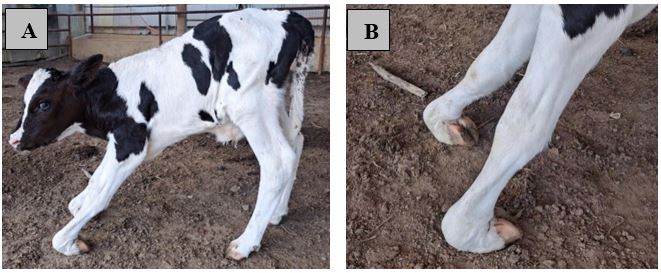
A. Clinical appearance of Knuckling; B. Bilateral flexion of fetlock joint.
Diagnosis
Based on clinical history and physical examination, the calf was diagnosed with bilateral Digital Flexural Deformity (DFD) commonly known as knuckling. No other congenital anomalies were detected and the calf remained otherwise clinically sound. Radiographic examination was not performed as the calf was examined and treated at the owner’s premises in a remote rural area where radiographic equipment was unavailable.
Therapeutic intervention
Considering bilateral involvement of digital flexor tendon, Z tenoplasty was performed as corrective procedure.
The calf was restrained in lateral recumbency. Mild sedation was done using diazepam (0.5 mg/kg, i.e., 3.5 mL of Inj. Sedil, 2mL vial) administered intravenously. A line block was then performed using 5ml of 2% lidocaine (Inj. G-Lidocaine, 50 mL vial) subcutaneously proximal to the incision site at both forelimbs.
Following aseptic preparation, a longitudinal skin incision was made on the medial aspects of the mid metacarpal region parallel to the affected tendon. The superficial digital flexor tendon was identified and exposed by blunt dissection (Figure 2A). A longitudinal cut was made on exposed tendon followed by two transverse cuts at opposite ends, creating a Z-shaped incision (Figure 2B). This allowed the tendons to be lengthened. The deep digital flexor tendon (DDFT) was treated the same way (Figure C, Figure D). The lengthened tendons were repositioned and sutured using a Pennington locking-loop pattern with 1-0 Prolene (Figure E). The skin was closed with a simple interrupted pattern using 1-0 Vicryl (Figure F). A plaster of Paris cast was then applied to both limbs to maintain alignment and provide immobilization.
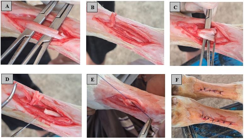
Correction procedure of Knuckling by Z-tenoplasty: A. Identification of Superior digital flexor tendon (SDFT) B. transecting SDFT C. Identification of Deep digital flexor tendon (DDFT) D. transecting DDFT E. Extension of tendon through Pennigton locking loop suture F. Closure of skin though simple interrupted suture.
As post-operative care, Ceftiofur sodium 1.1 mg/kg body weight (Inj. Tifur vet®, 500 mg vial), Flunixin Meglumine at 1.1 mg/kg bwt (Inj. Fixin vet®, 10 mL vial) and Pheniramine maleate at 1 mg/kg bwt Inj. Asta vet®, 10 mL vial) were administrated once intramuscularly for seven days.
Results
Immediately after surgery, substantial improvement in the alignment of both forelimbs was observed compared with preoperative position (Figure 3A). The plaster of Paris cast was removed on day 14. The calf was able to bear weight on both forelimbs with markedly improved limb positioning. At 28 days after surgery, the calf demonstrated restoration of standing posture normal gait compared with its preoperative condition (Figure 3B). No complications were seen, including wound infection, cast sores, excessive swelling, or tendon instability. The calf was able to stand and walk normally without assistance.
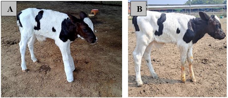
A. Immediate limb extension after surgery B. Restoration of normal posture and gait at follow up of 28 days.
Discussion
The present case demonstrated that bilateral digital flexural deformity (DFD) in a neonatal calf can be corrected using Z-tenoplasty and postoperative casting, even without hospital-based facilities. The calf regained functional weight bearing ability and walking within 28 days. The recovery timeline in this case falls within the range reported in comparable studies. Parashar (2017) documented functional recovery within similar postoperative period after flexor tenotomy in six bovine calves. Mc et al. (2024) also observed similar improvement in a day-old calf treated by tendon elongation, while Reddy and Sumiran (2021) reported favorable outcomes across eight calves managed by surgical correction of contracted flexor tendons. The 28 days recovery period of this report is therefore consistent with these reports and supports the reproducibility of Z-tenoplasty as a reliable surgical procedure.
The marked bilateral fetlock flexion and limited limb extension observed in this calf consistent with clinical characteristics of severe deformity described in neonatal calves, where impaired weight bearing and abnormal gait are commonly reported ; . In mild deformities, calves response to conservative approaches like physiotherapy, splitting and nutritional correction . Conversely, the pronounced tendon shortening and bilateral involvement observed in this calf required surgical correction to restore limb alignment . There are two core surgical methods for correction of DFD in neonatal calf: 1. Tenotomy and 2. Z-tenoplasty . Z-tenoplasty was preferred over tenotomy because tendon elongation allowed preservation of tendon continuity, thereby supporting more stable locomotion during growth, also documented at earlier studies ; ; .
Along with the tendon elongation, application of plaster of Paris cast after surgery played a supportive role by maintaining limb alignment and reducing pressure on the tendon. It facilitated progressive extension at healing .
In the present case, the calf was presented at three days, this early presentation combined with selection of Z-tenoplasty contributed to the good outcome. Thus, early age presentation, accurate diagnosis and appropriate selection of surgical technique are key determinants for good prognosis in calves with flexural deformity . This is because tendon and surrounding tissues are more pliable in neonatal life, making them easier to lengthen in caves treated later age .
The remote field conditions shaped several clinical decisions beyond the choice of procedure. Without access to radiography and ultrasonography, diagnosis depended entirely on clinical presentation and physical orthopedic evolution. Such method carries a higher risk of misclassifying tendon versus joint involvement compared with imaging support diagnosis. Similarly, without a controlled surgical environment, strict on-site asepsis and a full course of postoperative antibiotics were used to offset the added infection risk of the surgery.
Despite these constraints, the noticeable improvement in posture and gait within 28 days observed in this calf indicates that the combined surgical and immobilization protocol effectively restore the limb function. Such a protocol may be useful for veterinarians in clinical decision making and encourage to follow standardized treatment protocol for DFD in calves, particularly in resource limited area.
Conclusion
Z-tenoplasty combined with postoperative immobilization proved to be an effective surgical approach for correction of severe bilateral digital flexural deformity (DFD) in a three-day old calf. The protocol outlined in this report may serve as a practical surgical management approach for veterinarians managing similar cases in resource limited field condition in Bangladesh.
Acknowledgement
The authors acknowledge the owner of the calf for granting permission to report this case and for providing the consent for the surgical procedure and its use in publication.
Novelty statement
The novelty of the study lies in reporting standard management protocol of a common but rare congenital orthopedic disorder in neonatal calf. This case report documented a complete, replicable Z-tenoplasty protocol performed and managed entirely under remote field conditions, offering practical guidance for resource limited veterinary practice.
Authors’ contribution
Dr Abu Bakar and Dr Sajjad Hossain performed the surgical procedure. Dr Mehedi Hasan and Dr Thanim Islam drafted the manuscript and Dr Mizanur Rahman supervised the case and reviewed the manuscript. All authors read and approved the final manuscript.
Generative AI and AI-assisted technology statement
The authors declare that no generative AI and AI assisted technology was used in the creation of this manuscript.
Conflict of interests
The authors declare no conflict of interest.
References
- Anderson, D.E., Desrochers, A. and St. Jean, G., 2008. Management of Tendon Disorders in Cattle. Vet. Clin. N. Am. - Food Anim. Pract., 24(3): 551–566. https://doi.org/10.1016/j.cvfa.2008.07.008 [CrossRef]
- Chakraborty, S., Akter, M., Rahman, M., Yesmin, N., Juyena, N. and Alam, M., 2023. Congenital digital flexural deformity (knuckling): Epidemiology, the association of trace elements and surgical treatment in neonatal bovine calves. J. Adv. Vet. Anim. Res., 10(1): 88. https://doi.org/10.5455/javar.2023.j656 [CrossRef]
- Cooper, B. and Valentine, B., 2015. Jubb, Kennedy & Palmer’s Pathology of Domestic Animals, 6th ed., Vol. 1. Elsevier.
- Fazili, M.R., Bhattacharyya, H.K., Mir, M.U.R., Hafiz, A. and Tufani, N.A., 2014. Prevalence and effect of oxytetracycline on congenital fetlock knuckling in neonatal dairy calves. Onderstepoort J. Vet. Res., 81(1): 6 pages. [CrossRef]
- Fubini, S.L. and Ducharme, N.G., 2016. Farm Animal Surgery, 2nd ed., Elsevier Health Science.
- Mc, A., Karajagi, G. and Suresh, L., 2024. Surgical correction of congenital superficial and deep digital flexor tendon contracture in a day-old calf. 9(4): 117–119.
- Musa, G., Abdullah, K. and Caner, K., 2019. Degree of deformity and certain hormone, vitamin and trace element levels in the serum of calves with congenital flexural deformity. Medycyna Weterynaryjna, 75(6): 360–364. https://doi.org/10.21521/mw.6215 [CrossRef]
- Parashar, M.C., 2017. Surgical Management of Contracted Flexor Tendons—A Clinical Study of 6 Bovine calves. Intas Polivet, 18, 449–450.
- Rashmi, P.T. and Priyadharshini, 2018. Surgical management of congenital flexor tendon deformity in calves: A review of three cases. J. Entomol. Zool. Stud., 6(3): 544–546. [CrossRef]
- Reddy, K., Jagan M. and Sumiran, N., 2021. Surgical management of contracted flexor tendons at fetlock and carpal joint in calves- A Report of 8 Cases. Rumin. Sci., 10(2): 471–474.
- Sato, A., Kato, T. and Tajima, M., 2020. Flexor tendon transection and post-surgical external fixation in calves affected by severe metacarpophalangeal flexural deformity. J. Vet. Med. Sci., 82(10): 1480–1483. https://doi.org/10.1292/jvms.20-0057 [CrossRef]
- Schoiswohl, J., Eiter, J., Schwarzenbacher, H. and Kofler, J., 2019. Congenital flexural deformity in 93 calves—Appearance, treatment techniques and results of pedigree analysis. Schweizer Archiv Fur Tierheilkunde, 161(10): 677–688. https://doi.org/10.17236/sat00230 [CrossRef]

Services
Article usage statistics
Loading…
Related articles
- Virulence Profile of Enteropathogenic Escherichia Coli (Epec) Isolated From the Cases of Neonatal Calf DiarrheaAdvances in Animal and Veterinary Sciences · 2019
- Correction of the Immune Status of Cows by Using Aminoseleton During the Dry Period for Prevention of Antenatal Calf HypotrophyJournal of Animal Health and Production · 2019
- EFFECT OF CLIMATIC CONDITIONS ON WEED FREQUENCY OF WHEAT FIELDS AND COMPARISON OF GOLESTAN PROVINCE CITIES BY CLUSTER ANALYSIS (A Case study of IranPakistan Journal of Weed Science Research · 2024
- The Efficacy and Persistence of Spodoptera littoralis Nucleopolyhedrovirus (SpliMNPV) Applied in UV Protectants against the Beet Armyworm, Spodoptera exigua (Hubner) (Lepidoptera: Noctuidae) under Saudi Field ConditionsPakistan Journal of Zoology · 2018
- Suspected Bacillary Haemoglobinuria in a Calf: A Case ReportAdvances in Animal and Veterinary Sciences · 2016
- Neonatal Calf Diarrhea Caused by Escherichia coli: Pathogenic Mechanisms and Modern Control StrategiesVeterinary Sciences: Research and Reviews · 2026