
Pseudomonas aeruginosa biofilms pose a significant clinical problem in veterinary medicine, such as canine otitis externa, bovine mastitis, equine endometritis, and infections of veterinary medical devices. The alginate, Pel, and Psl polysaccharides, extracellular DNA (eDNA), and proteins form a physical and biochemical barrier that can contribute to antimicrobial resistance up to 1000 times greater than that of planktonic cells. Treatment of these infections is becoming increasingly problematic as conventional antibiotic treatment may fail to eradicate the biofilm-associated infections, and as multidrug-resistant (MDR) infections are becoming a growing therapeutic problem. The extracellular matrix of P. aeruginosa biofilms impedes antibiotic penetration, protects persisting cells, and protects bacteria from host immune responses, making conventional antibiotic treatment ineffective against biofilms. Bacteriophages, especially lytic, depolymerase-encoding phages, degrade exopolysaccharide components, multiply and penetrate the biofilm architecture, and lyse metabolically active bacterial cells. This process represents a mechanistically different approach to anti-biofilm. The aim of this review is to consolidate the existing experimental data regarding phage-based anti-biofilm approaches, phage-antibiotic synergy (PAS), phage-based therapeutic strategies, and their use in veterinary infection models. Preclinical results of in vitro, ex vivo, and in vivo experiments are analysed together with existing limitations, regulatory frameworks, and future research needs. Phage therapy is a scientifically viable approach to disrupt P. aeruginosa biofilms in veterinary applications, but it needs standardization, a strong safety profile, clinical testing in target veterinary species, and harmonized regulatory pathways.
Review · Volume 14, Issue 12, Pages 2689–2709, 4 November 2026 · Open Access
Bacteriophage-Mediated Biofilm Disruption Against Pseudomonas aeruginosa in Veterinary Medicine: A Review
· · · · ·
Affiliations & Notes ⌄
- 1Faculty of Animal Science and Technology, Universitas Brawijaya, Malang, 65145, Indonesia
- 2Department of Biology, Faculty of Science, Technology and Mathematics, Universitas Brawijaya, Malang, 65145, Indonesia
- *Correspondence | Veronica Margareta Ani Nurgiartiningsih
Article Info ⌄
- Journal:
- Advances in Animal and Veterinary Sciences
- Submitted Date:
- 1 August 2026
- Accepted Date:
- 24 September 2026
- Published Date:
- 4 November 2026
Abstract
INTRODUCTION
Pseudomonas aeruginosa (P. aeruginosa) is a Gram-negative opportunistic pathogen of major clinical significance in human and veterinary medicine. It is widespread in the environment and is recognized as a zoonotic pathogen that can spread between animal, human, and environmental reservoirs . The pathogen is highly adaptable and has a high degree of genomic flexibility, making it a formidable nosocomial threat and capable of inducing a wide range of infections, including soft-tissue lesions, bacteremia, and pneumonia ; . In veterinary medicine, P. aeruginosa infects many domestic animals, including dogs, mares, chinchillas, and dairy animals, which develop ear infections, pneumonia, septicemia, and enteritis ; . One of the most common inflammatory diseases is that of canine otitis externa, a condition that affects about 10-20% of dogs and is one of the most common conditions seen in veterinary practice ; .
The emergence and spread of multidrug resistant (MDR) P. aeruginosa isolates have limited treatment options for this organism. MDR P. aeruginosa is a high-priority pathogen for the World Health Organization and is a growing problem in clinical veterinary practice ; . The prevalence of MDR is reported to be widely variable across different parts of the world and different sources of isolates. In a single centre veterinary study of 155 clinical isolates of P. aeruginosa collected over seven years (2007–2013) at an academic veterinary hospital in South Africa, a high proportion of the isolates (92% of the 155 isolates) were identified as MDR with a particularly high resistance to amoxicillin-clavulanic acid (93%), penicillin-G (96%) and doxycycline (87%) and a comparatively low resistance to imipenem (6%), gentamicin (18%) and tobramycin (12%) . In contrast, one commonly quoted figure of 56% MDR among 50 P. aeruginosa isolates originated from human clinical laboratories in Egypt, and resistance was focused on aminoglycosides, fluoroquinolones, and monobactams; this study was based on human hospital isolates and is presented here for comparison, but not as an indication of veterinary resistance patterns . As in the South African study, results from any single hospital or region should not be extrapolated to the entire veterinary population without additional multi-site surveillance. Moreover, fluoroquinolone resistance has developed in bacteria via upregulation of efflux pumps and mutations in the gyrA and parC genes, and is causing complications in veterinary therapeutics ; . Treatment is becoming challenging due to acquired resistance by horizontal gene transfer and mutations, as well as intrinsic resistance through outer membrane barriers and multidrug efflux pumps ; .
Biofilm as a virulence factor
P. aeruginosa is mostly found in biofilms rather than as free-swimming planktonic cells ; . Biofilms are structured microbial communities in extracellular polymeric substances under permissive environmental conditions, which are composed of polysaccharides, proteins, lipids, and extracellular DNA (eDNA). These biofilms shield bacteria from environmental stresses and phagocytosis, favoring persistent infection . Biofilm-associated infections are persistent and hard to treat with standard antimicrobials, and are responsible for recurrent, treatment-refractory disease in veterinary species ; .
Hemolysins, rhamnolipids, and proteases are some of the virulence factors that play a role in the pathogenesis of P. aeruginosa beyond the role of the biofilm itself . In the context of biofilm, quorum sensing regulates the behavior of bacteria, thereby promoting the production of the matrix, evading host immunity, and helping resist antimicrobials. These resistance mechanisms, including β-lactamase production, efflux pumps, and biofilm formation, make treatment difficult and highlight the need for alternative therapeutic strategies ; .
Limitations of conventional veterinary antibiotics and antiseptics
P. aeruginosa has both intrinsic and acquired resistance to many antimicrobials. Intrinsic resistance mechanisms include chromosomally encoded pumps and low outer membrane permeability ; . Antibiotic resistance has been promoted by the indiscriminate use of antibiotics in both human and veterinary medicine, including the emergence of resistance to carbapenems, which are considered last-resort antimicrobial drugs . The treatment of P. aeruginosa infections is becoming a significant challenge as antibiotic resistance is increasing, giving clinicians limited options to treat the infection .
Biofilm resistance further complicates the issue because the biofilm-forming organisms are found to be resistant to concentrations of antibiotics, disinfectants, and biocides that would never be achieved clinically in systemic use . Even the use of traditional antiseptics for ear canal infections or wounds may not work against existing biofilms and traditional methods of wound management (debridement, lavage, topical antimicrobials) are also not effective against P. aeruginosa biofilms. Moreover, biofilms reform in 24 hours or less after treatment, indicating that the current standard of care for managing biofilm-related infections in veterinary medicine may have limited efficacy in eradicating established biofilms ; ; .
Why phages? Historical context and resurgence
The use of bacteriophages as therapeutic agents dates back to the early part of the twentieth century, prior to the development of antibiotics. In the 1910s, Twort and d’Hérelle described the bacteriophages independently, and in the 1920s and 1930s, phage preparations were marketed commercially and used in the treatment of human and veterinary diseases with significant scientific and commercial success, such as fowl typhoid and bovine hemorrhagic septicemia .
Therapy using phages was, however, mostly forgotten in North America and Western Europe in the 1940s for a number of intertwined reasons. Initial preparations were very rough, poorly titrated, and often contaminated, and the biological characteristics of phages were not well understood, such as the distinction between lytic and temperate phages, and the high host specificity of phages, so preparations were often used on bacteria that they were unable to lyse, and the results of clinical trials were erratic . Controlled trials were rare, and the ones performed were not conducted with randomization and control groups and had statistically inconclusive results, such as the British Indian government’s “Bacteriophage Inquiry.” Penicillin was produced in large quantities during the Second World War, and standardized and broad-spectrum antibiotics and sulphonamides were quickly developed, making them a convenient, effective, and reliable alternative, and scientific debates about d’Hérelle undermined trust. During the Cold War, the use of phage therapy was then limited to a few centers in the Soviet Union (the Eliava Institute, Tbilisi) and Poland (Hirszfeld Institute, Wrocław) .
Although the use of phages for therapeutic purposes has declined since the introduction and application of antibiotics, with the development of antimicrobial resistance (AMR) as a global issue, there has been a renewed interest in the scientific community . These historical limitations directly influence the current strategies of veterinary development: the failure in the past to standardize and control the production process is now addressed by good manufacturing practice production, the endotoxin removal and the full genome characterization of phages, excluding temperate and virulence-gene-carrying phages ; ; the historical problem of narrow and unpredictable host range is compensated with the use of phage banks, cocktail formulation and pre-treatment susceptibility tests of clinical isolates; the lack of controlled clinical evidence in the past has prompted the requirement for randomized, adequately powered field trials in target veterinary species before regulatory approval ; . The benefits of phage over antibiotics are a good safety profile, host specificity, antibiotic resistance and biofilm inhibition/disruption ; ; .
Objectives of the review
The goal of this review is to present a broad overview of the existing knowledge about the use of bacteriophages to disrupt P. aeruginosa biofilms in the context of veterinary medicine. In particular, the review discusses: (i) the range of P. aeruginosa biofilm-associated diseases in veterinary species; (ii) the structural and functional barriers posed by P. aeruginosa biofilms; (iii) how bacteriophages disrupt P. aeruginosa biofilms; (iv) preclinical and experimental evidence in veterinary-relevant models; (v) current challenges and limitations; and (vi) future research priorities required to progress P. aeruginosa phage therapy to veterinary clinical use.
The mechanism of phage-mediated biofilm disruption (matrix degradation, depolymerase activity, and persister cell dynamics) has been well characterized in the context of human and environmental diseases ; , but not in the specific veterinary disease contexts in which it is clinically applied. This review aims to fill this void by synthesizing evidence for veterinary otitis externa ; , 2025), bovine mastitis , equine genital tract infection , and veterinary device-associated infections ; , as well as the different delivery methods (topical, intra-mammary, intrauterine) and veterinary regulatory pathways that set apart clinical translation in animals from human medicine. There is no evidence that the biology of P. aeruginosa–phage is fundamentally different in human and animal hosts; if mechanistic data specific to veterinary isolates are not available, this is noted explicitly as a priority for future research, but not extrapolated uncritically from human studies.
Pseudomonas aeruginosa biofilm in animal diseases
Canine otitis externa
P. aeruginosa is a major pathogen in canine otitis externa. Veterinary otitis externa affects about 10–20% of dogs and is one of the most common presenting conditions ; . The proportion of P. aeruginosa among canine isolates from otitis externa has been reported to range from approximately 6% to more than 35%, with several recent studies reporting 19-20% , 2025). P. aeruginosa has been reported in up to 35% of chronic and recalcitrant cases of otitis externa ; , but is less prevalent in acute cases . Biofilm production has been identified in 40-95% of isolates (depending on the study) of P. aeruginosa collected from dogs with ear infections ; , and geographical differences are known, as there are differences in prevalence between isolates from the UK, Europe, and Asia . Breed-specific data in the current literature specific to P. aeruginosa-associated otitis is limited, other than the fact that breeds with pendulous ears or allergic skin disease are more susceptible to otitis externa generally . The main bacteria involved in exacerbating clinical signs and promoting disease progression in canine otitis externa are thought to be P. aeruginosa and Staphylococcus pseudintermedius. Both pathogens are commonly reported as being associated with chronic recurrent otitis in dogs .
The ability of P. aeruginosa to form biofilm is an important virulence factor and likely contributes to chronic otitis externa in dogs. Biofilms lead to antimicrobial tolerance, chronic infections, and diminish the effectiveness of traditional antimicrobial therapy . Both experimental and clinical studies have been conducted on the ability of bacteriophages to inhibit P. aeruginosa strains isolated from dogs with clinical otitis externa. One study showed that a specially developed phage cocktail eliminated 90% of P. aeruginosa strains in an in vivo experimental model of canine otitis externa .
In addition to otitis, P. aeruginosa is a significant pathogen in chronic deep pyoderma and wound infections in dogs. Fluoroquinolone-resistant clinical isolates have been recovered from canine skin diseases such as otitis externa, chronic deep pyoderma, and wound infections ; . P. aeruginosa has also been reported in canine urinary tract infections by Hattab et al. (2021) and Jasim and Hayyawi (2025), and it has also been reported as a pathogen of bacterial keratitis in dogs by Santos et al. (2011).
Bovine mastitis
P. aeruginosa is a cause of clinical bovine mastitis, leading to economic losses in dairy production . MDR P. aeruginosa strains obtained from milk samples of mastitis cows are a therapeutic challenge due to their resistance patterns and ability to form biofilms . P. aeruginosa has also been reported as a cause of mastitis in dairy animals by De Melo et al. (2019).
Equine endometritis
Chronic infectious endometritis is a major reproductive disease in mares, affecting 25–60% of barren mares and resulting in significant economic losses in the equine breeding industry . P. aeruginosa and Klebsiella pneumoniae are regarded as relevant equine genital bacteria due to their strong ability to form biofilms and high antibiotic resistance. The biofilm state is especially problematic in the uterine environment, as it limits the success of antibiotic treatment, and repeated infusions with antibiotics can also promote the development of antimicrobial resistance . P. aeruginosa can also be responsible for endometritis in mares after breeding or obstetric manipulations .
Infections in mink and chinchillas
The pathogen has been isolated from minks with hemorrhagic pneumonia, and phage vB_PaeM_LS1 targeting these strains has been shown to be effective at lysing all P. aeruginosa strains isolated from minks ; . Ear infection, pneumonia, septicemia, and enteritis are also clinical conditions that can result from P. aeruginosa infection in chinchillas .
Biofilm on medical devices
P. aeruginosa readily forms biofilms on different abiotic surfaces such as stainless steel, silicone, and different medical devices. The pathogen is well documented to form biofilms on indwelling urinary catheters, which lead to catheter associated urinary tract infections. Biofilms on the surfaces of catheters are more resistant to antibiotics due to the ability of the biofilm matrix to protect it, making it less accessible to the antimicrobials ; .
A specific application of phage therapy that is well studied in a human derived in vitro model is the control of urinary catheter biofilms, with the phage vB_Ps_ZCPS13 significantly reducing P. aeruginosa from the surface of urinary catheters in controlled experiments .
Structural and functional barriers of Pseudomonas aeruginosa biofilm
Biofilm matrix components: Alginate, Pel, Psl, and extracellular DNA
P. aeruginosa biofilms have a complex extracellular matrix (ECM) that consists of several exopolysaccharide components, extracellular DNA (eDNA), and proteins. Alginate is one of the major exopolysaccharides produced by P. aeruginosa and the major matrix component of mucoid strains, typical of chronic infections. In non-mucoid environmental and early infection isolates, both Pel and Psl polysaccharides contribute to structural and adhesion functions . All these components together produce an extracellular matrix that serves as a physical barrier to antibiotic penetration ; .
Extracellular DNA released by autolysis or active secretion is crucial for biofilm structure and cohesion. The collective matrix also offers better survival of embedded bacteria by acting as a physical barrier to antimicrobial agents ; . The depolymerase enzymes encoded by phages are especially pertinent to this barrier, as they can break alginate polymers and other biofilm components, thereby providing a mechanism by which the phages disrupt the biofilm in addition to their direct bactericidal action ; . The relative contribution of these matrix components is strain-phenotype dependent and does not set a fixed ratio; it is usually altered by the occurrence of a mutation in the mucA regulatory gene during chronic infection, resulting in a matrix composition shifted from a Pel/Psl dominated structure towards an alginate dominated structure ; . These changes have an immediate impact on phage susceptibility. Mucoid conversion may itself be a phage resistance mechanism: non-mucoid P. aeruginosa were exposed to a lytic/temperate phage and the resultant mucoid variants were obtained and found to be stably resistant, with the mucoid layer suggested to be responsible for the resistance to phage adsorption . This results in a partial counterbalance to the penetration described above mediated by a depolymerase that is not present in all phages and which cannot degrade the matrix component protecting a particular strain . The susceptibility was also regulated by biofilm maturation stage as in a recent study comparing two Psl-targeting P. aeruginosa phages both were able to prevent the initial attachment on the bacterial surface, but only one phage was capable of disrupting a mature biofilm in flow culture; in this study, phage pressure also quickly selected for escape mutants that lost the ability to adhere to surfaces, which may be a useful therapeutic strategy to shift the bacteria into a more antibiotic susceptible planktonic phase . All of these results suggest that mucoid/non-mucoid phenotype and biofilm maturation stage are not random factors, but rather active factors that affect the efficacy of phages and should be taken into consideration when selecting phages and designing phage cocktails ; .
Persister cells and phenotypic resistance
Persister cells are a sub-population of metabolically dormant bacteria within the biofilm that are phenotypically resistant to antibiotic action, but do not contain a genetic resistance mutation . Biofilms consist of nucleic acids, proteins, polysaccharides, lipids, and bacteria with reduced metabolic and replication rates, and these low-metabolism cells are not very susceptible to antibiotics that require active cellular processes to function ; . Biofilm formation plays a significant role in the resistance of bacteria to quinolones, aminoglycosides, and β-lactams, and facilitates long-term persistence in part by providing protection to persister sub-populations .
The chronic and relapsing nature of biofilm-associated infections is partly explained by the persistence phenomenon ; . Persister cells survive antibiotic treatment to repopulate the biofilm when the antibiotic is removed .
Physical shielding from antibiotics and host immunity
The polysaccharide matrix of P. aeruginosa biofilms limits antibiotic penetration, contributing to bacterial resistance . Not all antibiotics are equally effective in penetrating this barrier. Penetration of small, zwitterionic fluoroquinolones into the biofilm matrix is rapid and unhindered ; , whereas cationic aminoglycosides (e.g., gentamicin, tobramycin) are substantially retarded, binding electrostatically to the anionic polymers of the matrix, particularly extracellular DNA , which chelates cations and can induce up to 640-fold increased aminoglycoside resistance without affecting β-lactam or fluoroquinolone susceptibility . β-lactam penetration is also delayed, and this delay can be synergistic with the matrix-localised β-lactamase activity, giving the enzyme more time to degrade the antibiotic before it reaches its bacterial target . Recent studies with defined 3D biofilm models have further complicated this class-based picture, demonstrating that antibiotic diffusion through the alginate/eDNA matrix is not the predominant cause of tolerance for a number of the tested antibiotics and that the binding sites within the matrix are filled over time, irrespective of antibiotic class, indicating that matrix-independent adaptive mechanisms also play a significant role in biofilm antibiotic tolerance . These class-specific differences in matrix penetration have direct implications for phage-antibiotic combination strategies. Phage-derived depolymerases would be expected to be most beneficial in combination with the most matrix-dependent antibiotics (aminoglycosides and β-lactams) compared with the less matrix-dependent antibiotics (fluoroquinolones) ; . This specific hypothesis has not been directly tested to our knowledge, and is a concrete testable direction for future research on the synergy of phages and antibiotics. Antimicrobial resistance of biofilms can be up to 1,000-fold higher than that of planktonic cells ; . In addition to antibiotic protection, the biofilm matrix shields bacteria from host immune surveillance, including phagocytosis and other immune effector mechanisms ; . The presence of biofilm-associated resistance in already antibiotic-resistant bacteria adds to the therapeutic challenge of MDR P. aeruginosa infections . In other situations, embedded bacteria can survive phage attack even when the planktonic cells are susceptible to phages, highlighting the need for careful selection of phages for anti-biofilm therapy , (See Figure 1).
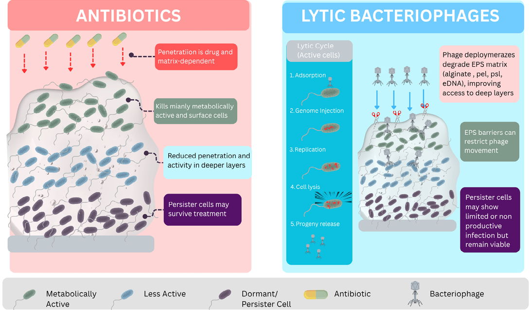
Comparison of antibiotic and lytic bacteriophage actions within Pseudomonas aeruginosa biofilms. Side-by-side comparison of the two therapeutic modalities discussed in this review. Left (Antibiotics): Penetration is drug- and matrix-dependent; antibiotics preferentially kill metabolically active surface cells, with reduced penetration and activity in deeper, less metabolically active layers, allowing dormant persister cell populations to survive treatment. Right (Lytic bacteriophages): Infection follows a receptor- and physiology-dependent five-step lytic cycle (adsorption, genome injection, replication, cell lysis, progeny release) restricted to metabolically active, susceptible cells. Phage-encoded depolymerases degrade the EPS matrix (alginate, Pel, Psl, eDNA), improving phage access to deeper biofilm layers. Dormant/persister cells generally limit productive phage infection: phage genome entry can be followed by non-productive “hibernation” rather than lysis, with the cell remaining viable until resuscitation. EPS matrix diffusion barriers can also restrict phage movement through the biofilm.
Bacteriophages as anti-biofilm agents
Lytic versus temperate phages: Why lytic phages are preferred
The difference between the lytic and lysogenic (temperate) phage life cycles is very significant in terms of therapeutic applications ; . Once a lytic phage infects its host, it kills the host and replicates itself . Temperate phages, on the other hand, can enter into the host’s chromosome and become a prophage, which replicates along with the host’s genome without immediately lysing it ; .
Therapeutic phages are usually virulent and strictly lytic, as temperate phages have the potential to transfer virulence or resistance genes horizontally and to become integrated into the bacterial genome ; . However, this is not a universal rule: temperate phages, in the form of obligately lytic derivatives, have been successfully used in human therapy, such as a ground-breaking study for disseminated Mycobacterium abscessus infection . This safety concern does not apply to endolysins from temperate phages because the purified protein is used, not the viable phages .
The formation of clear plaques on bacterial lawns is one of the features that can indicate the lytic activity, whereas turbid plaques indicate lysogeny . Several phages against P. aeruginosa have been characterized as lytic, such as vB_PaeP_PS28 , vB_PaeM_PKMS3 , PAJD-1 , phage AZ1 , and vB_PaeM_LS1 .
Mechanisms of biofilm disruption
Depolymerase degrading exopolysaccharides
Polysaccharide depolymerases are the anti-biofilm enzymes found in bacteriophages. These enzymes are usually attached to the tail spikes or tail fibers of phages and break down exopolysaccharide components, which degrade the structural framework of the biofilm matrix . These enzymes are not uniform in substrate specificity: There are alginate lyases specific to the mannuronate/guluronate polymer of alginate, which are found in P. aeruginosa and Azotobacter phages , and specific enzymes to Psl or Pel, as found in P. aeruginosa’s own matrix-remodelling enzymes PslG and PelA . Ideally, the dominant matrix component of the target strain should be taken into account when designing a phage cocktail, with alginate lyase-encoding phages selected for use against mucoid isolates and Psl/Pel-targeting phages selected for use against non-mucoid isolates, but tools to quickly identify the matrix component at the point of care are not yet available ; . Phages may encode extracellular polysaccharide depolymerases that aid biofilm penetration and degradation of the extracellular polysaccharide matrix, which can enhance phage-antibiotic synergism .
Halo formation around plaques in bacterial lawns can serve as a phenotypic marker for depolymerase activity, as the enzyme diffuses out of the plaque and degrades surrounding exopolysaccharides . Phage Motto was found to have efficient biofilm degrading activity , and phages with putative depolymerase activity tested in synthetic CF sputum medium were found to have antibiofilm activity stronger than that of high-concentration antibiotics . Mechanistically, depolymerase genes are important in successfully selecting phages for effective anti-biofilm activity . The absence of these genes has been suggested as an explanation for the failure of some phages to reduce established biofilms in vitro .
Penetration via phage multiplication
A key advantage of phage therapy over chemical anti-biofilm agents is the ability to amplify exponentially at the infection site. Phages multiply inside the infected host bacteria, leading to an increase in the number of phages at the infection site and facilitating the penetration and destruction of the biofilm ; . One of the important operational advantages is that the concentration of the phages can be raised at the infection site by auto-dosing . This property is especially useful in the biofilm environment, where the matrix restricts the access of externally added agents to all bacterial populations .
Therapeutic phage candidates, therefore, should have high replication rates. The latent period of Phage vB_PaeP_PS28 was 15 minutes, with a burst size of approximately 210 virions per infected bacterium . In the case of P. aeruginosa phages isolated from dogs with otitis, the latent periods were short, up to 30 minutes, and phage titers after one hour post-infection were 7.92–9.06 log PFU/mL . The latent period and burst size of Phage AZ1 were 33 minutes and 326 phages/infected cell, respectively . For PAJD-1, the latent period and burst size were 20 minutes and 223 PFU/infected cell, respectively . Biofilm architecture, however, has important constraints on the movement and replication of phages. The EPS matrix may disrupt the diffusion of phages in the biofilm, and dense cell clusters formed by strong cell-cell adhesion may restrict the diffusion of phages to the entire community . Nutrient gradients result in high nutrient availability in the outer layers, and lower availability as depth increases, with cells that are closer to the liquid interface growing at a higher rate, and cells that are deeper in the interior of the biofilm growing at a relatively lower rate . Bacteria in structured colonies can be sensitive but not be lysed, primarily because of a decrease in growth in the centre of the colonies and not loss of receptors . This spatial structure and metabolic stratification of the biofilm restricts the access to cells and the successful proliferation of phages, which is why some phages are able to reduce the biofilm only to a certain extent, and that is why combination treatments (phage-antibiotic synergy, depolymerase-producing phages) are often necessary for deeper biofilm eradication ; ; (See Figure 2).
Killing of metabolically active and persister cells
Lytic phage infection is usually associated with the growth rate of the host cell, and the fully growth-arrested cells are, in general, resistant to productive phage replication . Commonly studied lytic phages typically respond to a dormant host cell in one of two ways: Either failing to adsorb altogether, or adsorbing and entering a quiescent, non-productive “hibernation” state in which the phage genome persists without replicating or killing the cell until the host resuscitates naturally and sufficient metabolic activity resumes; only then can the phage complete its lytic cycle . The clinical importance of this distinction is that during dormancy, the hibernating phages do not actively kill persister cells, but rather await a “resuscitation” event that is not caused by the phages . One rare exception has been identified, a recently isolated P. aeruginosa phage, Paride, which was demonstrated to be capable of direct lytic replication and killing within antibiotic-tolerant, deep dormant cells without prior resuscitation, using the host’s own stress-response machinery; this phage completely sterilized the deep dormant population when combined with the carbapenem meropenem, but only killed ~99% of the dormant cells alone , and this ability seems to be very rare among characterised phages . Mechanistically, Paride’s ability to replicate in dormant cells relies on the hijacking of host stress-response pathways, not on any phage-intrinsic mechanism that is not directly related to host physiology, as is the case for antibiotic tolerance . These stress-response systems are widely shared amongst Gram-negative bacteria, and the principle may be applicable to other species beyond P. aeruginosa, but this has not been confirmed. Significantly, Paride was discovered by a targeted screen that was developed to detect phages that can infect dormant hosts, indicating that this characteristic may not be as unusual as it seems and that it may be more common if targeted searches are made for it. The exact genes that are responsible for this have not yet been identified, and it is unclear if this trait could be transferred to other phages. This review explicitly states this as an important open question for the study of phage therapy against persister populations associated with biofilms, as no study to date has directly monitored the dynamics of persister cell resuscitation and phage infection in real-time.
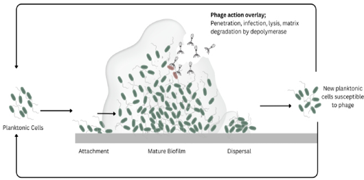
Biofilm life cycle and bacteriophage action sites.Schematic overview of Pseudomonas aeruginosa biofilm development and the stages at which Lytic bacteriophages act. (1) Planktonic phase: free-floating bacteria in the bulk fluid. (2) Attachment: bacteria adhere to a substratum, progressing from reversible to irreversible attachment. (3) Maturation: development of a structured biofilm with an EPS matrix (alginate, Pel, Psl, eDNA), water channels, and embedded microcolonies containing metabolically active cells, dormant/persister cells, and phage-resistant mutants, phage infection of susceptible cells, and depolymerase-mediated matrix degradation, which enhances further phage penetration and biofilm dispersion. (4) Dispersal: bacteria or small clusters detach from the biofilm and return to the planktonic state; (5) newly dispersed cells remain susceptible to phage infection, limiting the seeding of new infection sites.
The presence of various characterized phages that degraded mature biofilms, as shown by the scanning electron microscopy (SEM) of the destruction of the architecture of the biofilm after treatment ; and the crystal violet biomass assays, indirectly indicates activity against metabolically active cells within the biofilm population. Direct killing of bacteria and degradation of biofilm matrix work together to decrease the number of viable bacteria and biofilm biomass, although it is possible that not all the persister cells are eliminated in one application .
Phage-antibiotic synergy against biofilms
The phenomenon of Phage-Antibiotic Synergy (PAS) is the ability of sub-inhibitory or inhibitory concentrations of antibiotics to enhance the effect of the phage against the bacteria and biofilm . The mechanism of PAS is based on several complementary interactions: Phage mediated matrix degradation enhances antibiotic penetration into the biofilm ; phage-resistant mutants may compensate for receptor loss by downregulating the expression of efflux pumps and paradoxically making them more susceptible to antibiotics .
There is strong experimental evidence supporting PAS for P. aeruginosa biofilms. The anti-biofilm activity of combined ciprofloxacin and phages was significantly better against dual-species P. aeruginosa and MRSA biofilms than either agent alone. The dual-species biofilms were completely eradicated after staggered exposure to phages for 12 hours, followed by ciprofloxacin (1 mg/L), a concentration far below the one required to eradicate the biofilm alone. Importantly, phages were administered sequentially, followed by antibiotics, which proved more effective than simultaneous administration. The heat production (a metabolic indicator) was more than 90% lower in the simultaneous treatment with phage and antibiotic than in the untreated biofilms .
The advantage of phage-first sequencing is believed to be that the phage can replicate extensively, largely unhindered by the presence of an antibiotic, before the antibiotic interferes with the bacterial machinery that the phage relies on, or with bacterial replication itself, and thus antagonises the phage amplification . However, this finding does not translate across the board: The pre-exposure interval of the phage, the concentration of the antibiotic, and the type of antibiotic (synergism or antagonism) are all reported to vary by study, and no single optimal timing or dosing has yet been determined throughout the literature. The optimal interval has been reported as 12 hours pre-exposure to near-simultaneous administration in other systems, and this is likely to depend on the system being used. There is also a risk of antagonism, as sequencing can be different across phage-antibiotic-pathogen combinations, with antibiotics that inhibit host transcription/translation before the phage has completed enough rounds of replication, limiting the ability to amplify the phage rather than killing it .
The combination of phage PH826 and ciprofloxacin inhibited biofilm biomass formation by 89.44-92.79 % after 48 hours . Synergistic interactions between vB_PaeM_PS3 and traditionally used antibiotics were reported in the treatment of Pseudomonas infections , and between isolated equine phages and gentamicin, enrofloxacin, and trimethoprim-sulfadiazine were confirmed in a veterinary setting . The combination of phages and antibiotics may enhance bacterial susceptibility to antibiotics , and the phage OMKO1 targets the OprM protein of the MexAB and MexXY efflux pumps, which enhance antibiotic susceptibility in MDR strains through an evolutionary trade-off mechanism .
Phages-derived endolysins and therapeutic strategies
In addition to natural phage isolates, biotechnological engineering of phage products is a new frontier in the field of anti-biofilm therapeutics ; . Peptidoglycan-degrading enzymes encoded by phages, called endolysins, can be recombinantly produced and used outside of the phage replication cycle as purified proteins to lyse bacteria . The phage-derived endolysin PlyPAJD-1 showed high bacteriolytic activity and a wide antibacterial spectrum against MDR P. aeruginosa, indicating the potential of these enzymes as therapeutic agents .
Endolysin therapy, however, is also limited in practice for veterinary use because of the short half-life in vivo, requiring repeated doses, and immunogenicity risk on repeated systemic injections, as seen with other endolysins in preclinical and clinical studies . Another restriction of the current review is that endolysins are generally less active against Gram-negative bacteria (GNB) than against Gram-positive bacteria (GPB) because the outer membrane of GNB prevents endolysins from reaching the peptidoglycan substrate, which requires co-formulation with membrane-permeabilising agents or protein engineering . A direct, quantified comparison of the cost of production of recombinant endolysin versus phage was not found in the literature and is therefore left as an open question, rather than a speculation on cost.
Preclinical and experimental evidence in veterinary models
In vitro biofilm reduction on surfaces
In vitro biofilm reduction assays have been used to establish baseline evidence of anti-biofilm action of phages and have used a variety of P. aeruginosa strains, including clinical isolates from dogs, cattle, horses, and minks ; . Several phages were characterised and have shown good reduction of preformed mature biofilms ; . Much of this evidence has a methodological caveat: Most of the biomass reduction values reported below come from crystal violet staining and measure total biomass, including dead cells and matrix material. A decrease in stained biomass does not necessarily correspond to a decrease in live bacterial load and may be due to matrix degradation alone, and viable bacteria may remain within protected areas in the remaining biofilm . Of the studies summarised below, only the otitis-relevant biofilm model provides a paired viable-count outcome (log CFU/well) along with the biomass reduction; the other studies provide a biomass percentage or structural (SEM) evidence but not a viable-count outcome.
A further restriction is host source. Of the phages with quantitative biofilm or mechanistic data, some were characterized against veterinary-origin isolates. The others are from sewage or environmental sources and were characterized against human clinical isolates or reference strains like PAO1, and include most of the latent period, burst size, and depolymerase data referenced in this review. We retain this evidence because P. aeruginosa is a single species moving between human and animal hosts with no known host-dependent divergence in phage receptor biology , but the efficacy is not necessarily transferable between strains, and the host range of phages is dependent on the biofilm matrix composition of the strains.
The mature biofilms of strong biofilm-producing P. aeruginosa isolates were significantly reduced by phage vB_PaeP_PS28 in a dose-dependent manner, with the highest activity observed at the multiplicity of infection (MOI) of 10. The phage was able to break down biofilms and reduce biomass in all the tested strains . Phage AZ1 was able to reduce 48-hour-old biofilm biomass by about threefold compared to the untreated control (P < 0.05), with SEM also showing the disruption of the biofilm . Phage vB_PaeM_LS1 effectively disrupted preformed 48 hours biofilms, and SEM showed that the biofilm architecture was destroyed after treatment .
A six-phage cocktail for broad host-range coverage reduced the biofilm biomass by 63% in PAO1, 64% in PaPh5, 37% in PaPh32, and 65% in PAK-lumi strains, after 4 hours of treatment with the cocktail . Phage vB_PaeP_PZH3 degraded established biofilm biomass by 31–45%, depending on MOI, and inhibited planktonic bacterial growth at various MOIs . Overall, Phage vB_Ps_ZCPS13 exhibited strong biofilm inhibition and clearance capabilities, successfully removing P. aeruginosa from the surface of urinary catheters . Phage Motto exhibited anti-biofilm properties against strong biofilm producers with ≥2-fold biofilm biomass reduction within 24 hours . In an in vitro otitis-relevant biofilm model, a phage cocktail combined with glycerol, glycine, and Tween 20 reduced P. aeruginosa biofilms by about 5.5 log CFU/well .
In one characterization study by Dalponte et al. (2025) that focused on veterinary applications, three of seven characterised phages significantly decreased the biofilm biomass by up to 93.38%, and phage PH826 showed strong lytic activity against MDR P. aeruginosa at various MOIs . Phages PA-319, PA-575, and PA-711 exhibited strong antibiofilm activity and showed greater biofilm-disrupting activity than high concentrations of antibiotics in the synthetic CF sputum medium . Key phage characteristics are summarised in Table 1.
Ex vivo models
The ex vivo model is a tissue-based model, ranging between in vitro culture and whole animal experiments in terms of physiologic evidence. Two related but different systems were used by Dalponte et al. (2026). One was an ex vivo canine skin model based on excised skin, and another was a reconstructed canine epidermal equivalent consisting of cultured keratinocytes. In the ex vivo skin model, bacterial load was decreased by approximately 4 log units (P < 0.05), and SEM and immunofluorescence microscopy showed disruption of the biofilm structure. In the epidermal equivalent model, bacterial burden was also decreased from 8.33 ± 0.64 to 1.18 ± 0.64 log₁₀ CFU/mL (P = 0.001), and no bacteria were detected in 6 of 9 samples treated with phages, and CXCL8, an inflammatory marker secreted by keratinocytes, was also decreased.
As to the complexity of the models: the epidermal equivalent model was specifically confirmed to not affect the integrity of the skin barrier, however as an in vitro reconstructed keratinocyte-based system, it does not include the presence of resident immune cells, vasculature and systemic recruitment of immune cells; while genuine excised ex vivo skin does have the presence of skin-associated immune cells, it does not have the influx of circulating immune cells, as it is not perfused . This review does not know if native skin flora was included or excluded in either model, nor can it be sure from published data. Review explicitly states that both models were not ear-canal-specific, but rather used generalized canine skin or epidermis because the ear canal has its own microenvironment (cerumen composition, humidity, resident flora, anatomy) that was not captured by these skin models, and therefore their representativeness of chronic otitis externa should be interpreted with caution .
In vivo studies
Veterinary relevant mouse wound infection models
P. aeruginosa mouse models have been widely used to test the efficacy of phages in vivo. The results demonstrated that mice infected with P. aeruginosa and treated with vB_PaeP_PS28 exhibited significantly better survival compared to the infected mice that were not treated, with all the infected mice not treated dying within 24 hours, while the infected mice treated with vB_PaeP_PS28 had significantly reduced mortality, supporting the potential of phage therapy for managing P. aeruginosa infections in veterinary medicine ; . Phage treatment remarkably lowered bacterial counts in liver and spleen: The liver bacterial loads of 3,193 ± 12 CFU/g in phage-treated animals compared to 3.6 × 10⁵ ± 23 CFU/g in untreated animals, and the spleen bacterial loads of 5,134 ± 13 CFU/g in phage-treated animals versus 8.8 × 10⁵ ± 15 CFU/g in untreated animals .
Phage vB_PaeP_PZH3 also enhanced wound healing and repair in a mouse wound infection model, and the number of bacteria and wound area in the phage-treated mice were significantly reduced compared to the control infected mice . Phage vB_PaeM_PS3 resulted in a significant increase in survival rate and a marked decrease in the colonization of bacteria in the spleen and liver of infected mice . A six-phage cocktail was effective against acute respiratory infection in a murine model . In addition, phages have proven to be effective against murine models of systemic infection and mouse models of burn wound infection ; . In an otitis externa mouse model, other formulation ingredients (glycerol, glycine, and Tween 20) improved the efficacy of the phage cocktails against P. aeruginosa .
The mouse mastitis model has directly provided veterinary-translatable evidence: the intramammary injection of phage PAJD-1 decreased the number of bacteria and repaired the damage to the mammary gland in mice infected with MDR P. aeruginosa . The results obtained in mice should be interpreted with caution when extrapolating to the target veterinary species, as there are significant differences in immune cell subset composition, skin structure and wound-healing kinetics, and overall body size and physiology between mice and dogs, horses and cattle, which have previously led to efficacy
Table 1: Characterized bacteriophages with therapeutic and anti-biofilm potential against Pseudomonas aeruginosa.
| Phage/ Cocktail | Host origin and source of test isolates | Anti‑biofilm mechanism | Reported biofilm reduction/ efficacy | Reference |
| vB_PaeP_PS28 | Sewage / Human clinical isolates (systemic infection model) | Lytic phage (Podoviridae, N4‑like); rapid replication (15‑min latent period, ~210 virions/bacterium); genome free of resistance/lysogeny genes | 100% survival vs. 0% in untreated mice; ~100‑fold reduction in liver/spleen; significant biofilm degradation (MOI‑dependent) | |
| vB_PaeM_PKMS3 | Sewage (Kochi, India) / Human Clinical Pseudomonas aeruginosa isolates | Putative bacteriolytic and anti-biofilm proteins identified through genomic annotation | >60% biofilm clearance within 6 h; >90% within 24 h; ~3‑fold reduction; well tolerated in zebrafish model | |
| vB_PaeS-PAJD-1 | Dairy farm sewage / Bovine mastitis isolates (MDR P. aeruginosa from milk) | Lytic phage encodes the recombinant endolysin PlyPAJD‑1; rapid adsorption (~95% binding within 5 min) | Lysed 80% (16/20) of strains, incl. all 5 MDR farm isolates; latent period 20 min, burst size 223 PFU/cell; reduced bacterial load in mouse mastitis model. | |
| vB_PaeM_LS1 | Hospital sewage / Mink (farmed/exotic) and human respiratory isolates | Lytic phage (Myoviridae, Pbunavirus); direct lytic activity with architectural disruption | 99.7% biofilm reduction at 8 h; effective disruption of preformed 48‑h biofilms | |
| vB_PaeP_PZH3 | Hospital wastewater (China) / Human MDR clinical isolates (wound infection model) | Lytic activity against planktonic cells and established biofilms; MOI-dependent biofilm degradation | 31–45% established biofilm degradation (MOI 0.001–10); reduced bacterial counts and wound area in vivo | |
| vB_Ps_ZCPS13 | Sewage (Egypt) / Human UTI isolates (pan‑drug‑resistant, catheter‑associated) | Broad pH/temperature‑stable lytic phage (pH 3–11, <70 °C); myovirus, Pakpunavirus | Strong biofilm inhibition; near‑complete clearance from catheter surfaces; >93% host range; non‑cytotoxic to fibroblasts | |
| Motto | River sewage (Chennai, India) / Human clinical isolates (strong biofilm producers) | T1‑like/siphovirus; efficient biofilm‑degrading enzymes; ORF21 and ORF28 tail fiber proteins | ≥2‑fold biofilm reduction within 24 h; 90% survival in Caenorhabditis elegans model at MOI 10 | |
| Six‑phage cocktail (PYO2, DEV, E215, E217, PAK_P1, PAK_P4) | Sewage / Human cystic fibrosis (CF) clinical isolates | Complementary host ranges; rational cocktail design | 62% (PAO1), 64% (PaPh5), 37% (PaPh32), 66% (PAK-lumi) biofilm biomass reduction, all P<0.001, 4h; a 5th strain (AA43) showed only 19% reduction (not significant, P=0.6); Covers 77% of tested CF clinical isolates | |
| Otitis cocktail + glycerol/glycine/Tween 20 | Environmental sewage (typical) / Canine otitis externa isolates (chronic infections) | Phage cocktail (pPa_SNUABM_DT01 + pSp_SNUABM‑J) combined with matrix‑disrupting excipients | ~5.5 log CFU/well reduction for P. aeruginosa; ~3 log CFU/well for Staphylococcus pseudintermedius | |
| P5U5 | Dairy cow manure and uterine secretions / Canine ulcerative keratitis isolates (MDR ocular infections) | Broad host‑range lytic phage (Siphoviridae); stable pH 4–12 | Lysed 100% of 26 MDR isolates tested; combined with P2S2 covered all tested strains |
Notes: CF, cystic fibrosis; CFU, colony-forming units; MDR, multidrug-resistant; MOI, multiplicity of infection; PFU, plaque-forming units; UTI, urinary tract infection. Isolation source did not show a discernible relationship with anti-biofilm efficacy within this dataset: high-efficacy phages (e.g., vB_PaeP_PS28, P5U5) and lower-efficacy phages both originated from sewage as well as from animal-associated sources (dairy manure, uterine secretions), and no systematic comparison of source and efficacy has been conducted to our knowledge.
in mice not being observed in production animals which has been attributed, in part, to fundamental differences in the immune systems of rodents and production animals . The above mouse studies have not been directly confirmed in dogs, horses, or cattle in natural infections, and the review explicitly acknowledges this translational gap, rather than assuming the efficacy in the mouse to be directly applicable to veterinary patients. This further emphasizes the need for the companion-animal case evidence that was discussed in the next section, which is still limited, as a closer approximation to the target clinical population.
Galleria mellonella as an initial screening model
Greater wax moth (Galleria mellonella) larvae have become an economical and ethical alternative to vertebrate models for the initial safety and efficacy testing of phages . The immune system of the Galleria mellonella (G. mellonella) is an invertebrate system. Still, it has functional homologies with the innate immune system of vertebrates, thus providing a useful tool for preliminary evaluation of the potential of phages as therapeutics ; . This model has, however, been acknowledged as having some limitations that need to be taken into account when interpreting results. G. mellonella has no adaptive immune response (no antibodies, no lymphocyte-mediated immunity) and relies solely on innate immune response, so it cannot predict outcomes that rely on adaptive immunity, such as the neutralization of anti-phage antibodies over repeated doses ; . More fundamentally, there is no guarantee that a correlation between the outcomes of G. mellonella and mammalian or clinical outcomes will be found for all pathogens. This review does not state any phage that was effective in G. mellonella but not in a mammalian model in the literature, and notes that efficacy in G. mellonella should be viewed as a screening indicator, not as a validated predictor of efficacy in a mammalian model.
A six-phage broad-spectrum cocktail was able to treat bacteremia in G. mellonella larvae, and treatment with the cocktail prior to bacterial infection was able to give prophylactic protection . The G. mellonella model could be used as an economical alternative to vertebrate animal testing to assess the susceptibility of clinical isolates to phages before the test . In another invertebrate model, Caenorhabditis elegans, Motto phage treatment led to 90% survival at MOI 10 . A vertebrate safety model that falls between invertebrate screening and mammalian therapeutic testing is a safety model using a zebrafish (Danio rerio) that was validated with phage vB_PaeM_PKMS3, which was found to be well tolerated .
Small animal and companion animal studies
Although these models are useful for proof of concept, in veterinary medicine, it is essential to have evidence from the target species to achieve clinical translation. There is limited, but promising, existing evidence in companion animals. A formulated phage cocktail (comprising both P. aeruginosa and Staphylococcus pseudintermedius phages) was given to five dogs with chronic otitis externa, and by day 7, the bacterial load of P. aeruginosa was reduced by approximately 2.1 log CFU/g, and inflammatory clinical indicators such as discharge, erythema, odor, and ulceration were improved, which lasted until 1-year follow-up without any adverse effects reported . There are a number of important caveats to consider when interpreting this evidence. The study was conducted with a before/after study design in the same 5 dogs, without a concurrent control arm using antibiotics, which means that the improvement cannot be necessarily concluded to be due to the phage cocktail alone and not due to natural fluctuation or regression to the mean. The five dogs were chosen out of 36 dogs with otitis who were deemed to have basic eligibility criteria (co-infection with both target pathogens, antibiotic treatment failure, and owner consent), but the study does not report how these five were selected from the remaining eligible pool, which could introduce selection bias. Again, the study could not be generalized to all dogs because all five dogs were working German Shepherds, a single working population of the breed. However, the structural changes in the ear canal (occlusion, stenosis) were not improved with treatment, implying that the infectious and inflammatory component of disease was targeted by phage therapy, but not the established anatomical damage. This is still a useful early clinical indicator, though not as compelling as controlled trial data . A comprehensive summary of the experimental and clinical evidence across all these model types is provided in Table 2.
Safety and phage kinetics in target species
Safety assessment is a necessary step in any therapeutic development pathway. The safety indicators have been consistently good in several studies of the characterization of the phages. Control mice treated with phages had no clinical abnormalities and 100% survival (Abdelghafar et al., 2023a, b). Phages replicated only in the presence of host bacteria, as confirmed by higher phage titers in infected mice compared to uninfected controls, with phagesbeing quickly removed from the liver and spleen of healthy phage-injected mice, as desired for a self-dosing agent .
Table 2: Experimental and clinical evidence for bacteriophage therapy against Pseudomonas aeruginosa, organized by study model
| Model type | Species/ System | Phage(s) tested | Study design | Key outcomes | References |
| In vitro (biofilm biomass assay) | P. aeruginosa clinical isolates (PAO1, PaPh5, PaPh32, PAK‑lumi) | Six‑phage cocktail (PYO2, DEV, E215, E217, PAK_P1, PAK_P4) | Uncontrolled, single experiment | 37%–66% biofilm biomass reduction across strains; near‑complete elimination by 48 h | |
| In vitro (Negative/ non‑significant finding) | P. aeruginosa clinical isolate AA43 | Six‑phage cocktail | Uncontrolled, single experiment | 19% biofilm reduction; not statistically significant (P=0.6) | |
| In vitro (biofilm biomass assay) | MDR clinical isolates, varying MOI | vB_PaeP_PZH3 | Uncontrolled, single experiment; MOI range tested | 31–45% established biofilm degradation; (MOI 0.001–10) | |
| In vitro (device biofilm) | Urinary catheter‑associated isolates | vB_Ps_ZCPS13 | Uncontrolled, single experiment | Strong inhibition and clearance from catheter surfaces; ~3 log CFU/mL reduction | |
| Ex vivo (skin model) | Canine skin (Franz‑type diffusion cells) | JG003 + PTLAW1 cocktail | Controlled (untreated comparator) | ~4 log CFU/mL reduction in bacterial burden (P < 0.05) | |
| Ex vivo (tissue model) | Canine epidermal equivalent | JG003 + PTLAW1 cocktail | Controlled; n=9 samples stated | Bacterial burden decreased from 8.33 ± 0.64 to 1.18 ± 0.64 log₁₀ CFU/mL; no detectable keratinocyte cytotoxicity | |
| In vivo, mouse (systemic infection) | Murine sepsis / systemic infection model | vB_PaeP_PS28 | Controlled (untreated comparator) | 100% survival vs. 0% in untreated controls; ~100‑fold reduction in liver/spleen | |
| In vivo, mouse (mastitis) | Murine intramammary infection model | PAJD‑1 | Controlled (PBS comparator) | Reduced bacterial load (log 3.08 CFU/gland vs. log 5.79 CFU/gland in PBS control); repaired mammary gland tissue damage | |
| In vivo, mouse (wound infection) | Murine cutaneous wound infection model | vB_PaeP_PZH3 | Controlled (comparator) | Reduced bacterial counts and wound area vs controls | |
| In vivo, Invertebrate screening | Galleria mellonella larvae (bacteremia model) | Six‑phage cocktail | Controlled (comparator); invertebrate screening only | Effective treatment and prophylactic protection against bacteremia | |
| In vivo, Invertebrate screening | Caenorhabditis elegans infection model | Motto | Controlled comparator; invertebrate screening only | 90% survival at MOI 10 | |
| In vivo vertebrate (tolerability) | Danio rerio (zebrafish) | vB_PaeM_PKMS3 | Safety/tolerability only, not efficacy | Well tolerated; no significant toxicity observed; 100% survival at 96 h | |
| Companion animal case series (uncontrolled case series, distinct evidence level) | Dogs with chronic otitis externa (n = 5) | Formulated phage cocktail + excipients (pPa_SNUABM_DT01 + pSp_SNUABM‑J) | Uncontrolled before/after case series; n=5 | Reduced bacterial load (98.6% Day 4, 99.6% Day 7 for P. aeruginosa); sustained clinical improvement at 1‑year follow‑up; no adverse effects |
Note: CFU, colony‑forming units; MOI, multiplicity of infection; PBS, phosphate‑buffered saline. Outcome measures differ by study design and are not directly comparable across rows (biomass percentage reduction, log CFU reduction, and survival rate reflect different underlying endpoints appropriate to each model type). This table should be read as a qualitative summary of evidence across model types rather than a quantitative ranking of phage efficacy.
Modern phage characterisation routinely includes genome-level safety assessment. The therapeutic phage candidates should be free of any genes associated with drug resistance, pathogenicity, or lysogeny, as in the case of vB_PaeP_PS28 , PAJD-1 , vB_Ps_ZCPS13 and vB_PaeM_PKMS3 . The phage vB_Ps_ZCPS13 did not exhibit any toxicity against normal human skin fibroblast cell lines at high titer , and the phage vB_PaeM_PKMS3 exhibited excellent in vitro cytocompatibility with >60% biofilm clearance within 6 h and >90% within 24 h .
Environmental stability is also a practical consideration for veterinary formulations. The stability of Phage vB_Ps_ZCPS13 was observed between pH 3.0 and 11.0 and temperatures of less than 70°C . Phage AZ1 was found to be stable at pH 3 to 11 and at temperatures of 37–65°C . Canine purified phage preparations were found to be more than 10¹¹ PFU/mL and stable for at least 1.5 years, while the endotoxin content was below regulatory limits . The phage particles in PAJD-1 remained stable at several temperatures and pH conditions, and within 5 minutes, 95% of the phage particles bound to the host cells .
Challenges and limitations
Host range narrowness and the need for phage cocktails
Surface receptor expression varies considerably among P. aeruginosa isolates, and individual phages generally infect only a portion of the strains in the species ; . This limited host range is a basic obstacle that complicates empirical phage therapy application, as with antibiotics, where antimicrobial susceptibility testing is required prior to antibiotic prescription, but with phages, it is more limiting because the activity of phages is more strain-specific ; . The effectiveness of phage therapy is limited by its narrow host range, requiring the identification of the pathogen before its use for therapy . The host ranges of characterised phages vary considerably: BrSP1 infected 51.4% of P. aeruginosa strains from domestic animals ; vB_PaeM_PS3 infected >50% of included strains ; and PAJD-1 lysed 80% (16/20) of strains, including all five MDR farm isolates . The host ranges of the phages were between 9.8–68.6% for each phage .
The major method for dealing with the limitation of host range for any single phage is to use phage cocktails consisting of several phages with complementary host ranges. Phage cocktails are more effective in terms of their wider antibacterial spectrum , which can enhance the effectiveness of phage therapy . Rational cocktail design is an example of a six-phage cocktail that covers 77% of clinical cystic fibrosis isolates, which is better than the individual phage components . To create a customized phage cocktail and maximize the synergistic effect with antibiotics, diverse phage libraries are needed . There is no established clinical or regulatory threshold for acceptable phage cocktail coverage for veterinary use. Single phages in the above cited studies in this review covered between 9.8% and 80% of tested isolates , with a rationally designed 6-phage cocktail covering 77% of cystic fibrosis clinical isolates ; a number that has often been cited in the human phage therapy literature as a useful practical guide, but which has not been formally validated as a clinical cutoff. This range (around 70-80% of local clinical isolates) is recommended as a provisional, precedent-based target for veterinary phage cocktail design, but should not be considered as a predictive target for clinical outcome. Conventional phage susceptibility testing (spot testing or plaque assay) takes about 11–21 hours to give a result and is generally comparable, but not as rapid as, routine veterinary bacterial culture and susceptibility testing that is already in use, which may be too slow for treatment decisions in acute presentations. So, in-clinic phage susceptibility testing is still a gap in infrastructure and technology (See Figure 3).
Phage resistance evolution
Phage resistance can be acquired by bacteria in several ways, such as modification or downregulation of phage receptor molecules (usually cell surface molecules like the lipopolysaccharide components, pili, or outer membrane proteins), activation of restriction-modification systems, degradation of phage DNA by CRISPR-Cas systems, and production of antiphage systems . The appearance of phage-resistant bacterial variants is a major issue for continued therapeutic effectiveness .
Bacterial growth after phage treatment correlated with the appearance of bacteriophage-insensitive mutants (BIMs), arising at frequencies from 7.07 × 10⁻⁹ to 2.10 × 10⁻⁷ . Although the bacteria were initially killed by the cocktail, after overnight incubation, resistant variants were observed in cocktail-treated cultures, highlighting the selective nature of phage resistance as an inherent outcome of predation by phages .
But there are important countermeasures to phage resistance. Bacterial resistance to phages is often accompanied by evolutionary trade offs, including decreased biofilm formation, decreased antibiotic resistance, type III secretion, and motility, which can have therapeutic benefits. In a deliberate attempt to take advantage of this trade-off, phage OMKO1 has been shown to target the components
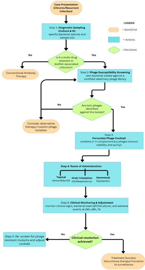
Decision flowchart for phage therapy in veterinary practice. Proposed clinical workflow for incorporating bacteriophage therapy into the management of chronic or recurrent P. aeruginosa infections in veterinary patients. Following case presentation, diagnostic sampling identifies the bacterial species and infection site. If the infection is not multidrug-resistant (MDR) or biofilm-associated, conventional antibiotic therapy is indicated. If MDR or biofilm-associated, the isolate undergoes phage susceptibility screening against a characterised veterinary phage library. Where no lytic phage is identified, alternative therapy or custom phage isolation is considered. Where lytic phages are identified, a cocktail of 2–4 complementary phages is formulated and administered via a route appropriate to the infection site (topical, oral/inhalation, or parenteral), with clinical monitoring of signs, bacterial load, and adverse events at defined intervals (24 h, 48 h, 7 days). Where clinical resolution is not achieved, re-screening for phage-resistant mutants and cocktail adjustment is recommended before further treatment cycles. This workflow is proposed based on the principles discussed in this review and has not been prospectively validated in clinical trials.
of efflux pumps, and as a result, phage-resistant mutants are more sensitive to antibiotics, as noted by Bowder et al. (2026). The study and exploitation of phage resistance trade-offs is thus an ongoing and promising field of research. The clinical duration of resistance development during therapy (as opposed to the in vitro mutation rates) was not defined by the data reviewed here. For the five-dog case series mentioned above . There was no testing for phage susceptibility at any point; this study does not provide any direct evidence either way. The capacity for rapid susceptibility testing, as mentioned above, is not available in veterinary practice and would be necessary for clinical monitoring of resistance emergence anyway.
Biofilm matrix hindering phage diffusion
The dense extracellular matrix of the biofilm creates physical barriers to diffusion that hinder the ability of the phage to reach embedded bacteria, even if the receptors are not modified . Not every phage can break down biofilms, and some phages are unable to infect bacteria in biofilms . Although phages are bactericidal in liquid culture, they may not have a significant effect on established biofilms in vitro, as was seen with the phages targeting the equine genital pathogens, which did not contain depolymerase genes .
Beyond the biofilm matrix, the architecture of biological structures further limits phage accessibility. Phages that inhibited biofilm biomass in microplate assays did not significantly inhibit the number of bacteria in ex vivo root canal models . Intact skin is a formidable barrier to phage penetration and bioavailability . The results highlight the need for the right delivery systems, such as topical formulations, hydrogels, or mechanical disruption adjuncts, to ensure that phages can gain access to bacteria in the biofilm in anatomically difficult areas ; .
Immune neutralization and phage clearance in animals
One of the challenges to the efficacy and pharmacokinetics of phage therapy is the host immune response. Phages, on repeated administration, are recognised as foreign antigens and can be neutralised by circulating antibodies, thus lowering the effective therapeutic titer of the phages. The immunogenicity of phage is regarded as a hurdle for its clinical applications. Immune cells, proteolytic enzymes, and local acidity at infection sites can decrease the infective phages and may be a factor in treatment failure .
Phage pharmacokinetic data in target animal species are still limited . Phages can be immunogenic, and neutralisation by antibodies may affect therapeutic outcomes . However, species-specific differences in immune responses to phages have not been carefully studied. Phages are quickly eliminated from the liver and spleen of healthy mice, but the clearance pattern in domestic animal species in clinical settings has yet to be carefully studied . Regarding specific mitigation strategies, none of these have been directly tested in the studies reviewed here for P. aeruginosa biofilm phage therapy. The self-dosing nature of phage therapy may modulate immune exposure: short treatment courses (e.g., 7 days) may not be significantly impacted by antibody-mediated clearance, whereas longer courses might be .
Lack of standardized veterinary formulations
One of the most significant practical challenges for implementing veterinary phage therapy is the lack of standardised and approved formulations that can be used in veterinary practice . Topical application for otitis externa in dogs, dermal wound healing, intramammary infusion for mastitis in cattle, intrauterine instillation for endometritis in horses, nebulization for respiratory infections, and possibly systemic use for bacteremia and deep-seated infections represent potential approaches at the different infection sites ; ; ; . Each route requires specific phage stability profiles and excipient compatibility, which have not been fully developed for veterinary use ; .
The development of methods for using phages and suitable storage conditions should consider environmental stability . The use of formulation strategies that combine phages with other excipients that disrupt biofilms, such as the glycerol, glycine, and Tween 20 formulation that was shown to increase the effectiveness of the phage cocktail in canine otitis models .This promising approach needs systematic optimisation and regulatory validation.
Regulatory void: No approved veterinary phage product
Although there have been decades of experimental evidence and several clinical case reports, none of the phages have been approved for veterinary clinical use in the treatment of biofilm infections by the major regulatory agencies, such as the FDA or EMA . This regulatory gap is a major obstacle to clinical application. Although there are limited established regulatory pathways for the use of phage therapy, veterinary practitioners still have no approved phage treatment options in severe MDR infections where they are unable to use conventional antibiotics ; .
One of the applicable US pathways is the Minor Use and Minor Species (MUMS) Act, which allows for conditional approval of the product for safety, manufacturing quality, and a “reasonable expectation of effectiveness” for up to five years until further studies are conducted (FDA Center for Veterinary Medicine, 2023). It does not apply uniformly to all of the applications described here, such as dogs, cats, cattle, and horses, which are considered to be major species; and conditional approval would require that the use is infrequent or geographically limited, which is not the case for common applications like treating canine otitis externa or bovine mastitis (FDA Center for Veterinary Medicine, 2023). The pathway is more appropriate for the truly minor species mentioned elsewhere in this review, for example, mink and chinchilla (FDA Center for Veterinary Medicine, 2023). For major species or other indications, an alternative US route is the Investigational New Animal Drug (INAD) exemption under 21 CFR Part 511, which permits a sponsor to ship an unapproved phage product for clinical investigation after submission of a Notice of Claimed Investigational Exemption to FDA’s Center for Veterinary Medicine; for food-producing species, this also requires showing that edible tissues would not be unsafe before slaughter or milk withdrawal (U.S. .
In the European Union, the products of phage therapy belong to the category of “novel therapies” and are not subject to national approval for marketing, but must be approved through the EMA’s centralized procedure. However, the pathway has been defined in principle, as of the latest EMA guidance on the product class, none of the available bacteriophage products have been marketed as veterinary medicines or feed additives in the EU (European .
Future directions and research priorities
High-throughput screening of anti-biofilm phages
Systematic, high throughput screening to identify phages with strong and specific anti-biofilm activity against different P. aeruginosa isolates are needed. In one study, 61 phages were isolated and tested against 64 human and animal clinical isolates, yielding 8 phages with a broad host range and high lytic activity , which shows the effectiveness of isolating many phages from the environment. The authors highlighted the importance of isolating new specific phages and studying their biological properties .
Future direction should specifically target veterinary environmental sources, such as barn effluent, farm water systems, and veterinary hospital drains, which might contain phages, and should be specifically targeted in future screening programs. Standardized biofilm-reduction assays, automated plaque scoring, and genome sequencing pipelines should be incorporated into screening workflows to expedite identification of candidates that have depolymerase genes, broad host ranges, and genomic safety profiles.
Combination and formulation strategies
Combining phage therapy with biofilm-dispersing enzymes is a promising approach. In the case of combined therapies, optimization of the dose and timing of phage and antibiotic exposure is important .
The use of phage therapy for veterinary applications could be enhanced by formulation strategies and other biofilm-disrupting elements, as well as by using phage cocktails. For example, addition of glycerol, glycine, and Tween 20 to a phage cocktail improved efficacy in canine otitis models , and a systematic study of optimal combinations of these adjuvants for every infection context is warranted.
Phage delivery systems for veterinary applications
To translate this laboratory efficacy to clinical effectiveness, advanced delivery systems that protect the viability of the phage, control the release kinetics, and allow access to the infection site are critical ; . Technologies already developed for other labile biologics for topical use in veterinary medicine provide promising starting points for adaptation: hydrogels and gel-based otic formulations are already used in veterinary medicine and could be adapted for sustained delivery of phage into the ear canal ; ; liposomal encapsulation, which has been developed to protect other labile biologics from degradation, could be adapted to protect the phage from local proteolytic and immune inactivation ; ; and implantable or depot formulations have been developed for prolonged delivery of veterinary antimicrobials and could be adapted for intramammary or intrauterine delivery of phage . The different routes have specific stability requirements, such as thermal stability for field storage, pH tolerance for the gastrointestinal or urogenital tract, and ionic/osmotic compatibility with the target tissue, which have not been systematically characterized for the different phage-specific formulations reviewed here.
Longitudinal field trials in livestock and companion animals
Controlled field trials in the target species under natural clinical conditions are the crucial link between laboratory promise and regulatory approval. There is currently limited evidence from large animal-controlled trials to assess phage therapy in bovine mastitis caused by MDR P. aeruginosa, but this therapy is a promising alternative . Further research is needed to assess the safety and efficacy of phage therapy before it can be used in the clinic and to investigate the potential of phage-antibiotic combinations against challenging equine pathogens .
A field trial of phage therapy for bovine mastitis should be carried out using the standard conventions of mastitis trials: the level of randomization should be the mammary quarter or cow level, as the intra mammary infusion is clearly visible and therefore would not be a blinding step; and bacterial cure (culture-negative at a fixed post-treatment time point) should be the primary endpoint to maintain blinding, because clinical signs may resolve without bacterial clearance. Treatment failure should be confirmed by culture rather than by symptoms only at specific times, and a concurrent control arm is needed to distinguish spontaneous bacteriological clearance from treatment effect in mastitis .
Economic analysis versus conventional therapy
In veterinary medicine, especially in production-animal settings where margins are thin, economic viability is likely to be a key determinant of the eventual adoption of phage therapy as an alternative to conventional antimicrobials. To our knowledge, there is no published cost-effectiveness analysis that directly compares phage therapy with traditional antibiotic therapy in the veterinary field, so the discussion below should be interpreted as a framework for future cost-effectiveness studies, not a synthesis of existing data.
In the future, a more thorough analysis would have to factor in the costs of the phage therapy, such as phage production and purification, diagnostic testing for phage-bacteria matching, regulatory requirements, and duration of the treatment, as well as the costs of conventional therapy, including treatment failure, re-infection, spread of antibiotic resistance, and, in food-producing animals, discards and associated production losses. In addition to the individual treatment economics, any such framework should reflect the wider societal value of the reduction of antimicrobial use and antimicrobial resistance transmission from a One Health perspective.
CONCLUSION
Bacteriophage-mediated disruption of P. aeruginosa biofilms represents a scientifically promising and mechanistically well-grounded therapeutic strategy with particular promise for veterinary medicine. The data presented here show that lytic phages, particularly those with depolymerase activity, can break down exopolysaccharide components of biofilm matrices, multiply within mature biofilms, decrease the viable bacterial population, and synergise with antibiotics to enhance biofilm disruption. Strong in vitro evidence shows that several phages reduced biofilm biomass across various veterinary isolates of P. aeruginosa. These findings are supported by ex vivo canine skin models and in vivo data, including mouse mastitis, wound infection, and systemic infection models, and small-scale canine companion animal case series.
However, significant challenges remain. The narrow host range requires phage cocktail strategies and matched phage selection, while the emergence of phage resistance needs to be addressed by rational cocktail design and evolutionarily exploiting trade-offs. Delivery system innovation is needed for biofilm matrix barriers, immune neutralization, and tissue penetration limitations. There are significant gaps in the critical evidence for large animal species (Equine and Bovine) for which controlled clinical evidence remains very limited. There is no standard veterinary phage formulation, and regulatory pathways for veterinary phage products remain a major translational challenge.
Collaborative investment is needed to develop phage therapy to the point of veterinary approval. Veterinarians need to bring their clinical knowledge and field trial facilities; microbiologists need to establish effective phage isolation, characterization, and banking systems; bioengineers need to design optimised delivery systems; and regulatory scientists need to collaborate with the regulatory agencies to create fit-for-purpose regulatory systems for biological phage products. The global antimicrobial resistance crisis, the One Health approach to linking animal and human health outcomes, and the growing body of evidence supporting the anti-biofilm activity of phages all make this an important investment now.
ACKNOWLEDGEMENT
This research received no external funding. The authors would like to acknowledge reviewers whose constructive comments helped improve the manuscript.
NOVELTY STATEMENT
This is, to our knowledge, the first review to synthesize phage-mediated P. aeruginosa biofilm disruption specifically across multiple veterinary species, including canine, bovine, equine, mink, and chinchilla, and to integrate veterinary-specific delivery routes and regulatory pathways (MUMS, INAD, EU Regulation 2019/6) that distinguish clinical translation in animals from human medicine.
AUTHORS’ CONTRIBUTION
Conceptualization was carried out by SA and VMA. Investigation and literature search were performed by KS and SA. Data curation was performed by SA. SA wrote the original draft, while KS, YDJ, and VMA contributed to writing, review, and editing. Visualization and critical revision were carried out by SS and IN. YDJ and VMA provided supervision. Funding acquisition is not applicable, as this research received no external funding. All authors reviewed and approved the final manuscript.
Generative AI and AI assisted technology statement
AI-assisted technology is only used to improve readability and grammar. No generative AI tool was used in the generation of figures or the interpretation of scientific content.
Conflict of interest
The authors have declared no conflict of interest.
References
- Abdelghafar A, El-Ganiny A, Shaker G, Askoura M (2023a). A novel lytic phage exhibiting a remarkable in vivo therapeutic potential and higher antibiofilm activity against Pseudomonas aeruginosa. Eur. J. Clin. Microbiol. Infect. Dis., 42: 1207-1234. https://doi.org/10.1007/s10096-023-04649-y [CrossRef]
- Abdelghafar A, El-Ganiny A, Shaker G, Askoura M (2023b). Isolation of a bacteriophage targeting Pseudomonas aeruginosa and exhibits a promising in vivo efficacy. AMB Express, 13: 79. https://doi.org/10.1186/s13568-023-01582-3 [CrossRef]
- Abdelrahman F, Easwaran M, Daramola OI, Ragab S, Lynch S, Oduselu TJ, Khan FM, Ayobami A, Adnan F, Torrents E, Sanmukh S, El-Shibiny A (2021). Phage-encoded endolysins. Antibiotics, 10: 124. https://doi.org/10.3390/antibiotics10020124 [CrossRef]
- Afonso AC, Sousa M, Pinto AR, Cotovio M, Simões M, Saavedra MJ (2023). Biofilm production by critical antibiotic-resistant pathogens from an equine wound. Animals, 13: 1342. https://doi.org/10.3390/ani13081342 [CrossRef]
- Amyx-Sherer K, Awasthi LC, Zheng A, Johannesman A, LeRoux M, Reichhardt C (2025). Two unrelated Pseudomonas aeruginosa phages require the exopolysaccharide PSL for infection. NPJ Biofilms Microbiomes, 11: 211. https://doi.org/10.1038/s41522-025-00841-4 [CrossRef]
- Billings N, Ramirez Millan M, Caldara M, Rusconi R, Tarasova Y, Stocker R, Ribbeck K (2013). The extracellular matrix component Psl provides fast-acting antibiotic defense in Pseudomonas aeruginosa biofilms. PLoS Pathog., 9: e1003526. https://doi.org/10.1371/journal.ppat.1003526 [CrossRef]
- Bowder M, Kapoor H, Carrell SJ, Danelishvili L (2026). Phage isolation and functional characterization reveal strong antibiofilm activity against Pseudomonas aeruginosa in a cystic fibrosis sputum model. Front. Cell. Infect. Microbiol., 16: 1753740. https://doi.org/10.3389/fcimb.2026.1753740 [CrossRef]
- Breidenstein EBM, de la Fuente-Núñez C, Hancock REW (2011). Pseudomonas aeruginosa: All roads lead to resistance. Trends Microbiol., 19: 419-426. https://doi.org/10.1016/j.tim.2011.04.005 [CrossRef]
- Ciofu O, Tolker-Nielsen T (2019). Tolerance and resistance of Pseudomonas aeruginosa biofilms to antimicrobial agents how P. aeruginosa can escape antibiotics. Front. Microbiol., 10. https://doi.org/10.3389/fmicb.2019.00913 [CrossRef]
- Dalponte A, Filor V, Lübke-Becker A, Fulde M, Alter T, Müsken M, Bäumer W (2025). Characterization and purification of Pseudomonas aeruginosa phages for the treatment of canine infections. BMC Microbiol., 25: 289. https://doi.org/10.1186/s12866-025-04005-4 [CrossRef]
- Dalponte A, Filor V, Nerlich A, Müsken M, Fulde M, Bäumer W (2026). Evaluation of bacteriophage efficacy against Pseudomonas aeruginosa in ex vivo and in vitro canine skin systems. Sci. Rep., 16: 7167. https://doi.org/10.1038/s41598-026-40091-8 [CrossRef]
- De Melo ACC, Da Mata Gomes A, Melo FL, Ardisson-Araújo DMP, De Vargas APC, Ely VL, Kitajima EW, Ribeiro BM, Wolff JLC (2019). Characterization of a bacteriophage with broad host range against strains of Pseudomonas aeruginosa isolated from domestic animals. BMC Microbiol., 19: 134. https://doi.org/10.1186/s12866-019-1481-z [CrossRef]
- Dedrick RM, Guerrero-Bustamante CA, Garlena RA, Russell DA, Ford K, Harris K, Gilmour KC, Soothill J, Jacobs-Sera D, Schooley RT, Hatfull GF, Spencer H (2019). Engineered bacteriophages for treatment of a patient with a disseminated drug-resistant Mycobacterium abscessus. Nat. Med., 25: 730-733. https://doi.org/10.1038/s41591-019-0437-z [CrossRef]
- Elbehiry A, Marzouk E, Edrees HM, Ibrahem M, Alzahrani S, Anagreyyah S, Abualola H, Alghamdi A, Alzahrani A, Jaber M, Abu-Okail A (2026). Understanding Pseudomonas aeruginosa biofilms: Quorum sensing, c-di-GMP signaling, and emerging antibiofilm approaches. Microorganisms, 14: 109. https://doi.org/10.3390/microorganisms14010109 [CrossRef]
- Eliasi UL, Sebola D, Oguttu JW, Qekwana DN (2020). Antimicrobial resistance patterns of Pseudomonas aeruginosa isolated from canine clinical cases at a veterinary academic hospital in South Africa. J. S. Afr. Vet. Assoc., 91: a2052. https://doi.org/10.4102/jsava.v91i0.2052 [CrossRef]
- European Medicines Agency (2023). Guideline on quality, safety and efficacy of veterinary medicinal products specifically designed for phage therapy. https://www.ema.europa.eu/en/scientific-guidelines/quality-safety-efficacy-bacteriophages-veterinary-medicines. Accessed on September 5, 2026.
- FDA Center for Veterinary Medicine (2023). Minor use and minor species. https://www.fda.gov/animal-veterinary Accessed on September 5, 2026.
- Fernández-Billón M, Llambías-Cabot AE, Jordana-Lluch E, Oliver A, Macià MD (2023). Mechanisms of antibiotic resistance in Pseudomonas aeruginosa biofilms. Biofilm, 5: 100129. https://doi.org/10.1016/j.bioflm.2023.100129 [CrossRef]
- Forti F, Roach DR, Cafora M, Pasini ME, Horner DS, Fiscarelli EV, Rossitto M, Cariani L, Briani F, Debarbieux L, Ghisotti D (2018). Design of a broad-range bacteriophage cocktail that reduces Pseudomonas aeruginosa biofilms and treats acute infections in two animal models. Antimicrob. Agents Chemother., 62: e02573-17. https://doi.org/10.1128/AAC.02573-17 [CrossRef]
- Furusawa T, Iwano H, Higuchi H, Yokota H, Usui M, Iwasaki T, Tamura Y (2016). Bacteriophage can lyse antibiotic-resistant Pseudomonas aeruginosa isolated from canine diseases. J. Vet. Med. Sci., 78: 1035-1038. https://doi.org/10.1292/jvms.15-0310 [CrossRef]
- Ghafoor A, Hay ID, Rehm BHA (2011). Role of exopolysaccharides in Pseudomonas aeruginosa biofilm formation and architecture. Appl. Environ. Microbiol., 77: 5238-5246. https://doi.org/10.1128/AEM.00637-11 [CrossRef]
- Guzman E, Montoya M (2018). Contributions of farm animals to immunology. Front. Vet. Sci., 5: 307. https://doi.org/10.3389/fvets.2018.00307 [CrossRef]
- Haenni M, Bour M, Châtre P, Madec JY, Plésiat P, Jeannot K (2017). Resistance of animal strains of Pseudomonas aeruginosa to carbapenems. Front. Microbiol., 8. https://doi.org/10.3389/fmicb.2017.01847 [CrossRef]
- Hattab J, Mosca F, Di Francesco CE, Aste G, Marruchella G, Guardiani P, Giorgio Tiscar P (2021). Occurrence, antimicrobial susceptibility, and pathogenic factors of Pseudomonas aeruginosa in canine clinical samples. Vet. World, 14: 978-985. https://doi.org/10.14202/vetworld.2021.978-985 [CrossRef]
- Horiuk Y, Tsymbalisty V, Kukhtyn M, Horiuk V, Kozhyn V, Chuhno V (2025). New bacteriophages for treating canine external otitis caused by Pseudomonas aeruginosa and Staphylococcus pseudintermedius. World’s Vet. J., 15: 389-398. https://doi.org/10.54203/scil.2025.wvj39 [CrossRef]
- Hu Z, Chen W, Chen X, Li P, Li M, Zhou L, Du H, Liu Y, Zhang W, Tan Z (2024). Combination of phage PH826 and antibiotics inhibits biofilm formation of multidrug-resistant Pseudomonas aeruginosa. Acta Microbiol. Sin., 64: 473-488.
- Jamal M, Andleeb S, Jalil F, Imran M, Nawaz MA, Hussain T, Ali M, Das CR (2017). Isolation and characterization of a bacteriophage and its utilization against multi-drug resistant Pseudomonas aeruginosa-2995. Life Sci., 190: 21-28. https://doi.org/10.1016/j.lfs.2017.09.034 [CrossRef]
- Jasim SE, Hayyawi SM (2025). Isolation and detection of biofilm producing Pseudomonas aeruginosa from suspected urinary tract infections in dogs and its resistance to antibiotics. Iraqi J. Vet. Med., 49: 45-54. https://doi.org/10.30539/ggeaaq50 [CrossRef]
- Jurado-Martín I, Sainz-Mejías M, McClean S (2021). Pseudomonas aeruginosa: an audacious pathogen with an adaptable arsenal of virulence factors. Int. J. Mol. Sci., 22: 3128. https://doi.org/10.3390/ijms22063128 [CrossRef]
- Kaiser SJ, DeRosa A, Ewers C, Günther F (2021). Phenotypical comparison of Pseudomonas aeruginosa isolated from human and veterinary samples; impact of host adaptation on infection pathogenesis. Research Square (Preprint). https://doi.org/10.21203/rs.3.rs-1170308/v1 [CrossRef]
- Köhne M, Hüsch R, Peh E, Hirnet J, Tönissen A, Müsken M, Plötz M, Kittler S, Sieme H (2025). Newly isolated bacteriophages show efficacy and phage-antibiotic synergy in vitro against the equine genital pathogens Klebsiella pneumoniae and Pseudomonas aeruginosa. BMC Vet. Res., 21: 568. https://doi.org/10.1186/s12917-025-04989-1 [CrossRef]
- Kwon J, Kim SG, Kim SW, Kim HJ, Kang JW, Jo SJ, Giri SS, Jeong WJ, Bin Lee S, Kim JH, Park SC (2025). Tailoring formulation for enhanced phage therapy in canine otitis externa: A cocktail approach targeting Pseudomonas aeruginosa and Staphylococcus pseudintermedius. Vet. Microbiol., 301: 110354. https://doi.org/10.1016/j.vetmic.2024.110354 [CrossRef]
- Latino L, Essoh C, Blouin Y, Vu Thien H, Pourcel C (2014). A novel Pseudomonas aeruginosa bacteriophage, Ab31, a chimera formed from temperate phage PAJU2 and P. putida lytic phage AF: characteristics and mechanism of bacterial resistance. PLoS One, 9: e93777. https://doi.org/10.1371/journal.pone.0093777 [CrossRef]
- Latka A, Maciejewska B, Majkowska-Skrobek G, Briers Y, Drulis-Kawa Z (2017). Bacteriophage-encoded virion-associated enzymes to overcome the carbohydrate barriers during the infection process. Appl. Microbiol. Biotechnol., 101: 3103-3119. https://doi.org/10.1007/s00253-017-8224-6 [CrossRef]
- Lewis K (2001). Riddle of biofilm resistance. Antimicrob. Agents Chemother., 45: 999-1007. https://doi.org/10.1128/AAC.45.4.999-1007.2001 [CrossRef]
- Liu CG, Green SI, Min L, Clark JR, Salazar KC, Terwilliger AL, Kaplan HB, Trautner BW, Ramig RF, Maresso AW (2020). Phage-antibiotic synergy is driven by a unique combination of antibacterial mechanism of action and stoichiometry. mBio, 11: 4. https://doi.org/10.1128/mBio.01462-20 [CrossRef]
- Ma J, Pang X, Zhang R, Ding C, Zheng Y, Zhao Y, Qian L, Ni J, Ji J, Kong X, Hu W, Chang F, Chen L (2025). vB_PaeP_PZH3, a novel bacteriophage for the treatment of MDR Pseudomonas aeruginosa in a mouse wound infection model. J. Glob. Antimicrob. Resist., 44: 197-206. https://doi.org/10.1016/j.jgar.2025.06.019 [CrossRef]
- Maffei E, Woischnig AK, Burkolter MR, Heyer Y, Humolli D, Thürkauf N, Bock T, Schmidt A, Manfredi P, Egli A, Khanna N, Jenal U, Harms A (2024). Phage Paride can kill dormant, antibiotic-tolerant cells of Pseudomonas aeruginosa by direct lytic replication. Nat. Commun., 15: 175. https://doi.org/10.1038/s41467-023-44157-3 [CrossRef]
- Manohar P, Loh B, Turner D, Tamizhselvi R, Mathankumar M, Elangovan N, Nachimuthu R, Leptihn S (2024). In vitro and in vivo evaluation of the biofilm-degrading Pseudomonas phage Motto, as a candidate for phage therapy. Front. Microbiol., 15: 1344962. https://doi.org/10.3389/fmicb.2024.1344962 [CrossRef]
- Marongiu L, Burkard M, Lauer UM, Hoelzle LE, Venturelli S (2022). Reassessment of historical clinical trials supports the effectiveness of phage therapy. Clin. Microbiol. Rev., 35: e00062-22. https://doi.org/10.1128/cmr.00062-22 [CrossRef]
- Mohamed AA, El-Zayat EM, El-Shibiny A (2025). Efficacy of phage vB_Ps_ZCPS13 in controlling pan-drug-resistant Pseudomonas aeruginosa from urinary tract infections (UTIs) and eradicating biofilms from urinary catheters. Virol. J., 22: 236. https://doi.org/10.1186/s12985-025-02848-x [CrossRef]
- Mohan N, Sritha KS, Jose J, Menachery SJ, Mohan CC, Isshack AT, Gopinathan H, Koyyappurath S, Abhitha K, Dalvi YB, Bhat SG (2026). Characterization, genomic insights and anti-biofilm potential of phage vB_PaeM_PKMS3, a lytic Pbunavirus capable of infecting clinical Pseudomonas aeruginosa isolates. Antonie van Leeuwenhoek, 119: 74. https://doi.org/10.1007/s10482-026-02282-1 [CrossRef]
- Mulcahy H, Charron-Mazenod L, Lewenza S (2008). Extracellular DNA chelates cations and induces antibiotic resistance in Pseudomonas aeruginosa biofilms. PLoS Pathog., 4: e1000213. https://doi.org/10.1371/journal.ppat.1000213 [CrossRef]
- Osondu-Chuka GO, Schandl S, Scheberl A, Guillaume O, Ovsianikov A, Reimhult E (2026). Beyond the matrix: rethinking antibiotic tolerance in CF biofilms using 3D models. NPJ Biofilms Microbiomes, 12: 3. https://doi.org/10.1038/s41522-025-00869-6 [CrossRef]
- Park Y, Oh J, Park S, Sum S, Song W, Chae J, Park H (2020). Antimicrobial resistance and novel mutations detected in the gyrA and parC genes of Pseudomonas aeruginosa strains isolated from companion dogs. BMC Vet. Res., 16: 111. https://doi.org/10.1186/s12917-020-02328-0 [CrossRef]
- Phee A, Bondy-Denomy J, Kishen A, Basrani B, Azarpazhooh A, Maxwell K (2013). Efficacy of bacteriophage treatment on Pseudomonas aeruginosa biofilms. J. Endod., 39: 364-369 https://doi.org/10.1016/j.joen.2012.10.023. [CrossRef]
- Pye CC, Yu AA, Weese JS (2013). Evaluation of biofilm production by Pseudomonas aeruginosa from canine ears and the impact of biofilm on antimicrobial susceptibility in vitro. Vet. Dermatol., 24: 446-e99. https://doi.org/10.1111/vde.12040 [CrossRef]
- Sahoo K, Meshram S (2024). The evolution of phage therapy: A comprehensive review of current applications and future innovations. Cureus, 16: e70414. https://doi.org/10.7759/cureus.70414 [CrossRef]
- Santos TMA, Ledbetter EC, Caixeta LS, Bicalho MLS, Bicalho RC (2011). Isolation and characterization of two bacteriophages with strong in vitro antimicrobial activity against Pseudomonas aeruginosa isolated from dogs with ocular infections. Am. J. Vet. Res., 72: 1079-1086. https://doi.org/10.2460/ajvr.72.8.1079 [CrossRef]
- Secker B, Shaw S, Atterbury RJ (2023). Pseudomonas spp. in canine otitis externa. Microorganisms, 11: 2650. https://doi.org/10.3390/microorganisms11112650 [CrossRef]
- Secker B, Shaw S, Hobley L, Atterbury RJ (2025). Genomic and phenotypic characterisation of Pseudomonas aeruginosa isolates from canine otitis externa reveals high-risk sequence types identical to those found in human nosocomial infections. Front. Microbiol., 16: 1526843. https://doi.org/10.3389/fmicb.2025.1526843 [CrossRef]
- Seth AK, Geringer MR, Gurjala AN, Hong SJ, Galiano RD, Leung KP, Mustoe TA (2012). Treatment of Pseudomonas aeruginosa biofilm-infected wounds with clinical wound care strategies: A quantitative study using an in vivo rabbit ear model. Plast. Reconstr. Surg., 129: 2. https://doi.org/10.1097/PRS.0b013e31823aeb3b [CrossRef]
- Testa S, Berger S, Piccardi P, Oechslin F, Resch G, Mitri S (2019). Spatial structure affects phage efficacy in infecting dual-strain biofilms of Pseudomonas aeruginosa. Commun. Biol., 2: 405. https://doi.org/10.1038/s42003-019-0633-x [CrossRef]
- Thi MTT, Wibowo D, Rehm BHA (2020). Pseudomonas aeruginosa biofilms. Int. J. Mol. Sci., 21: 8671. https://doi.org/10.3390/ijms21228671 [CrossRef]
- Tkhilaishvili T, Wang L, Perka C, Trampuz A, Gonzalez Moreno M (2020). Using bacteriophages as a Trojan horse to the killing of dual-species biofilm formed by Pseudomonas aeruginosa and methicillin resistant Staphylococcus aureus. Front. Microbiol., 11: 695. https://doi.org/10.3389/fmicb.2020.00695 [CrossRef]
- Tuon FF, Dantas LR, Suss PH, Tasca Ribeiro VS (2022). Pathogenesis of the Pseudomonas aeruginosa biofilm: a review. Pathogens, 11: 300. https://doi.org/10.3390/pathogens11030300 [CrossRef]
- U.S. Food and Drug Administration (2024). Requirements for investigational new animal drug exemptions. https://www.ecfr.gov/current/title-21/chapter-I/subchapter-E/part-511. Accessed on September 5, 2026.
- Vashchyk Y, Morozenko D, Seliukova N, Zakhariev A, Dotsenko R, Zemlianskyi A, Shapovalova O, Dotsenko E (2021). Pseudomonas aeruginosa as a priority group representative of bacteria with multiple antibiotic resistance. ScienceRise: Biol. Sci., 3: 33-40. https://doi.org/10.15587/2519-8025.2021.241238 [CrossRef]
- Vetrivel A, Ramasamy M, Vetrivel P, Natchimuthu S, Arunachalam S, Kim GS, Murugesan R (2021). Pseudomonas aeruginosa biofilm formation and its control. Biologics, 1: 312-336. https://doi.org/10.3390/biologics1030019 [CrossRef]
- Wang Z, Xue Y, Gao Y, Guo M, Liu Y, Zou X, Cheng Y, Ma J, Wang H, Sun J, Yan Y (2021). Phage vB_PaeS-PAJD-1 rescues murine mastitis infected with multidrug-resistant Pseudomonas aeruginosa. Front. Cell. Infect. Microbiol., 11: 689770. https://doi.org/10.3389/fcimb.2021.689770 [CrossRef]
- Yuan Y, Qu K, Tan D, Li X, Wang L, Cong C, Xiu Z, Xu Y (2019). Isolation and characterization of a bacteriophage and its potential to disrupt multi-drug resistant Pseudomonas aeruginosa biofilms. Microb. Pathog., 128: 329-336. https://doi.org/10.1016/j.micpath.2019.01.032 [CrossRef]
- Zulk JJ, Patras KA, Maresso AW (2024). The rise, fall, and resurgence of phage therapy for urinary tract infection. EcoSal Plus, 12: 1. https://doi.org/10.1128/ecosalplus.esp-0029-2023 [CrossRef]

Services
Article usage statistics
Loading…
Related articles
- Isolation and Characterization of a Pseudomonas aeruginosa Bacteriophage from Agriculture Drain WaterJournal of Virological Sciences · 2024
- The effect of Lactobacillus acidophilus as a probiotic against Pseudomonas aeruginosa growth and biofilm formationNovel Research in Microbiology Journal · 2019
- Prescence and Verification of LasI and LasR Genes in Pseudomonas aeruginosa Causing Urinary Tract Infection in DogsJournal of Animal Health and Production · 2025
- Bacteriological and Histopathological Evaluation of Infectious Lymphadenitis Caused by Pseudomonas aeruginosa in Awasi SheepAdvances in Animal and Veterinary Sciences · 2019
- Emerging Pathogenic Methicillin-Resistant Staphylococcus aureus (MRSA) Strains in Veterinary Medicine (Narrative Review)Advanced Analytical Pathology · 2025
- Inhibitory Effects of Carvacrol on BlaTEM and Exos Genes Expression in ESβL Producing Pseudomonas aeruginosa Isolated from Kidney Lesions of Broiler ChickensAdvances in Animal and Veterinary Sciences · 2021