
Severe neurologic listeriosis in small ruminants is typically associated with a guarded to poor prognosis, particularly in recumbent patients. This report describes an unusual recovery in a 3.5-year-old Nigerian Dwarf doe with acute neurologic dysfunction following abortion, characterized by lateral recumbence, severe torticollis, and vertical nystagmus. Hematological and biochemical analyses supported inflammation, including leukocytosis and elevated creatine kinase activity, while culture of fetoplacental fluids identified Listeria welshimeri with in vitro resistance to benzylpenicillin. The organism was not isolated from the central nervous system, and its causal association with the neurologic disease could not be established. Cerebrospinal fluid analysis was not performed; therefore, neurologic listeriosis remained a presumptive diagnosis based on clinical, environmental, and microbiological findings. The patient was treated with penicillin G potassium (approximately 44,000 IU/kg), thiamine, anti-inflammatories, fluid therapy, nutritional support, and intensive nursing care. The doe improved from recumbence to independent ambulation within 12 days. Leukocytosis resolved (approximately 32 to 16 K/µL) and creatine kinase decreased (1512 to 218 U/L). At discharge, the goat was ambulatory with a mild residual head tilt, with feeding and rumination resumed. Post-discharge management included trimethoprim-sulfamethoxazole and monitoring. This case illustrates a favorable clinical response despite in vitro benzylpenicillin resistance in the fetoplacental L. welshimeri isolate; however, the isolate’s role in the neurologic disease remains uncertain. The outcome likely reflected early recognition, multimodal therapy, and supportive care. This case contributes to limited evidence regarding atypical Listeria species and their clinical relevance in small ruminants.
Case Report · Volume 14, Issue 12, Pages 2710–2716, 4 November 2026 · Open Access
Listeriosis Recovery in a Nigerian Dwarf Goat
· · · ·
Affiliations & Notes ⌄
- 1Department of Large Animal Medicine and Surgery, Tuskegee University, College of Veterinary Medicine, 1200 W Montgomery Rd, Tuskegee, AL, USA
- *Correspondence | Ferdinand Bizimana; Email: fbizimana@tuskegee.edu
Article Info ⌄
- Journal:
- Advances in Animal and Veterinary Sciences
- Submitted Date:
- 9 July 2026
- Accepted Date:
- 9 September 2026
- Published Date:
- 4 November 2026
Abstract
Introduction
Listeriosis is a globally significant zoonotic disease caused by bacteria of the genus Listeria, of which Listeria monocytogenes is the primary pathogen affecting both humans and animals ; . The genus has expanded to include multiple species, including L. ivanovii and L. welshimeri, although L. monocytogenes remains the principal cause of clinical disease . First described by Murray et al. (1926) and later renamed by Pirie (1940), Listeria spp. are small Gram-positive, non-spore-forming, facultatively intracellular rods that are catalase-positive, oxidase-negative, β-hemolytic, and motile at lower temperatures .
Listeriosis has a worldwide distribution and affects a broad range of hosts, including humans, ruminants, pigs, and wildlife, with more than 40 species reported to be susceptible . In humans, it is a major foodborne disease with high case mortality rates, particularly amongst immunocompromised individuals, pregnant women, and neonates . In livestock, ruminants; especially sheep, goats, and cattle, are most frequently affected, with clinical syndromes including encephalitis, abortion, and septicemia . The encephalitic form is the most common presentation in small ruminants and results from invasion of the central nervous system following ingestion of contaminated feed, particularly poorly fermented silage or spoiled forage .
Clinically, neurologic listeriosis is characterized by asymmetric cranial nerve deficits, head tilt, circling, nystagmus, dysphagia, and progression to recumbence, which is widely associated with a guarded prognosis ; . Disease progression is often rapid, and mortality remains high despite treatment, particularly in advanced cases. Standard therapeutic protocols include high-dose penicillin or ampicillin, often combined with thiamine and supportive care; however, treatment success is variable due to factors such as delayed diagnosis, limited antimicrobial penetration across the blood–brain barrier, and the intracellular nature of the pathogen ; .
Listeria monocytogenes is notable for its environmental resilience and ability to survive under adverse conditions, including refrigeration temperatures, high salt concentrations, and acidic environments, as well as its capacity to form biofilms that enhance persistence and resistance to disinfectants . Although antimicrobial resistance in Listeria spp. has historically been uncommon, emerging reports suggest increasing variability in susceptibility profiles, raising questions about the predictive value of in vitro testing for clinical outcomes . Interpretation may be particularly challenging when a non-monocytogenes Listeria species is isolated from a non-CNS site, because such species generally have limited recognized pathogenicity and antimicrobial susceptibility varies among Listeria spp. ; . Transmission to humans may occur through ingestion of contaminated food or direct contact with infected animals or tissues, posing an occupational risk to veterinarians and livestock handlers .
This case is notable for describing severe neurologic disease consistent with listeriosis in a recumbent goat, from which Listeria welshimeri was isolated. Despite the reported in vitro antimicrobial resistance, the goat experienced a favorable clinical outcome. However, L. welshimeri was isolated from feto-placental fluids rather than the CNS, making its etiologic relationship to the neurologic disease uncertain. This creates a clinical dilemma when in vitro resistance conflicts with the use of penicillin, a standard therapy for listeriosis . Reports involving non-monocytogenes Listeria species in neurologic disease of small ruminants are exceedingly rare, and their clinical relevance remains poorly defined. This case therefore highlights the need to interpret non-classical Listeria isolates and antimicrobial susceptibility results in conjunction with the clinical presentation and source of isolation.
Patient information
A 3.5-year-old Nigerian Dwarf doe (Boba Fetta) weighing 10.9 kg (24 lb) presented with acute onset neurologic dysfunction following abortion during her second pregnancy. The interval between abortion and onset of neurologic signs was approximately 2 days. Primary clinical concerns included lateral recumbence, severe torticollis, and vertical nystagmus, suggestive of central vestibular involvement (Figure 1). Neurologic examination included assessment of mentation, posture, ocular position and movement, menace response, pupillary light and palpebral reflexes, facial symmetry, swallowing, and tongue movement. Severe postural dysfunction was evident, with inability to maintain an upright position or ambulate independently, while no additional cranial nerve abnormalities were recorded. The patient originated from a small herd of six goats with no similar clinical signs reported. Environmental history revealed exposure to wet hay used as bedding following recent weather fluctuations (cold stress, heavy rainfall, and subsequent heat), representing a significant risk factor for listerial contamination. There was no prior history of neurologic disease, and the doe had previously completed a normal gestation. No recent dietary changes, toxin exposure, or trauma were reported.
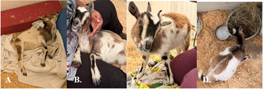
Sequential clinical progression from acute neurologic presentation to recovery. (A) recumbence; (B) assisted support; (C) partial recovery; (D) standing and feeding.
Clinical findings
Over the first 48 hours of hospitalization, the patient remained recumbent but stable. By day five of hospitalization, vital parameters were within acceptable ranges for the species, including a temperature of 100.4°F, heart rate of 120 bpm, respiratory rate of 30 breaths per minute, capillary refill time of 2 seconds, and pink, moist mucous membranes. Body condition score was 3/9. Neurologic abnormalities persisted. Neurologic examination revealed central vestibular signs, including head tilt, vertical nystagmus, and loss of balance, without evidence of peripheral ear disease. Mentation was dull but responsive, and cranial nerve deficits included abnormalities consistent with brainstem involvement, notably vestibular dysfunction (cranial nerve VIII), with suspected involvement of adjacent cranial nerves (V and VII), although detailed cranial nerve testing was limited by recumbence. Gastrointestinal function remained intact, with defecation and eructation observed; urination was also normal. Despite a guarded prognosis, the patient demonstrated gradual clinical improvement.
Diagnostic assessment
Comprehensive diagnostic evaluation was undertaken to identify the underlying cause of acute neurologic dysfunction following recent abortion. Because cerebrospinal fluid (CSF) analysis was not performed and Listeria was not isolated from the central nervous system, the etiologic diagnosis remained presumptive.
Abdominal ultrasonography
Abdominal ultrasonography was performed at presentation due to the recent history of abortion to evaluate for retained fetal or placental tissues. No abnormalities were detected, reducing concern for retained fetal or placental material, although this finding did not establish the cause of the neurologic signs.
Hematologic and biochemical analysis
Serial hematologic and serum biochemical evaluations revealed marked leukocytosis indicating systemic inflammation but not specifically supporting CNS infection. The recent abortion and associated physiologic stress were also considered potential contributors to the leukocytosis. Elevated creatine kinase (CK) concentrations were noted, likely secondary to muscle damage associated with prolonged recumbence. Additional findings included mild anemia, elevated aspartate aminotransferase (AST) indicative of tissue injury, and hypocalcemia (Table 1).
Bacterial culture and antimicrobial susceptibility testing
Bacteriological analysis was performed using standard aerobic culture techniques on selective media for Listeria spp. (Oxford/Palcam agar), followed by incubation at 35-37 °C. The sample used for bacterial culture was obtained from fetoplacental fluids/tissues. Therefore, isolation of Listeria from this site provided evidence of reproductive tract exposure/infection but could not establish that the organism was responsible for the concurrent neurologic disease. Presumptive colonies were identified based on characteristic morphology and confirmed using an automated identification system, the Matrix-Assisted Laser Desorption/Ionization Time-of-Flight (MALDI-TOF) mass spectrometry, which identified Listeria welshimeri with high confidence . CSF collection was not performed during the acute clinical period. Consequently, CSF cytology, protein analysis, and bacterial culture were unavailable to support CNS infection. This represents an important diagnostic limitation of the case. Antimicrobial susceptibility testing was conducted using a standardized Kirby-Bauer disk diffusion method and interpreted according to the Clinical and Laboratory Standards Institute (CLSI) guidelines for Listeria spp. . The isolate demonstrated in vitro resistance to benzylpenicillin, while susceptibility to trimethoprim-sulfamethoxazole was observed. Interpretation of antimicrobial susceptibility was extrapolated from available guidelines, as standardized breakpoints for Listeria welshimeri are not well established .
Differential diagnoses and diagnostic interpretation
Differential diagnoses considered for the acute central vestibular/brainstem signs included listerial encephalitis, polioencephalomalacia, caprine arthritis encephalitis (CAE), intracranial abscessation, and toxic encephalopathy. The acute onset, vertical nystagmus, severe postural
Table 1: Selected hematologic and biochemical findings.
| Parameter | Initial 3/5/2026 | Follow-up 3/9/2026 | Reference range | Interpretation |
| WBC (K/µL) | ≈ 32 | ≈ 16 | 6–19 | Marked leukocytosis → improving |
| Hematocrit (%) | 33 | 21.8 | 22–39 | Developing anemia |
| CK (U/L) | 1512 | 218 | 28–130 | Muscle damage → improving |
| AST (U/L) | 488 | 446 | 122–321 | Tissue injury |
| Ca2+ (mg/dL) | 6.9 | 6.9 | 8.2–9.8 | Hypocalcemia |
WBC: white blood cell count; K/µL: ×10³ cells per microliter; CK: creatine kinase; U/L: units per liter; AST: aspartate aminotransferase; Ca2+: ionized calcium; mg/dL: milligrams per deciliter.
Table 2: Comprehensive multimodal therapeutic protocol for case management and clinical rationale.
| Therapeutic category | Agent/ intervention | Dosage, route, frequency | Duration | Clinical rationale |
| Antimicrobial therapy | Penicillin G potassium | ~44,000 IU/kg, SC, q6-8h | During hospitalization | First-line therapy for Listeria spp.; administered at high doses to enhance central nervous system penetration and maintained despite in vitro resistance based on expected pharmacologic efficacy and favorable clinical response. |
| Trimethoprim–sulfamethoxazole | ~22 mg/kg, PO, q12h | 7 days post-discharge | Continued antimicrobial coverage following clinical improvement | |
| Neurologic support | Thiamine (Vitamin B1) | ~50 mg/kg, SC, q6-8h | During hospitalization | Neuroprotective support; addresses neuronal energy deficits and potential concurrent polioencephalomalacia |
| Anti-inflammatory therapy | Dexamethasone | 0.5-0.9 mg/kg, IV, q24h | Tapered during hospitalization | Reduces neuroinflammation and cerebral edema associated with brainstem encephalitis |
| Meloxicam | ~0.5 mg/kg, PO, q24h | 10 days (discontinued prior to discharge) | Analgesic and anti-inflammatory support | |
| Sedation/ Anxiolysis | Midazolam | ~0.1-0.2 mg/kg, IV, PRN | As needed | Facilitates handling and reduces stress in neurologically compromised patient |
| Ocular therapy | Ofloxacin ophthalmic | 1 drop OU, q24h | During hospitalization | Prevents secondary ocular infection due to reduced blink reflex |
| Flurbiprofen ophthalmic | 1 drop OU, q24h | During hospitalization | Reduces ocular inflammation and discomfort | |
| Supplementation | Vitamin B12 | 0.5 mL, SC, q24h | During hospitalization | Supports hematologic recovery |
| Vitamin A and D | 0.3 mL, SC, q24h | During hospitalization | Supports immune and epithelial function | |
| Selenium (Bo-Se) | As per label | Single/as indicated | Antioxidant and neuromuscular support | |
| Supportive care | IV fluids, dextrose, calcium | As indicated | During hospitalization | Maintains hydration, energy balance, and corrects metabolic derangements |
| Nursing care (repositioning, assisted feeding, bedding) | Continuous | Throughout hospitalization | Prevents decubital injury, aspiration, and secondary complications |
IU/kg: international units per kilogram; SC: subcutaneous; IV: intravenous; PO: per os (oral); q6-8h: every 6-8 hours; q12h: every 12 hours; q24h: every 24 hours; PRN: as needed; CNS: central nervous system; OU: oculus uterque (both eyes); Bo-Se: selenium and vitamin E injectable preparation.
dysfunction, history of abortion, and environmental exposure to wet hay increased clinical suspicion for listeriosis; however, these findings were not pathognomonic. In the absence of CSF analysis, advanced neuroimaging, or histopathology, the alternative neurologic differentials could not be definitively excluded.
Accordingly, neurologic listeriosis was considered a presumptive clinical diagnosis based on neurologic localization, environmental history, temporal association with abortion, and isolation of L. welshimeri from fetoplacental material. The fetoplacental isolate cannot be considered definitive evidence of CNS infection, and its causal relationship to the neurologic syndrome remains uncertain.
Timeline
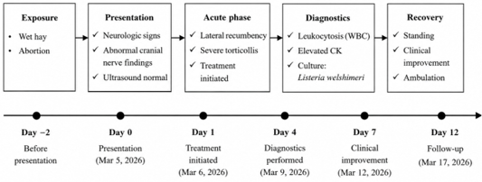
illustrates the progression from exposure through diagnosis, treatment, and recovery, with reference to white blood cell count (WBC), creatine kinase (CK), ultrasonography.
Therapeutic intervention
As detailed in Table 2 below, an integrated approach targeting infection control, neuroprotection, and clinical recovery, included antimicrobial, neurologic, anti-inflammatory, and supportive therapies with defined dosing regimens.
Follow-up and outcomes
The patient demonstrated progressive clinical improvement during hospitalization, transitioning from lateral recumbence to independent ambulation, with restoration of voluntary feeding, drinking, and normal rumination by discharge. Clinical improvement was evaluated based on restoration of posture, ability to stand unassisted, normalization of appetite, and resolution of nystagmus. A structured neurological assessment of mentation, posture, nystagmus, ambulation, and feeding showed normalization of four of five domains at discharge, with only a mild residual head tilt.
Neurologic assessment revealed marked resolution of deficits, with only mild residual head tilt. Treatment adherence was ensured through continuous in-hospital monitoring and structured nursing care, while post-discharge compliance was facilitated by clear owner instructions; the therapeutic regimen was well tolerated, with no evidence of gastrointestinal or systemic intolerance. At 2–3-week follow-up, independent ambulation and normal feeding and rumination were maintained, with no recurrence of neurologic.
Discussion
This case highlights several important clinical and pathophysiological aspects of neurologic listeriosis in small ruminants. The likely source of infection, wet hay used as bedding following rapid environmental changes, aligns with well-established epidemiologic risk factors, as Listeria spp. proliferate in poorly fermented or spoiled forage under fluctuating temperature and moisture conditions ; . Following ingestion, the organism may invade damaged oral mucosa and ascend to the brainstem via cranial nerves, particularly the trigeminal nerve, resulting in the characteristic neurologic manifestations of listerial encephalitis ; .
The observed neurologic signs, particularly torticollis and vertical nystagmus, are consistent with brainstem encephalitis and central vestibular dysfunction, which are hallmarks of listerial infection in goats and sheep ; . Although Listeria welshimeri was isolated, its role as the primary causative agent remains uncertain, as this species is typically considered non-pathogenic and may represent environmental contamination or an opportunistic organism. This observation contrasts with the established understanding that L. welshimeri is predominantly an environmental species with minimal pathogenic potential and lacking key virulence determinants required for neuroinvasion, such as internalins and listeriolysin O ; . Listerial encephalitis typically results from ingestion of contaminated feed or bedding, followed by invasion of damaged oral or pharyngeal mucosa and retrograde axonal migration along cranial nerves, particularly the trigeminal nerve, to the brainstem, leading to inflammation and characteristic vestibular signs .
Nevertheless, emerging genomic and experimental evidence suggests that certain non-monocytogenes Listeria spp., including L. welshimeri, may possess limited or atypical virulence potential under specific conditions, possibly through acquisition or expression of partial virulence-associated factors ; . These findings raise the possibility that, although uncommon, such species may occasionally be associated with clinical presentations indistinguishable from classical listeriosis. Therefore, although a possible association cannot be excluded, the isolation of L. welshimeri from fetoplacental material does not establish it as the cause of the neurologic disease.
Clinical recovery occurred despite the reported in vitro penicillin resistance; however, the diagnostic uncertainty prevents attributing this response specifically to treatment of L. welshimeri. This apparent discordance between antimicrobial susceptibility testing and clinical outcome has been increasingly recognized with intracellular pathogens such as Listeria, where pharmacokinetic and pharmacodynamic factors; particularly high-dose administration and tissue penetration into the central nervous system, play a critical role ; . Penicillin remains the first-line therapy for listeriosis due to its bactericidal activity and ability to achieve therapeutic concentrations when administered aggressively, and clinical response may not always correlate with laboratory susceptibility profiles . The case suggests that clinical response should be considered alongside in vitro susceptibility findings, although it provides limited evidence because the causative organism was not definitively established.
The favorable outcome observed is particularly notable given that prolonged lateral recumbence is typically associated with poor prognosis in small ruminant listeriosis ; . Early intensive therapy and supportive care may have contributed to recovery, although their specific effects cannot be determined. Similar reports emphasize that survival in advanced neurologic cases depends not only on antimicrobial therapy but also on meticulous nursing care and maintenance of physiologic stability . Overall, the favorable clinical outcome is noteworthy, but the absence of definitive CNS confirmation requires cautious interpretation of both the etiologic diagnosis and treatment response. Definitive diagnosis was limited by the absence of cerebrospinal fluid analysis, CNS isolation of Listeria, or postmortem confirmation, representing a major limitation of this case. Therefore, the diagnosis remained presumptive and based on clinical presentation, neurologic findings, environmental exposure, and isolation of L. welshimeri from fetoplacental material rather than the CNS.
Conclusion
This case describes the clinical recovery of a goat from severe neurologic signs of uncertain etiology, with a presumptive diagnosis of listeriosis. The isolation of Listeria welshimeri and its in vitro antimicrobial resistance profile highlights the complexity of interpreting laboratory findings in relation to clinical response. However, the absence of definitive CNS confirmation limits conclusions regarding the pathogenic role of L. welshimeri and the clinical significance of its antimicrobial resistance.
Acknowledgement
The authors thank the microbiology and biochemistry laboratory technicians at Tuskegee University College of Veterinary Medicine for performing the diagnostic analyses, and the large animal hospital technicians and veterinary students for their assistance with patient care and clinical monitoring.
Novelty Statement
This report documents recovery from lateral recumbence to independent ambulation within 12 days in a Nigerian Dwarf doe with presumptive neurologic listeriosis, alongside isolation of Listeria welshimeri reported as benzylpenicillin-resistant from fetoplacental material. The case highlights a diagnostic and therapeutic dilemma: how to interpret an atypical isolate and its susceptibility profile when the sampling site differs from the suspected site of disease. Although central nervous system infection was not confirmed, the documented recovery during multimodal treatment provides a clinically informative account of managing severe neurologic disease under diagnostic uncertainty.
Author Contribution
VH: Writing, review and editing, writing original draft, resources, methodology, formal analysis, conceptualization. DM: Writing review and editing, writing supervision. AE, BOO: Writing review and editing. FB: Writing review and editing, writing original draft, supervision, methodology, conceptualization.
Clinical outcome
From the owner’s perspective, the doe showed progressive recovery from severe neurologic dysfunction, transitioning from recumbence to normal feeding and mobility. The owner reported good compliance with treatment and expressed satisfaction with the outcome despite the initially guarded prognosis.
Funding
No funding was received for this manuscript.
Data availability statement
The data that support the findings are available on request from the corresponding author.
Generative AI and AI assisted technology statement
The authors declare that no generative AI and AI assisted technology was used in the creation of this manuscript.
Conflicts of interest
The authors have declared no conflicts of interest.
References
- Bizzini A, Greub G (2010). Matrix-assisted laser desorption ionization time-of-flight mass spectrometry, a revolution in clinical microbial identification. Clin. Microbiol. Infect., 16(11): 1614–1619. https://doi.org/10.1111/j.1469-0691.2010.03311.x [CrossRef]
- Carpentier B, Cerf O (2011). Persistence of Listeria monocytogenes in food industry equipment and premises. Int. J. Food Microbiol., 145(1): 1–8. https://doi.org/10.1016/j.ijfoodmicro.2011.01.005 [CrossRef]
- Cheong S, Rothwell JG, Schlesener C, Weimer BC, Miramontes C, Pereira RV, Pagliari P, Pires AF (2026). Genomic characterization of Listeria monocytogenes isolated from small ruminants in integrated crop-livestock systems. BMC Microbiol., 26(1): 81. https://doi.org/10.1186/s12866-025-04626-9 [CrossRef]
- Constable PD, Hinchcliff KW, Done SH, Grünberg W (2016). Veterinary medicine: A textbook of the diseases of cattle, horses, sheep, pigs and goats. Elsevier health sciences. Ocytogenes, L. innocua, and L. welshimeri. Microbial Drug Resist., 15(1): 27–32.
- Davis, J. A., and Jackson, C. R. (2009). Comparative antimicrobial susceptibility of Listeria monocytogenes, L. innocua, and L. welshimeri. Microbial Drug Resistance, 15(1), 27-32. https://doi.org/10.1089/mdr.2009.0863 [CrossRef]
- Ersdal C (2026). Listeria infection in small ruminants. In encyclopedia of livestock medicine for large animal and poultry production. Springer. pp. 750–757. https://doi.org/10.1007/978-3-031-61549-8_213 [CrossRef]
- Hudzicki J (2009). Kirby-Bauer disk diffusion susceptibility test protocol. Am. Soc. Microbiol., 15(1): 1–23.
- Huff T, Weisbrod LJ, Daly DT (2024). Neuroanatomy, cranial nerve 5 (trigeminal). StatPearls Publishing.
- Koopmans MM, Brouwer MC, Vázquez-Boland JA, Van De Beek D (2023). Human listeriosis. Clin. Microbiol. Rev., 36(1): e00060-19. https://doi.org/10.1128/cmr.00060-19 [CrossRef]
- Korsak D, Szuplewska M (2016). Characterization of nonpathogenic Listeria species isolated from food and food processing environment. Int. J. Food Microbiol., 238: 274–280. https://doi.org/10.1016/j.ijfoodmicro.2016.08.032 [CrossRef]
- Low J, Donachie W (1997). A review of Listeria monocytogenes and listeriosis. Vet. J., 153(1): 9–29. https://doi.org/10.1016/S1090-0233(97)80005-6 [CrossRef]
- Murray EGD, Webb RA, Swann MBR (1926). A disease of rabbits characterised by a large mononuclear leucocytosis, caused by a hitherto undescribed bacillus Bacterium monocytogenes (n. Sp.). J. Pathol. Bacteriol., 29(4): 407–439. https://doi.org/10.1002/path.1700290409 [CrossRef]
- Orsi RH, Wiedmann M (2016). Characteristics and distribution of Listeria spp., including Listeria species newly described since 2009. Appl. Microbiol. Biotechnol., 100(12): 5273–5287. https://doi.org/10.1007/s00253-016-7552-2 [CrossRef]
- Pirie JH (1940). Listeria: Change of name for a genus bacteria. Nature, 145(3668): 264–264. https://doi.org/10.1038/145264a0 [CrossRef]
- Plumb D (2015). Plumb’s veterinary drug handbook. https://www.vet-library.com/ebook/plumbs-veterinary-drug-handbook-10th-edition/
- Radoshevich L, Cossart P (2018). Listeria monocytogenes: Towards a complete picture of its physiology and pathogenesis. Nat. Rev. Microbiol., 16(1): 32–46. https://doi.org/10.1038/nrmicro.2017.126 [CrossRef]
- Radostits OM, Gay CC, Hinchcliff KW, Constable PD (2006). Veterinary medicine E-Book: Veterinary medicine E-Book. Elsevier Health Sciences.
- Sharma S, Gandhi A, Sah S, Singh MP, Sharma GD, Verma A (2025). Listeria infections: The unexpected risks in everyday foods. Clinical Infection in Practice, 26, 100489. https://doi.org/10.1016/j.clinpr.2025.100489 [CrossRef]
- Swaminathan B, Gerner-Smidt P (2007). The epidemiology of human listeriosis. Microbes Infect., 9(10): 1236–1243. https://doi.org/10.1016/j.micinf.2007.05.011 [CrossRef]
- Troxler R, Von Graevenitz A, Funke G, Wiedemann B, Stock I (2000). Natural antibiotic susceptibility of Listeria species: L. grayi, L. innocua, L. ivanovii, L. monocytogenes, L. seeligeri and L. welshimeri strains. Clin. Microbiol. Infect., 6(10): 525–535. https://doi.org/10.1046/j.1469-0691.2000.00168.x [CrossRef]
- Vázquez-Boland JA, Kuhn M, Berche P, Chakraborty T, Domı́nguez-Bernal G, Goebel W, González-Zorn B, Wehland J, Kreft J (2001). Listeria pathogenesis and molecular virulence determinants. Clin. Microbiol. Rev., 14(3): 584–640. https://doi.org/10.1128/CMR.14.3.584-640.2001 [CrossRef]
- Wei P, Bao R, Fan Y (2020). Brainstem encephalitis caused by Listeria monocytogenes. Pathogens, 9(9): 715. https://doi.org/10.3390/pathogens9090715 [CrossRef]
- Wilson A, Gray J, Chandry PS, Fox EM (2018). Phenotypic and genotypic analysis of antimicrobial resistance among Listeria monocytogenes isolated from Australian food production chains. Genes, 9(2): 80. https://doi.org/10.3390/genes9020080 [CrossRef]
- Zakrzewski AJ, Chajęcka-Wierzchowska W, Zadernowska A, Podlasz P (2020). Virulence characterization of Listeria monocytogenes, Listeria innocua, and Listeria welshimeri isolated from fish and shrimp using in vivo early zebrafish larvae models and molecular study. Pathogens, 9(12): 1028. https://doi.org/10.3390/pathogens9121028 [CrossRef]
- Zhang L, Nagel M, Olson WP, Chesler AT, O’Connor DH (2024). Trigeminal innervation and tactile responses in mouse tongue. Cell Rep., 43(9). https://doi.org/10.1016/j.celrep.2024.114665 [CrossRef]

Services
Article usage statistics
Loading…
Related articles
- Effect of Systemic Ivermectin on Epidermal Tunnel Size, Severe Asthetic Appearance of Skin, Haematological and Biochemical Inbalances Caused by <i>Sarcoptes</i> <i>scabie</i> <i>var</i> <i>caprae</i> in West African Dwarf Goats, in Ibadan, NigeriaThe Journal of Advances in Parasitology · 2015
- Effects of Lesion Distribution on the Hematology and some Biochemical Indices of Sarcoptes scabiei Infested West African Dwarf GoatThe Journal of Advances in Parasitology · 2016
- Tithonia diversifolia Leaf Meal as a Supplement for West African Dwarf GoatsAdvances in Animal and Veterinary Sciences · 2026
- Efficacy of Vaginal Electrical Resistance Measurement in West African Dwarf Goats Synchronized using Ovsynch and Double PG ProtocolAdvances in Animal and Veterinary Sciences · 2024
- Effect of Seasonal Variation on the Hormonal Profile of West African Dwarf (WAD) GoatsJournal of Animal Health and Production · 2023
- Growth Performance and Carcass Characteristics of West African Dwarf Goats Fed Cassava Peel-based Ration Supplemented with EnzymesJournal of Animal Health and Production · 2024