
General anesthesia commonly used in feline practice can impair gastrointestinal (GI) motility, increasing risk of postoperative ileus. Ketamine, a dissociative anesthetic with sympathomimetic effects, is often paired with a sedative premedicant acepromazine or diazepam to offset its adverse effects, yet comparative data on their differential GI effects in cats remain limited. This study used contrast radiography to compare the effects of ketamine-acepromazine and ketamine-diazepam on intestinal amplitude and GI transit time in cats undergoing general anesthesia. Eighteen adult male domestic cats were randomly allocated to three treatment groups: an unanesthetized control group and two treatment groups receiving ketamine combined with either acepromazine (T1) or diazepam (T2), with atropine premedication in both treatment groups. Barium sulfate was administered orally, and ventrodorsal (VD) radiographs were obtained at set intervals. Intestinal amplitude was measured with ImageJ, and transit time was scored from barium distribution across GI zones. Data were analyzed using repeated-measures ANOVA and Kruskal-Wallis tests, with Tukey’s HSD and Mann-Whitney U post-hoc comparisons. Intestinal amplitude did not differ significantly among groups. GI transit was significantly slower in both anesthetized groups than in controls, although ketamine-acepromazine showed a numerical trend toward faster transit than ketamine-diazepam, the difference between the two protocols was not statistically significant. Both protocols were safe, producing no significant effect on intestinal amplitude. Study interpretation is limited by small sample size, non-significant differences in amplitude and transit between protocols, and a short postanesthetic observation window.
Research Article · Volume 14, Issue 12, Pages 2810–2819, 10 November 2026 · Open Access
Contrast Radiography Analysis of Gastrointestinal Motility in Cats (Felis catus) Under General Anesthesia: Ketamine-Acepromazine Versus Ketamine-Diazepam
· · · · · · ·
Affiliations & Notes ⌄
- 1Profession Program of Veterinary Medicine, Faculty of Veterinary Medicine, Universitas Airlangga, Campus C Mulyorejo, Surabaya, 60115, East Java, Indonesia
- 2Veterinary Clinical Division, Department of Veterinary Science, Faculty of Veterinary Medicine, Universitas Airlangga, Campus C Mulyorejo, Surabaya 60115, Indonesia
- 3Division of Basic Veterinary Medicine, Department of Veterinary Science, Faculty of Veterinary Medicine, Universitas Airlangga, Campus C Mulyorejo, Surabaya, East Java, 60115, Indonesia
- *Correspondence | Hardany Primarizky; Email: hardany-p@fkh.unair.ac.id
Article Info ⌄
- Journal:
- Advances in Animal and Veterinary Sciences
- Submitted Date:
- 23 July 2026
- Accepted Date:
- 31 October 2026
- Published Date:
- 10 November 2026
Abstract
INTRODUCTION
Anesthesia is indispensable in veterinary surgical practice, providing the immobilization, analgesia, and muscle relaxation required for invasive procedures. Anesthetic agents impair gastrointestinal (GI) function through several converging mechanisms, including disruption of autonomic nervous system balance and direct impairment of intestinal smooth muscle contractility . In the postoperative period, GI hypomotility occurring in the absence of mechanical obstruction constitutes postoperative ileus (POI), a condition associated with abdominal distension, delayed recovery, and increased complication rates that, in severe cases, which may progress to bowel rupture, peritonitis, and septic shock ; ; . Cats possess unique gastrointestinal physiological traits, including a high susceptibility to stress-induced sympathetic activation and pronounced opioid- and anesthetic-associated dysmotility ; , making POI a particularly critical concern during feline abdominal imaging and surgical procedures . Although the present study did not involve surgery, anesthesia-induced GI hypomotility shares underlying autonomic and pharmacological mechanisms with surgical POI, and is directly relevant to any feline patient undergoing general anesthesia for diagnostic or minor procedures, not exclusively those undergoing invasive surgery.
Ketamine hydrochloride (HCl) is a dissociative anesthetic widely used in feline practice for its rapid onset, wide safety margin, and versatility ; . Through actions mediated by N-methyl-D-aspartate (NMDA) receptor antagonism and inhibition of voltage-gated Ca²⁺ channels, ketamine exerts sympathomimetic effects that inhibit smooth muscle contractility and slow GI transit ; ; .
To mitigate these adverse GI effects, ketamine is routinely combined with a sedative premedicant, most commonly acepromazine or diazepam. Acepromazine, a phenothiazine tranquilizer, and diazepam, a benzodiazepine, act through distinct pharmacological pathways dopamine D2 receptor antagonism and GABAA receptor modulation, respectively that can differentially influence gastrointestinal motility when combined with ketamine ; . Given these divergent pharmacological pathways, the net effect of each premedicant on GI motility when combined with ketamine may differ substantially.
Atropine sulfate is also routinely administered as an anticholinergic premedicant in feline anesthetic protocols to prevent anesthesia-induced bradycardia and hypersalivation. However, its parasympatholytic action is not confined to the cardiovascular and respiratory systems: by blocking muscarinic M1 and M3 receptors in the gastrointestinal tract, atropine also exerts a well-documented suppressive effect on GI motility ; , an effect that must be considered alongside those of the sedative premedicants themselves.
Contrast radiography using a barium sulfate (BaSO4) suspension offers a non-invasive and clinically practical method for evaluating GI motility in cats, enabling sequential assessment of luminal content transit across anatomically defined zones . Two complementary parameters can be derived from this technique: Intestinal luminal diameter, which reflects the amplitude of peristaltic contraction and relaxation, and GI transit time, which reflects the overall rate of aboral content movement . It should be acknowledged that contrast radiography provides an indirect assessment of GI motility, inferred from the movement of luminal content, rather than a direct measure of smooth muscle electrical or mechanical activity such as would be obtained via manometry or electromyography.
While individual sedative profiles are documented, comparative objective data on ketamine-acepromazine versus ketamine-diazepam on feline GI contractility remain lacking. We hypothesized that ketamine-acepromazine would result in less GI transit inhibition than ketamine-diazepam due to the antidopaminergic effects of acepromazine.
Despite the widespread clinical use of both ketamine-acepromazine and ketamine-diazepam in feline anesthesia, direct comparative data on their differential effects on GI motility remain scarce. The present study therefore aimed to evaluate and compare the effects of these two anesthetic combinations on GI motility in adult domestic cats using contrast radiography during and after anesthesia, with intestinal amplitude and GI transit time as the primary outcome measures.
MATERIALS AND METHODS
Ethical approval and animal selection
This study was conducted in accordance with animal welfare principles. Institutional Animal Care and Use Committee (ACUC) approval (Ref: 2.KE.022.02.2019) was granted for the broader experimental protocol within which the present radiograph acquisition and analysis were performed, and all procedures complied with ethical standards throughout the study period. All experimental procedures were carried out at the Radiology Unit of the Veterinary Teaching Hospital, Faculty of Veterinary Medicine, Universitas Airlangga, Surabaya, Indonesia. Eighteen adult male domestic cats (Felis catus), client-owned animals admitted for elective, non-gastrointestinal minor procedures (e.g., routine castrations), with body weights ranging from 2.5 to 3.5 kg, were enrolled from consenting owners who provided written informed consent; no monetary compensation was offered . All animals were clinically examined and confirmed to be in good health prior to inclusion. Male cats were selected exclusively to eliminate fluctuations in GI motility associated with estrous-cycle hormonal variation (progesterone/estrogen effects on smooth muscle); this male-only cohort is acknowledged as a limitation, as it excludes female physiological variables. All cats underwent a strict 12-hour food fast to standardize baseline gastric volume, and water was withheld for 2 hours prior to premedication to prevent gastric dilution while avoiding dehydration; cats were then housed individually in stainless-steel cages equipped with litter boxes for 2 hours prior to induction to observe spontaneous defecation and ensure distal colon evacuation, thereby minimizing intestinal content artifact on radiographs and reducing the risk of aspiration pneumonia during anesthesia . All guidelines for the care and use of laboratory animals were strictly followed to ensure animal welfare and minimize suffering.
Experimental design and treatment groups
A total of 18 male cats were randomly allocated to three treatment groups (n = 6 per group): The control group (C), Treatment group 1 (T1, ketamine-acepromazine), and Treatment group 2 (T2, ketamine-diazepam). The control group received no anesthetic agents. Control cats underwent identical handling, restraint, and orogastric tube placement, including sham administration of the vehicle prior to BaSO₄, to control for stress-induced sympathetic inhibition of motility. Cats in T1 were premedicated with acepromazine (0.2 mg/kg body weight, IM) and atropine sulfate (0.02 mg/kg BW, IM), followed by induction with ketamine HCl (20 mg/kg BW, IM) . Cats in T2 were premedicated with diazepam (0.5 mg/kg BW, IV) and atropine sulfate (0.02 mg/kg BW, IM), followed by ketamine HCl (20 mg/kg BW, IM) . Diazepam was administered intravenously while acepromazine was administered intramuscularly to mirror standard clinical veterinary practice, as IV diazepam provides rapid, predictable induction without the tissue irritation associated with intramuscular diazepam formulations. Atropine (0.02 mg/kg IM) was included in both T1 and T2 to maintain standardized clinical anesthetic protocol conditions; its shared presence across both treatment groups controls for anticholinergic effects, allowing between-group comparisons to isolate the influence of the sedative premedicant. Atropine was omitted from the control group because this group was intended to represent baseline, non-anesthetized physiology rather than a pharmacological comparator; a dedicated atropine-only arm, while valuable for isolating its independent effect on GI motility, was beyond the scope of the present clinically oriented comparison.
Contrast administration and radiographic protocol
Following anesthesia induction (or without anesthesia in group C), a 10 Fr red rubber feeding tube was placed orogastrically, with correct positioning verified via negative-pressure aspiration and palpation. BaSO₄ suspension was then administered at a dose of 10 mL/kg BW (30% w/v) representing the standard diagnostic volume required to achieve adequate mucosal coating and luminal distension in feline contrast radiography ; the tube was removed immediately following infusion, and no regurgitation occurred in any animal. Ventrodorsal (VD) abdominal radiographic images were obtained immediately after BaSO₄ administration and at 5-, 15-, and 30-minutes post-administration during anesthesia, and at 0-, 15-, and 30-minutes post-anesthesia. Time 0 post-anesthesia was defined as the exact point of extubation and return to sternal recumbency. All images were acquired with the animal positioned in sternal recumbency, centered over the 2nd–3rd lumbar vertebrae (L2–L3) and encompassing the area from the caudal border of the costal arch to the pubis, as shown in Figure 1. The fixed barium volume (10 mL/kg) is itself a potential confounder, as gastric distension from a comparatively large luminal volume could independently influence transit regardless of anesthetic status; this volume was held constant across all groups, including the unanesthetized control, to minimize this concern, although a direct volume-effect could not be fully excluded (see Limitations). Post-anesthetic recovery was determined solely by extubation and return to sternal recumbency; a formal, validated recovery scoring system (e.g., assessment of righting reflex, standing, or ambulation) was not employed, which is acknowledged as a methodological simplification.
Outcome measures
Two primary outcome parameters were assessed: (i) Intestinal amplitude: The luminal diameter of the small intestine was measured on VD radiographs using ImageJ software (version 1.53, NIH, USA), expressed in millimeters (mm). Amplitude was defined as the maximum deviation between contracted and relaxed intestinal diameter, representing the extent of peristaltic wave displacement ; ImageJ was calibrated using an external radiopaque scale placed on the cassette, and three standardized intestinal segments (duodenum, proximal jejunum, distal jejunum) were measured per radiograph.
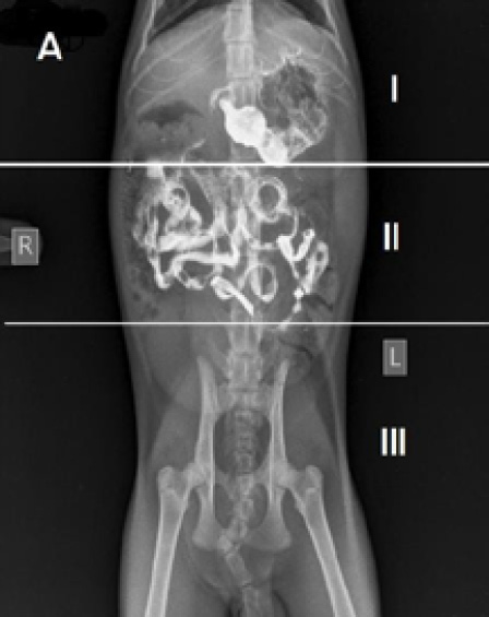
The zone division of cat’s gastrointestinal tract in ventrodorsal (VD) position. Zone 1: gastric area, zone 2: small intestine area, zone 3: colon area.
(ii) Gastrointestinal transit time: Transit time was evaluated using a validated four-point scoring system, modified from Thrall (2002), based on the percentage distribution of BaSO₄ opacity across three anatomical zones in the VD view: Zone 1 (gastric region), Zone 2 (small intestinal region), and Zone 3 (colonic region). Scores of 1–4 were assigned according to the proportion of contrast medium visible in each zone at each time point. All image evaluations for both outcome parameters were performed by two board-certified veterinary radiologists blinded to group allocation, with visual score allocation for transit performed independently by each radiologist using a scale adapted from validated diagnostic criteria ; inter-observer reliability was assessed using the intra-class correlation coefficient (ICC= 0.91). The scoring method used to evaluate BaSO₄ transit is summarized in Table 1. In brief, the threshold defining each of the four scores was specific to each fixed radiographic interval, with higher scores reflecting more advanced aboral progression of contrast relative to the distribution expected at that time point. For example, at the 5-minute interval, a score of 1 corresponded to contrast fully retained within Zone 1 (100%), whereas a score of 4 corresponded to a predominant shift into Zone 2 (75%, with the remainder in Zone 1); by the 30-minute interval, the reference thresholds for each score shifted further aborally to account for the greater contrast progression expected by that time.
Statistical analysis
Data normality was assessed using the Shapiro-Wilk test, and intestinal amplitude data, for which normality was formally assessed, were analyzed using a two-way repeated-measures analysis of variance (ANOVA) across all time points. Post-hoc pairwise comparisons following significant ANOVA effects (p < 0.05) were conducted using Tukey’s honestly significant difference (HSD) test with Bonferroni correction for multiple time-point comparisons. GI transit time scoring data, which were non-parametric, were analyzed using the Kruskal-Wallis test (p < 0.05), followed by Mann-Whitney U post-hoc comparisons with Bonferroni-adjusted p-values. All statistical analyses were performed using IBM SPSS Statistics (version 22.0).
RESULTS
Intestinal amplitude
The mean (± SD) small intestinal amplitudes recorded at each time point across all three treatment groups are presented in Table 2. Statistical analysis by two-way repeated-measures ANOVA revealed no significant differences in intestinal amplitude among groups C, T1, and T2 at any time point (p > 0.05), as indicated by identical superscript letters in Table 2. This two-way repeated-measures ANOVA showed no significant main
Table 1: The scoring of barium sulfate (BaSO₄) movement rate on radiograph ventrodorsal view (VD) in percentage (%) during anesthesia and post-anesthesia stage.
| Score | During anesthesia | Post-anesthesia | ||||||||||||||||
| 5 min | 15 min | 30 min | 0 min | 15 min | 30 min | |||||||||||||
| Zone 1 | Zone 2 | Zone 3 | Zone 1 | Zone 2 | Zone 3 | Zone 1 | Zone 2 | Zone 3 | Zone 1 | Zone 2 | Zone 3 | Zone 1 | Zone 2 | Zone 3 | Zone 1 | Zone 2 | Zone 3 | |
| 1 | 100 | 0 | 0 | 75 | 25 | 0 | 50 | 50 | 0 | 50 | 50 | 0 | 25 | 75 | 0 | 0 | 100 | 0 |
| 2 | 75 | 25 | 0 | 50 | 50 | 0 | 25 | 75 | 0 | 25 | 75 | 0 | 0 | 100 | 0 | 0 | 75 | 25 |
| 3 | 50 | 50 | 0 | 25 | 75 | 0 | 0 | 100 | 0 | 0 | 100 | 0 | 0 | 75 | 25 | 0 | 50 | 50 |
| 4 | 25 | 75 | 0 | 0 | 100 | 0 | 0 | 75 | 25 | 0 | 75 | 25 | 0 | 50 | 50 | 0 | 25 | 75 |
Zone 1: gastric area; Zone 2: small intestine area; Zone 3: colonic area.
Table 2: The small intestine amplitude across three treatment groups during anesthesia stage (mean ± SD).
| Group | Small intestine amplitude Mean ± SD (mm) | ||
| 5 min | 15 min | 30 min | |
| Control (C) | 3.55a ± 2.16 Median: 3.00 (2.41–3.30) |
7.52a ± 10.92 Median: 2.85 (2.36–6.00) |
2.89a ± 1.27 Median: 2.87 (1.81–3.89) |
| T1 | 9.28a±13.31 Median: 4.16 (2.08–19.05) |
2.84a±0.97 Median: 2.65 (2.06–3.72) |
5.16a±7.47 Median: 2.10 (1.29–10.58) |
| T2 | 2.85a±0.80 Median: 2.82 (2.06–3.66) |
3.08a±0.48 Median: 2.91 (2.71–3.62) |
2.46a±1.98 Median: 2.83 (0.31–4.23) |
Note: Values with the same superscript letter (a) within a column are not significantly different (p > 0.05; two-way repeated-measures ANOVA with Tukey’s HSD post-hoc test, Bonferroni-corrected).
effect of group (p = 0.730), no significant main effect of time (p = 0.579), and no significant group×time interaction (0 = 0.343).
During the anesthesia stage, the T1 group exhibited numerically higher amplitude values at 5 min (9.28 ± 13.31 mm) and 30 min (5.16 ± 7.47 mm) compared with C and T2, whereas at 15 min, T1 amplitude (2.84 ± 0.97 mm) was lower than both C and T2. The T2 group maintained the lowest amplitude values across most time points, suggesting a mild but non-significant suppressive effect on intestinal contractility as shown in Table 2. These data were audited in light of the large standard deviations observed at these intervals; the extreme variance was found to be driven by a single outlier cat exhibiting transient peristaltic surges immediately following BaSO₄ administration. Median (IQR) values, which are less sensitive to this single extreme observation, are reported alongside mean ± SD in Table 2. Specifically, the elevated variance was traced to a single animal exhibiting markedly increased amplitude at 5 minutes (32.96 mm) and 30 minutes (18.46 mm), while remaining within the typical range at 15 minutes (4.40 mm); the remaining four T1 animals showed amplitude values consistently between 1.4 and 5.2 mm across all three intervals.
In the post-anesthesia stage, a similar pattern was observed: no significant difference in amplitude was detected among groups at 0-, 15-, or 30-min, indicating that GI smooth muscle contractility was not significantly altered by either anesthetic protocol, as shown in Table 3.
Gastrointestinal transit time
The mean (± SD) BaSO4 transit time scores for each group at each radiographic interval are presented in Table 4 (during-anesthesia stage) and Table 5 (post-anesthesia stage). During the anesthesia stage, no significant difference in BaSO4 transit time was observed among the three groups (p > 0.05); however, at 15 and 30 min, the transit time scores for cats receiving diazepam (0.5 mg/kg BW) and ketamine (20 mg/kg BW) were numerically lower than those for cats receiving acepromazine (0.2 mg/kg BW) and ketamine (20 mg/kg BW) as shown in Table 4.
Table 3: The small intestine amplitude across three treatment groups in the post-anesthesia stage (mean ± SD).
| Group | Small intestine amplitude Mean ± SD (mm) | ||
| 5 min | 15 min | 30 min | |
| Control (C) | 2.60a ± 0.36 | 2.04a ± 0.22 | 2.26a ± 0.38 |
| T1 | 2.04a ± 0.22 | 2.76a ± 0.49 | 2.76a ± 1.06 |
| T2 | 2.50a ± 0.75 | 2.70a ± 0.65 | 2.51a ± 0.76 |
Note: Values with the same superscript letter (a) within a column are not significantly different (p > 0.05; two-way repeated-measures ANOVA with Tukey’s HSD post-hoc test, Bonferroni-corrected).
Table 4: The BaSO₄ gastrointestinal transit time scores (mean ± SD) across treatment groups and time points (during anesthesia stage).
| Treatment group | BaSO₄ transit time score (mean ± SD) | ||
| 0 min | 15 min | 30 min | |
| Control (C) | 1.00a ± 0.408 | 1.00a ± 0.753 | 1.50a ± 1.049 |
| T1 (Ketamine Acepromazine) | 1.00a ± 0.516 | 1.00a ± 0.983 | 1.00a ± 0.753 |
| T2 (Ketamine Diazepam) | 1.00a ± 0.837 | 0.50a ± 0.816 | 0.50a ± 0.816 |
Different superscript letters within a column indicate significant differences (p < 0.05; Kruskal-Wallis with Mann-Whitney U post-hoc test).
Table 5: The BaSO₄ gastrointestinal transit time scores (mean ± SD) across treatment groups and time points (in post-anesthesia stage).
| Treatment group | BaSO₄ transit time score (mean ± SD) | ||
| 0 min | 15 min | 30 min | |
| Control (C) | 1.50a ± 1.049 | 2.50a ± 1.049 | 2.33a ± 1.211 |
| T1 (Ketamine Acepromazine) | 1.00a ± 0.837 | 1.00b ± 0.548 | 0.00b ± 0,000 |
| T2 (Ketamine Diazepam) | 0.50a ± 0.816 | 0.50b ± 0.548 | 0.00b ± 0.408 |
Different superscript letters within a column indicate significant differences (p < 0.05; Kruskal-Wallis with Mann-Whitney U post-hoc test).
Table 5 showed the statistical analysis of the BaSO₄ transit scoring data across all treatments. A statistically significant difference was observed at the 15- and 30-minute intervals in the post-anesthesia stage. At 15 minutes, the control group showed a statistically significant difference (p < 0.05) compared with both T1 and T2, but T1 and T2 did not differ significantly from one another. At 30 minutes post-anesthesia, the control group again showed a statistically significant difference (p < 0.05) compared with both T1 and T2, while T1 and T2 again did not differ significantly. Post-hoc pairwise comparisons confirmed that T1 and T2 did not differ significantly at any time point during either the anesthesia or post-anesthesia stage (p > 0.05).
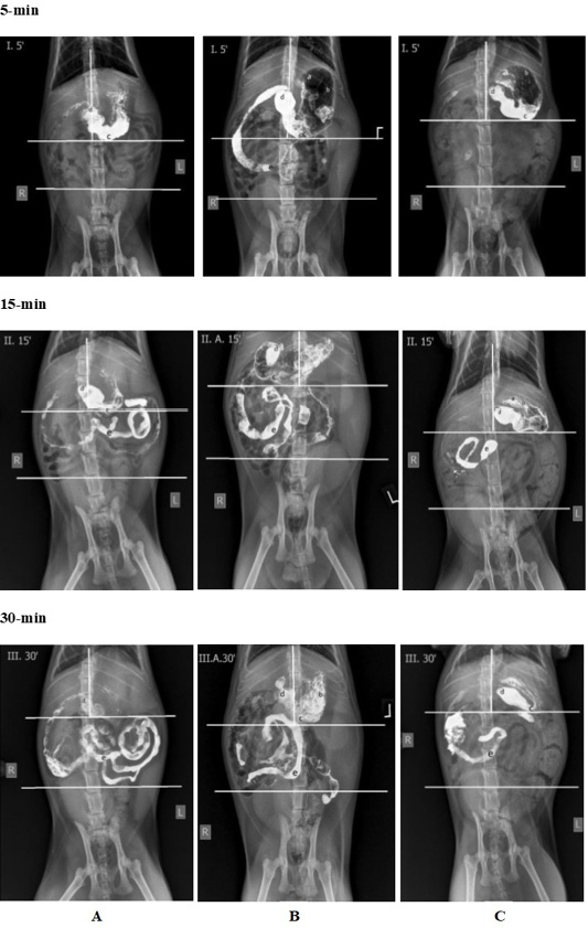
Radiograph of the ventrodorsal view (VD) of the gastrointestinal organ in 0-, 15- and 30-minute. A: control group, B: T1 group, and C: T2 group, with small annotation showing the contrast agent BaSO4 position in gastro-intestinal tract; a: gastric cardia, b: gastric fundus, c: gastric corpus, d: gastric pylorus, e: small intestine.
BaSO4 transit was assessed via evaluation of the abdominal radiographs obtained from each group, performed independently by two blinded radiologists. Radiographs from groups C, T1, and T2 were evaluated individually according to the standard VD view and then compared in tabular form; on this view, the abdomen was divided into three anatomical zones, as illustrated in Figure 2.
At 0 minutes post-anesthesia, no visual difference was apparent among the C, T1, and T2 groups; in all three, most of the BaSO4 remained within Zone 1, spanning the stomach to the pyloric region. By 15 minutes post-anesthesia, BaSO4 in the control group had progressed into the small intestine and begun entering the colon. In the T1 group, BaSO4 was visible in the duodenum and small intestine but had not yet reached the colon, whereas in the T2 group, BaSO4 remained largely confined to the pylorus and was only beginning to enter the small intestine. By 30 minutes post-anesthesia, BaSO4 in the control group persisted in both the small intestine and colon; in the T1 group, it was confined to the small intestine; and in the T2 group, it remained detectable in both the stomach and small intestine. Radiographs were acquired at 0, 15, and 30 minutes post-anesthesia are presented in Figure 3.
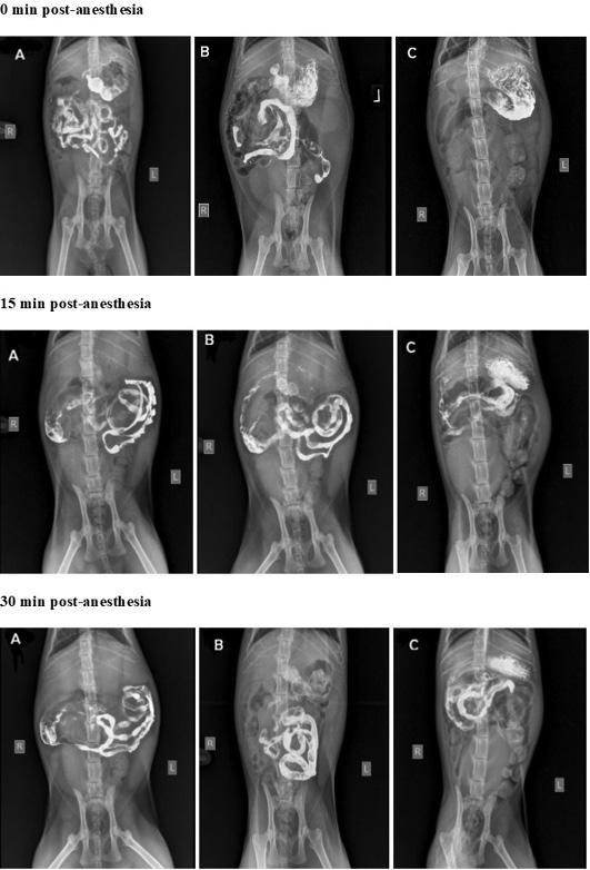
Radiograph of the ventrodorsal view (VD) of the gastrointestinal organ at the interval of 0-, 15-, and 30 minutes in post anesthesia stage. A: Control group, B: T1 Group, C: T2 Group.
DISCUSSION
The present study evaluated and compared the effects of two widely used feline anesthetic protocols ketamine-acepromazine and ketamine-diazepam on GI motility, as assessed by contrast radiography. Two principal findings emerged: first, neither anesthetic combination produced a statistically significant alteration in small intestinal amplitude relative to the unanesthetized control; and second, both combinations significantly slowed GI transit time relative to the control, although ketamine-acepromazine showed a numerical trend toward faster transit, compared with ketamine-diazepam, a difference that did not reach statistical significance.
The absence of significant between-group differences in intestinal amplitude is consistent with the physiological basis of segmental contraction, which involves cyclic activation of circular smooth muscle driven by phasic elevations in intracellular Ca²⁺ , while peristaltic propagation the coordinated sequence of longitudinal contraction followed by circular relaxation depends on intact enteric neural circuitry ; . Although ketamine inhibits Ca²⁺ influx through NMDA receptor blockade and atropine competitively antagonizes muscarinic receptors, thereby blocking cholinergic neurotransmission at postganglionic parasympathetic sites ; , neither effect appeared sufficient to produce a statistically detectable change in the diameter-based amplitude parameter at the doses employed here. The considerable inter-individual variability in amplitude measurements reflected in the large standard deviations observed in T1 at 5 and 30 minutes may have further limited the statistical power to detect such differences. A data audit indicated that this variability was driven primarily by a single outlier animal exhibiting transient peristaltic surges immediately following BaSO₄ administration; median (IQR) values are therefore reported alongside mean ± SD in Table 2 to better characterize this variability.
The significantly slower GI transit observed in both anesthetized groups relative to the unanesthetized control is attributable to the cumulative pharmacological actions of ketamine and atropine on enteric neural pathways. Ketamine’s sympathomimetic action, mediated through inhibition of neuronal catecholamine reuptake, activates adrenergic receptors that suppress GI motility ; and because NMDA receptors are highly concentrated in vagal afferent neurons projecting to the stomach and proximal small intestine , these regions are particularly susceptible to ketamine-induced motility suppression. Given the prolonged elimination half-life of ketamine in cats (approximately 2–3 hours; , clinically relevant plasma concentrations likely persisted throughout the post-anesthetic observation window, sustaining GI inhibition well beyond clinical recovery of consciousness. Atropine potentiates this effect by competitively antagonizing muscarinic M1 and M3 receptors in the GI tract, thereby reducing both secretory activity and motility ; .
The trend toward faster GI transit in the ketamine-acepromazine group compared with the ketamine-diazepam group can be explained by the divergent pharmacological profiles of the two sedatives with respect to GI smooth muscle. Acepromazine acts as a selective antagonist of dopamine D2 receptors; because D2 receptor activation normally inhibits voltage-dependent Ca²⁺ channels and suppresses acetylcholine release from postganglionic cholinergic neurons , blocking these receptors removes this inhibitory constraint, allowing enhanced Ca²⁺ influx into smooth muscle cells and potentiating peristaltic activity ; . Acepromazine further augments parasympathetic tone, which directly stimulates GI contractility . This antidopaminergic mechanism therefore partially counteracts the motility-suppressing effects of ketamine, resulting in relatively better-preserved GI transit. It should be noted that this parasympathetic-enhancing effect has not been directly measured in the present study and is inferred from acepromazine’s known antidopaminergic pharmacology; the claim of a direct stimulatory action on GI contractility should therefore be regarded as a proposed mechanism rather than a confirmed finding.
Diazepam’s action on the ENS, by contrast, is more complex and predominantly inhibitory in the context of GI motility. As a positive allosteric modulator of GABAA receptors , diazepam enhances the activity of inhibitory motor neurons that release nitric oxide (NO) and vasoactive intestinal polypeptide (VIP); both mediators relax intestinal smooth muscle by suppressing Ca²⁺ signaling and promoting hyperpolarization, thereby directly decelerating peristaltic propagation ; ; . It should be noted that while these enteric GABAergic inhibitory pathways are well-documented in rodent models, direct translational evidence in feline enteric tissue remains largely hypothetical and warrants further species-specific investigation. The net effect of diazepam is therefore to augment the inhibitory influence of ketamine on GI motility, producing a greater overall reduction in transit time than the acepromazine combination. This proposed augmentation of ketamine’s inhibitory effect is likewise inferred from known receptor pharmacology rather than directly measured in the present study, as GABA, NO, and VIP levels were not assessed; this mechanistic interpretation should therefore be regarded as provisional.
The role of atropine as a pre-anesthetic agent shared by both treatment groups warrants specific consideration. By competitively antagonizing acetylcholine at muscarinic receptors throughout the GI tract, atropine reduces glandular secretion, decreases smooth muscle tone, and impairs peristaltic coordination . This shared motility-inhibitory background, independent of the specific sedative employed, may partly explain why both T1 and T2 exhibited significantly slower transit than the unanesthetized control despite the partial motility-sparing effect of acepromazine in T1 . Because both T1 and T2 received atropine, a substantial component of the reduction in GI transit relative to Control is likely driven by muscarinic receptor blockade common to both groups, which may obscure any true baseline pharmacodynamic differences between acepromazine and diazepam on GI motility.
From a clinical perspective, these findings carry direct implications for anesthetic protocol selection in cats undergoing GI evaluation or surgery. Clinicians should anticipate delayed BaSO₄ transit with either anesthetic combination when performing diagnostic GI imaging, since both protocols significantly slow transit relative to the unanesthetized state; because the difference between ketamine-acepromazine and ketamine-diazepam was not statistically significant in the present study, neither combination can currently be recommended over the other specifically for preserving GI transit assessment accuracy. Nevertheless, both protocols remain clinically acceptable from a safety standpoint, as neither produced a significant alteration in the amplitude of intestinal contractions.
These findings are broadly consistent with prior feline motility literature. A previous contrast-radiography study evaluating tiletamine-zolazepam anesthesia in local domestic cats similarly reported altered gastrointestinal transit relative to the unanesthetized state , and comparable GI dysmotility have been described for other peri anesthetic agent such as α2-agonist, dexmedetomidine, giving notion that suppression and peri operative stress could positively synergized with depressants effect toward GI smooth muscles . Taken together with the present results, these observations reinforce that GI transit delay is a broadly shared feature of feline general anesthesia rather than a property specific to any single drug combination, underscoring the importance of accounting for anesthetic exposure when interpreting diagnostic GI imaging obtained under sedation or anesthesia.
This study has several limitations. The sample size of six cats per group limits statistical power to detect small but potentially clinically meaningful differences in amplitude. The relatively short observation window (maximum 30 minutes post-BaSO4 administration) may not capture later phases of GI recovery, and an independent parallel-group design was used rather than a crossover design because prior exposure to barium sulfate could plausibly leave residual contrast or alter baseline motility in the same animal, which would confound repeated within-animal comparisons across anesthetic protocols. Additionally, this study lacked single-agent control groups (e.g., ketamine alone or atropine alone), limiting the ability to fully isolate the individual contribution of each drug to the observed GI motility changes. The male-only cohort, while controlling for estrous-related hormonal variability, excludes female physiological variables and limits generalizability to female cats. Handling and injection-related stress may also have differed systematically between the control group (minimal handling, no injections) and the treatment groups (restraint and multiple injections), and such stress-induced sympathetic activation could independently influence GI motility irrespective of the anesthetic agents themselves; this potential confounder was not separately assessed. Similarly, the fixed barium volume (10 mL/kg) was constant across all groups but could not be fully excluded as an independent contributor to the observed transit times. Future studies incorporating larger sample sizes, longer post-anesthetic follow-up, and additional motility parameters such as electromyography or scintigraphy would help clarify these relationships.
CONCLUSION
In conclusion, ketamine-acepromazine and ketamine-diazepam combinations (with atropine) both induce significant suppression of gastrointestinal transit time in adult male domestic cats compared to unanesthetized controls, without significantly altering small intestinal contraction amplitude. Although ketamine-acepromazine demonstrated a numerical trend toward faster transit, the difference between the two protocols was not statistically significant. Clinicians should anticipate delayed contrast transit with both anesthetic combinations during diagnostic GI imaging. Larger studies are needed to determine whether the numerical trend toward faster transit with ketamine-acepromazine represents a clinically meaningful advantage.
Acknowledgements
The authors thank the staff of the Radiology Unit, Veterinary Teaching Hospital, Faculty of Veterinary Medicine, Universitas Airlangga, for their technical support during this study.
Novelty Statement
To our knowledge, this is the first study to directly compare ketamine-acepromazine and ketamine-diazepam, two of the most common feline anesthetic combinations, on GI motility in cats using barium contrast radiography. Both protocols significantly delayed contrast transit without altering intestinal contraction amplitude. These findings help veterinarians interpret GI imaging under anesthesia better and support welfare-centered anesthetic choices for cats.
Author’s ContrIBUTIon
HP and ISY conceived the idea and planned the manuscript. IAC, NSM, ISY and HP wrote the manuscript and contributed to original draft preparation and analysis. NT, BS, WMY and LM have made significant scientific support and also contributed to the interpretation of the results, review and editing. All authors provided significant contributions by giving feedback and help shape the manuscript.
Ethıcal statement
All experimental protocols and procedures involving animals in this study were reviewed and legally approved by the Animal Care and Use Committee (ACUC) of the Faculty of Veterinary Medicine, Universitas Airlangga (Approval No: 2.KE.022.02.2019).
Generative AI and AI assisted technology statement
The authors declare that no generative AI and AI assisted technology was used in the creation of this manuscript.
Conflıct of ınterest
The authors have declared no conflict of interest.
References
- Aditya R (2013). Efek Anestesi Ketamine-Acepromazine Terhadap Motilitas Saluran Pencernaan Kucing Melalui Studi Radiografi Kontras. Undergraduate’s thesis, IPB University, Bogor, Indonesia.
- Berridge MJ (2008). Smooth muscle cell calcium activation mechanisms. J. Physiol., 586(21): 5047–5061. [CrossRef]
- Bickelhaupt S, Pazahr S, Chuck N, Blume I, Froehlich JM, Cattin R, Raible S, Bouquet H, Bill U, Rogler G, Frei P, Boss A, Patak MA (2013). Crohn’s disease: Small bowel motility impairment correlates with inflammatory-related markers C-reactive protein and calprotectin. Neurogastroenterol. Motil., 25(6): 467–e363. [CrossRef]
- Czaja K, Ritter RC, Burns GA (2006). Vagal afferent neurons projecting to the stomach and small intestine exhibit multiple N-methyl-ᴅ-aspartate receptor subunit phenotypes. Brain Res., 1119(1): 86–93.
- Gaschen F (2011). Gastrointestinal motility: Why it matters. In: Proceedings of the international congress of the Italian association of companion animal veterinarians. Louisiana State University School of Veterinary Medicine, Louisiana, USA. pp. 534–537.
- Grasa L, Rebollar E, Arruebo MP, Plaza MA, Murillo MD (2004). The role of Ca²⁺ in the contractility of rabbit small intestine in vitro. J. Physiol. Pharmacol., 55(3): 639–650.
- Grimm KA, Lamont LA, Tranquilli WJ, Greene SA, Robertson SA (2015). Veterinary anesthesia and analgesia. In: The 6th Edition of Lumb and Jones. John Wiley & Sons, New Jersey, USA. pp. 246–253. [CrossRef]
- Han J, Kim N, Joo H, Kim E (2003). Ketamine blocks Ca²⁺-activated K⁺ channels in rabbit cerebral arterial smooth muscle cells. Am. J. Physiol. Heart Circ. Physiol., 285(3): H1347–H1355.
- Hellstrom EA, Ziegler AL, Blikslager AT (2021). Postoperative ileus: Comparative pathophysiology and future therapies. Front. Vet. Sci., 8: 714800. [CrossRef]
- Kandynesia A (2012). Studi radiografi kontras pengaruh anestesi tiletamin-zolazepam terhadap motilitas saluran pencernaan kucing lokal. Undergraduate’s thesis, IPB University, Bogor, Indonesia.
- Kochar RT, Gouda BP, Shah RS (2011). Retained portion of the appendix following laparoscopic appendicectomy causing peritonitis and ileus. J. Minim. Access Surg., 7(2): 154–155.
- Krantis A (2000). GABA in the mammalian enteric nervous system. Physiology, 15(6): 284–290. [CrossRef]
- Little SE (2015). Anesthesia for the aging cat. In: August’s consultations in feline internal medicine. Elsevier, St. Louis, MO, USA. 7: 986–994. [CrossRef]
- Mangel AW, Evans ED (2011). Depolarization-stimulated contractility of gastrointestinal smooth muscle in calcium-free solution: A review. ISRN Gastroenterol., 692528: 1–3. [CrossRef]
- Papich MG (2016). Saunders handbook of veterinary drugs, small and large animal, 4th edn. Elsevier, St. Louis, MO, USA.
- Plumb DC (2017). Plumb’s veterinary drug handbook, 9th edn. John Wiley & Sons. Blackwell, Iowa, USA.
- Shabana AM, Nasr ES, Moawad HE (2012). Effect of ketamine on intraoperative nausea and vomiting during elective caesarean section under spinal anaesthesia: A placebo-controlled, prospective, randomized, double-blinded study. Egypt. J. Anaesth., 28(2): 169–174. [CrossRef]
- Smith ME, Morton DG (2010). System of the body, the digestive system, basic science and clinical conditions, 2nd edn. Churchill Livingstone Elsevier, St. Louis, MO, USA, pp. 107–128.
- Steagall P, Robertson S, Taylor P (2018). Injectable anesthetics and induction of anesthesia. In: Feline anesthesia and pain management. Wiley-Blackwell, John Wiley & Sons. Hoboken, NJ, USA. pp. 49–57. [CrossRef]
- Steiner JM (2010). Small animal gastroenterology. Schlütersche, Germany. pp. xvii–266.
- Thrall DE (2002). Textbook of veterinary diagnostic radiology: 4th edn. Saunders Elsevier, St. Louis, MO, USA. pp. 74–87.
- Thrall DE (2018). Textbook of veterinary diagnostic radiology: 6th edn. Saunders Elsevier, St. Louis, MO, USA. pp. 769–811.
- Tonini M (1996). Recent advances in the pharmacology of gastrointestinal prokinetics. Pharmacol. Res., 33(4-5): 217–226. [CrossRef]
- Tranquilli WJ, Thurmon JC, Grimm KA (2007). Lumb and Jones’ veterinary anesthesia and analgesia, 4th edn. Blackwell Publishing, State Avenue, Ames, Iowa, USA.
- Van Geldre LAV, Lefebvre RA (2004). Interaction of NO and VIP in gastrointestinal smooth muscle relaxation. Curr. Pharm. Des., 10(20): 2483–2497. [CrossRef]
- Vather R, O’Grady G, Bissett IP, Dinning PG (2014). Postoperative ileus: Mechanisms and future directions for research. Clin. Exp. Pharmacol. Physiol., 41(5): 358–370. [CrossRef]
- Washabau RJ, Day MJ (2012). Canine and feline gastroenterology. Saunders Elsevier, St. Louis, MO, USA. pp. 257–266.
- Whitehead K, Cortes Y, Eirmann L (2016). Gastrointestinal dysmotility disorders in critically ill dogs and cats. J. Vet. Emerg. Crit. Care, 26(2): 234–253. [CrossRef]
- Zizzo MG, Mulè F, Serio R (2007). Functional evidence for GABA as modulator of the contractility of the longitudinal muscle in mouse duodenum: Role of GABAA and GABAC receptors. Neuropharmacology, 52(8): 1685–1690. [CrossRef]

Services
Article usage statistics
Loading…
Related articles
- Morphological and Morphometrical Comparative Study of Thyroid and Adrenal Glands Between Suckling and Adult Local Male Cats (Felis catus)Advances in Animal and Veterinary Sciences · 2024
- Comparison of Back and Loin Locomotor Bony Structures in Cat (Felis domestica) and Rabbit (Oryctolagus cuniculus) : (Thoracic and Lumbar Vertebrae)Advances in Animal and Veterinary Sciences · 2019
- Gastrointestinal Obstruction due to Linear Foreign Body in a Cat: A Case ReportAdvances in Animal and Veterinary Sciences · 2017
- Optimization of Polymerase Chain Reaction for Molecular Detection of Babesia Felis in Domestic CatsThe Journal of Advances in Parasitology · 2018
- Histopathological Analysis of the Male White Mice Liver After Exposure to Halothane AnesthesiaJournal of Animal Health and Production · 2024
- Evaluation of Ultrasonography, Progesterone Analysis, and Relaxin-Based Methods for Pregnancy Detection in Domestic CatsJournal of Animal Health and Production · 2025