
Entamoeba species are common intestinal protozoa infecting humans and many animals, including wildlife. However, limited molecular data are available on infections in captive ungulates in Iraq. This study aimed to assess the prevalence of Entamoeba using microscopy and to identify the species at the molecular level through partial 18S rRNA gene sequencing and phylogenetic analysis. A total of 180 fresh fecal samples were collected from four captive ungulate species belonging to the families Cervidae (Axis axis and Dama mesopotamica) and Bovidae (Gazella gazella and Gazella marica) in Baghdad, Hilla, and Karbala, Iraq. Samples were examined microscopically using direct wet-mount and formalin–ether concentration techniques. A stratified selection of ten microscopy-positive samples was further analyzed by PCR targeting the partial 18S rRNA gene, followed by DNA sequencing, BLAST comparison, and phylogenetic analysis. Microscopic examination revealed Entamoeba cysts in 90 of 180 samples (50.0%), with significant regional differences (P < 0.05): 41.38% in Baghdad, 52.38% in Hilla, and 70.00% in Karbala. Molecular analysis identified two Entamoeba species, E. histolytica (GenBank accession numbers PQ661235.1–PQ661236.1) and E. coli (PQ661237.1–PQ661239.1). The sequences shared over 99% identity with reference sequences in GenBank, and phylogenetic analysis placed the Iraqi isolates within the respective E. histolytica and E. coli clades. This is the first study to molecularly identify Entamoeba spp. in captive ungulates in Iraq, providing baseline epidemiological and genetic data for these parasites in wildlife. While the results indicate the presence of potentially zoonotic Entamoeba species, further research with larger molecular samples, higher-resolution genetic markers, and integrated human, livestock, and environmental sampling is necessary to better understand transmission dynamics within a One Health framework.
Research Article · Volume 15, Issue 1, Pages 66–75, 14 November 2026 · Open Access
Genetic Characterization of Entamoeba spp. in Captive Ungulates from Iraq Using 18S rRNA Gene Analysis
,
Affiliations & Notes ⌄
- 1Iraq Natural History Research Centre and Museum, University of Baghdad, Baghdad, Iraq
- *Correspondence | Mareym M. Alkhaiat; Email: maryam.m@nhm.uobaghdad.edu.iq
Article Info ⌄
- Journal:
- Journal of Animal Health and Production
- Submitted Date:
- 12 May 2026
- Accepted Date:
- 14 September 2026
- Published Date:
- 14 November 2026
Abstract
Introduction
Entamoeba species are intestinal amoeboid protozoa that infect humans and a wide range of domestic and wild animals worldwide. While most species are considered commensal, Entamoeba histolytica is an important pathogenic protozoan responsible for amoebiasis, a disease of considerable public health and veterinary importance. By contrast, Entamoeba coli is generally regarded as a non-pathogenic indicator of fecal contamination. Transmission occurs primarily via the faecal–oral route, with environmentally resistant cysts contaminating food, water, or the environment. Despite advances in sanitation and disease management, amoebiasis continues to be a significant public health concern in many low- and middle-income countries. Since Entamoeba species can infect both humans and animals, studying their epidemiology is important from a One Health perspective (World ; .
Recent molecular research has significantly advanced our knowledge of the epidemiology, host range, and genetic diversity of Entamoeba species in wildlife. These surveys have detected Entamoeba infections in captive and wild ungulates, zoo animals, wild boars, and other wildlife across Asia, Australia, Europe, and Africa, illustrating the wide host range of these protozoa ; ; . In Australia, Huaman et al. (2022) found Entamoeba spp. in wild deer and cattle, indicating potential sharing of parasites among sympatric hosts. Likewise, Huang et al. (2022) identified E. histolytica and E. dispar in farmed sika deer in China. Overall, these findings underscore the importance of molecular surveillance for precise identification of Entamoeba species in wildlife and for enhancing our understanding of host distribution and molecular epidemiology.
Recent molecular studies in Iraq have documented Entamoeba infections in humans and domestic animals (Al-Sudani, 2023; Al-Jubori et al., 2024; Hussain and Mahdi, 2024). However, comparable molecular investigations of captive wildlife have not been conducted. Consequently, information on the prevalence, species composition, genetic diversity, and phylogenetic relationships of Entamoeba spp. infecting captive ungulates in Iraq remains unavailable. This absence of molecular epidemiological and phylogenetic data limits current understanding of Entamoeba diversity in captive ungulates in Iraq and highlights the need for molecular characterization of circulating isolates.
Captive wildlife populations serve as unique ecological interfaces where close contact among animals and enclosure management may promote parasite persistence. Nonetheless, no molecular research has explored Entamoeba species infecting captive ungulates in Iraq.
This study aimed to assess the prevalence of Entamoeba infection in captive ungulates in central Iraq through microscopic examination and to genetically characterize the identified isolates via partial 18S rRNA gene sequencing, followed by phylogenetic analysis. It also aimed to establish baseline molecular epidemiological data on Entamoeba spp. in captive wildlife in Iraq.
Materials and Methods
Study area and sampling sites
The study was conducted between October 2023 and August 2024 in three provinces of central Iraq: Baghdad (33°20′00.0″N, 44°24′00.0″E), Babil (Hilla) (32°28′00.0″N, 44°25′00.0″E), and Karbala (32°36′00.0″N, 44°00′00.0″E). Sampling was performed throughout the study period in protected wildlife enclosures managed by the Iraqi Ministry of Environment.
The taxonomic classification of the deer species examined in the present study were shown in Figure 1. While, the geographical distribution of the study sites was shown in Figure 2. The sites comprise protected wildlife enclosures managed by regional authorities under the Iraqi Ministry of Environment and are located in central Iraq.
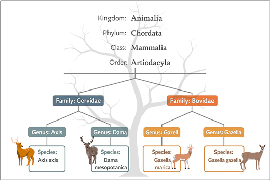
Taxonomic classification of the deer species examined in the present study in Iraq.
Sample collection and preservation
A total of 180 fresh fecal samples were collected from captive ungulates aged 6 months to 3 years between October 2023 and August 2024. Samples were collected immediately after spontaneous defecation to minimize environmental contamination and to avoid repeated sampling of the same individual whenever possible. Each sample was transferred into a sterile screw-capped container containing 2.5% potassium dichromate solution and labeled with a unique identification code indicating the host species, sampling location, approximate age class, and date of collection. The samples were transported to the laboratory under cooled conditions and stored at 4 °C until microscopic examination, after which aliquots designated for molecular analysis were preserved at −20 °C prior to DNA extraction. The sample size was calculated according to the accessibility and availability of captive deer within the participating conservation enclosures during the study period.
Microscopic examination and morphological diagnosis
Each fecal sample was examined using direct wet mounts and the formalin–ether concentration technique (FECT), following protocols from Cheesbrough (2006) and Allen and Ridley (1970). For microscopic analysis, saline and Lugol’s iodine smears were prepared and examined under a light microscope at 100× and 400× magnification. The FECT procedure involved mixing 1 g of faeces with 10 mL of 10% formalin, filtering through gauze, adding 3 mL of diethyl ether, and centrifuging at 2000 rpm for 5 minutes. The sediment was examined for Entamoeba cysts, noting their size and the number of nuclei in mature cysts. Microscopic identification was based on the morphological criteria described by Williams (1998), Cheesbrough (2006), and Garcia (2016), including cyst size, number of nuclei, and the morphology of chromatoidal bodies. Figure 3 depicts these features, aiding in cyst identification.
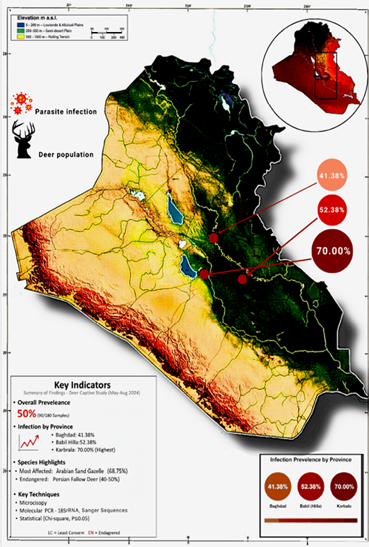
Map showing the sampling locations across three central Iraqi provinces (Baghdad, Babil/Hilla, and Karbala), indicating protected deer breeding enclosures and the observed epidemiological gradient.
DNA extraction, amplification, sequencing, and phylogenetic analyses
Comprehensive DNA analyses were conducted to investigate genetic variation among Entamoeba species, revealing evolutionary patterns across diverse ecological niches. DNA was extracted, amplified, and sequenced to test the central hypothesis and explore genetic diversity and phylogenetic relationships.
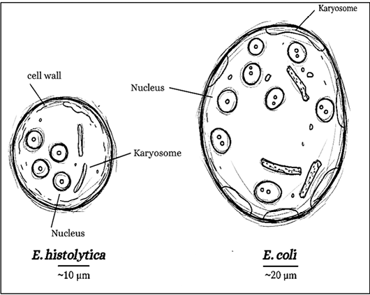
Schematic illustration of typical Entamoeba cysts and their principal morphological characteristics.
DNA extraction
For molecular analysis, ten microscopically confirmed positive samples were selected. The selected samples represented all available PCR-positive specimens from different host species and geographical locations. To ensure host diversity, all four deer species were represented. Infection intensity was also considered by including samples with both high- and low-intensity infections. Additionally, the species were represented by both E. histolytica and E. coli, based on initial cyst morphology. DNA quality was assessed by electrophoresis on 1% agarose gel prior to PCR amplification.
Genomic DNA was extracted from these ten fecal samples, which were stored at –20 °C with minimal freeze-thaw cycles to maintain integrity. The Intron Biotechnology DNA Extraction Kit (Korea) was used according to the manufacturer’s instructions, yielding DNA of suitable quality for subsequent molecular applications.
Target locus amplification and PCR settings
A ~585 base pair (bp) segment of the 18S rRNA gene was amplified via polymerase chain reaction (PCR) using a standard thermocycler. The primer pair described by Verweij et al. (2003) was used for molecular identification. The forward primer was 5′-GTTGATCCTGCCAGTATTATATG-3′, and the reverse primer was 5′-CACTATTGGAGCTGGAATTAC-3′.
Each PCR (polymerase chain reaction) was prepared in a 25 µL volume containing 12.5 µL of GoTaq Green Master Mix (2×, Promega, USA), which includes DNA polymerase, dNTPs, magnesium chloride, and buffers. Additionally, 1 µL of each primer (10 pmol/µL), 1.5 µL of DNA template, and 9 µL of nuclease-free water were added. The thermocycling protocol commenced with an initial denaturation at 95 °C for 5 minutes, followed by 35 cycles of 95 °C for 45 seconds (denaturation), 56 °C for 45 seconds (annealing), and 72 °C for 1 minute (extension). A final extension at 72 °C for 5 minutes concluded the process. The annealing temperature of 56 °C was selected based on prior calculations of primer melting temperature and gradient PCR experiments.
Each PCR run included a positive control (previously confirmed Entamoeba DNA), a no-template negative control containing nuclease-free water, and an extraction blank to monitor possible contamination during DNA extraction. DNA extraction, PCR preparation, and post-amplification procedures were performed in physically separated laboratory areas using aerosol-resistant filter tips.
DNA sequencing and sequence processing
Ten PCR-positive amplicons representing different host species and sampling locations were selected for sequencing to confirm species identity and obtain representative molecular data from the study population. The selected amplicons encompassed the available PCR-positive specimens from the different host species and geographic regions included in this study. PCR products were purified using the StrataPrep PCR Purification Kit (Agilent Technologies, USA) to remove residual primers, nucleotides, and other reaction components prior to sequencing. The purified amplicons were then submitted to Macrogen Inc. (Seoul, South Korea) for bidirectional Sanger sequencing. The resulting nucleotide sequences were aligned using the ClustalW algorithm implemented in MEGA version 12, and species identity was verified by comparison with reference sequences in the NCBI GenBank database using the BLAST algorithm ; . Chromatograms were visually inspected, and ambiguous nucleotide calls were manually edited before sequence alignment.
Phylogenetic analysis and data submission
Phylogenetic relationships among the obtained sequences and selected reference sequences retrieved from the NCBI GenBank database were inferred using the Maximum Likelihood (ML) method implemented in MEGA version 12. The Tamura–Nei model was selected as the best-fit substitution model according to the lowest AIC value K2P. Node support was evaluated using 1,000 bootstrap replicates (Kumar et al., 2018).
The use of additional markers was beyond the scope of the present study and is recommended for future investigations requiring strain-level discrimination.
Statistical analysis
Prevalence (%) was calculated as the number of positive samples divided by the total number of examined samples. Associations between infection status and geographical location were evaluated using Pearson’s Chi-square (χ²) test. Statistical significance was set at P < 0.05. All analyses were performed using IBM SPSS Statistics version 25 (IBM Corp., Armonk, NY, USA).
Results
Microscopic examination
Microscopic examination of 180 fecal samples revealed Entamoeba cysts in 90 samples, corresponding to an overall prevalence of 50.0%. Based on established morphological criteria ; , the observed cysts were spherical to oval, measured approximately 10–20 µm in diameter, and contained 1–4 nuclei with characteristic chromatoidal bodies (Table 1). Mature E. coli cysts were generally larger and contained up to eight nuclei, whereas mature E. histolytica cysts measured approximately 10–20 µm and contained up to four nuclei (Figure 4A, B). Because microscopy alone cannot reliably distinguish E. histolytica from morphologically similar species, molecular analysis was subsequently performed to confirm species identity.
Table 1: Morphological characteristics of cystic stages of Entamoeba species identified in the present study.
| Species | Size range (µm) | Nuclei (mature cyst) | Chromatoidal bar appearance | Pathogenic status |
| Entamoeba histolytica | 10–20 | 1–4 | Smooth, rounded ends | Potentially Pathogenic |
| Entamoeba coli | 10–35 | 1–8 | Splintered or irregular | Commensal |
The prevalence differed of Entamoeba infection varied significantly among the three study provinces (χ² = 8.45, P = 0.015). The highest prevalence was recorded in Karbala (70.0%; 21/30), followed by Babil (52.38%; 33/63) and Baghdad (41.38%; 36/87) (Table 2; Figure 5). The spatial distribution of infection is illustrated in the epidemiological map (Figure 2).
Differences in infection prevalence were also observed among the host species examined. Gazella marica showed an observed prevalence, particularly in Karbala (68.75%), whereas Dama mesopotamica showed prevalence values ranging from 40% to 50%. The prevalence recorded for each host species is presented in Table 2.
Molecular characterization and phylogenetic relationships
PCR amplification of the partial 18S rRNA gene yielded the expected 585 bp amplicon in 10 of the microscopically positive samples, confirming the presence of Entamoeba DNA (Figure 6). Ten representative PCR-positive amplicons selected from different host species and sampling locations were subjected to sequencing.
Table 2: Distribution of Entamoeba infection among deer species across selected Iraqi provinces.
| Province | Deer species | No. of samples | Positive | % positive | IUCN status |
| Baghdad | Gazella gazella | 32 | 14 | 43.75 | LC |
| Axis axis | 30 | 12 | 40.00 | LC | |
| Gazella marica | 25 | 10 | 50.00 | LC | |
| Subtotal | 87 | 36 | 41.38 | ||
| Babil (Hilla) | Gazella marica | 24 | 13 | 54.17 | LC |
| Gazella gazella | 29 | 15 | 51.72 | LC | |
| Dama mesopotamica | 10 | 4 | 40.00 | EN | |
| Subtotal | 63 | 33 | 52.38 | ||
| Karbala | Gazella marica | 16 | 11 | 68.75 | LC |
| Axis axis | 8 | 5 | 62.50 | LC | |
| Dama mesopotamica | 6 | 3 | 50.00 | EN | |
| Subtotal | 30 | 21 | 70.00 | ||
| Total | 180 | 90 | 50.00 |
LC= Least Concern; EN= Endangered (based on the IUCN Red List, 2024). Infection rate = (Positive/Total samples) × 100.
Sequence analysis and comparison with reference sequences in the NCBI GenBank database identified two Entamoeba species: E. histolytica and E. coli. The obtained sequences shared nucleotide identities ranged from 99.57% to 99.76% with corresponding reference sequences deposited in GenBank (Table 3). Specifically, E. histolytica isolates (PQ661235.1 and PQ661236.1) showed 99.57% identity with reference isolate KJ870203.1 from Cameroon, whereas E. coli isolates (PQ661237.1–PQ661239.1) shared 99.76% identity with reference isolate MH133210.1 from China.
Gene bank submissions
The nucleotide sequences generated in the present study were deposited in the NCBI GenBank database under accession numbers PQ661235.1–PQ661239.1 (Table 3). These represent the first publicly available molecular records of Entamoeba spp. obtained from captive ungulates in Iraq.
Phylogenetic analysis
Phylogenetic analysis of the partial 18S rRNA gene using the Maximum Likelihood method with 1,000 bootstrap replicates divided the sequences into two well-supported clades, representing E. histolytica and E. coli (Figure 7). Iraqi isolates grouped within their respective species clades alongside homologous reference sequences from GenBank. Pairwise sequence comparisons yielded p-distance values below 0.005 among the analyzed sequences.
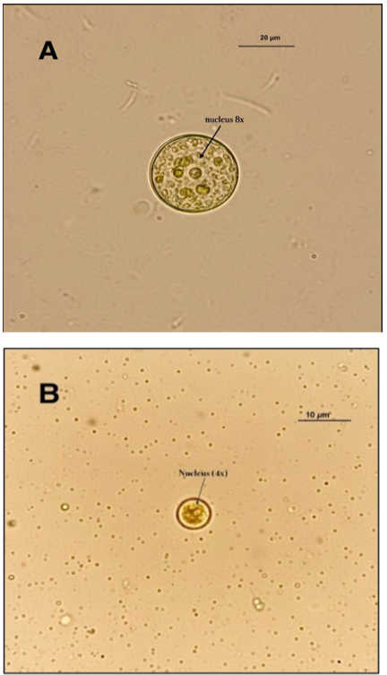
Representative mature cysts of Entamoeba species: (A) Entamoeba coli cyst with eight nuclei; (B) Entamoeba histolytica cyst with four nuclei.
Table 3: Molecular identification and BLAST analysis of Entamoeba isolates obtained in this study.
| Local accession No. | Species | Closest match (Accession) | Identity | Clinical significance |
| PQ661235.1 | E. histolytica | Cameroon (KJ870203.1) | 99.29% | Pathogenic |
| PQ661236.1 | E. histolytica | Cameroon (KJ870203.1) | 99.57% | Pathogenic |
| PQ661237.1 | E. coli | China (MH133210.1) | 99.76% | Commensal |
| PQ661238.1 | E. coli | China (MH133210.1) | 99.57% | Commensal |
| PQ661239.1 | E. coli | China (MH133210.1) | 99.57% | Commensal |
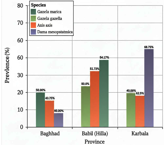
Prevalence of intestinal protozoan infections among captive deer across the three central Iraqi provinces.
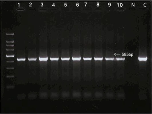
Agarose gel electrophoresis showing PCR amplification of the partial 18S rRNA gene (~585 bp) from faecal samples of captive deer.
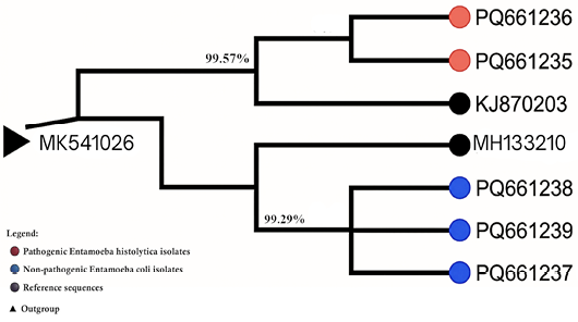
Phylogenetic relationships of Entamoeba species isolated from captive deer in Iraq based on partial 18S rRNA gene sequences.
Discussion
The current study differentiates molecularly between Entamoeba spp. infecting captive ungulates in Iraq and builds on prior microscopic surveys of the country’s wildlife. Microscopy showed a prevalence of 50.0%, and molecular analysis identified the presence of E. histolytica and E. coli. Although E. histolytica is recognized as a pathogenic species, the present study confirmed only species identity and did not evaluate clinical disease or pathogenicity in the sampled animals. These results broaden our understanding of intestinal protozoan infections in Iraqi wildlife and provide molecular data for future epidemiological research. While Al-Khaiat et al. (2025) previously documented intestinal parasites in captive deer, including Entamoeba spp., this study builds on those findings by providing molecular confirmation and phylogenetic analysis, thereby enhancing the accuracy of parasite identification.
The overall prevalence found in this study (50.0%) aligns with figures reported for both captive and wild ungulates across various countries, although notable variation exists among different studies. These prevalence rates are affected by factors such as host species, sample size, location, climate, management practices, and diagnostic techniques. Molecular methods generally yield more accurate species identification than microscopy alone, as morphological similarities among Entamoeba species can hinder distinction based solely on cyst appearance. Similar findings have been observed in captive wildlife from Egypt, India, and other regions, where Entamoeba infections are often detected in herbivorous mammals kept in captivity ; ; .
Significant geographical variation in infection prevalence was observed across the three study provinces, with the highest prevalence recorded in Karbala. Although the present study was not designed to determine the causes of these regional differences, variations in enclosure management, differences in husbandry practices, sanitation practices, water quality, environmental conditions, and animal contact rates may have contributed to the observed differences. Similar environmental and husbandry-related factors have previously been associated with variation in intestinal protozoan infections among captive wildlife populations ; . However, additional epidemiological studies incorporating environmental sampling and management-related variables are required to clarify the factors influencing parasite distribution under Iraqi conditions.
Microscopic examination enabled preliminary recognition of Entamoeba cysts based on established morphological criteria, including cyst size, number of nuclei, and chromatoidal body morphology ; . Nevertheless, microscopic examination alone cannot reliably distinguish E. histolytica from morphologically similar species such as E. dispar and E. moshkovskii. Consequently, molecular confirmation using PCR amplification and sequencing of the partial 18S rRNA gene was essential to accurately determine species identity. The combined application of microscopy and molecular techniques therefore increased the diagnostic reliability of the present investigation and reduced the possibility of species misidentification.
Sequence analysis demonstrated that the Iraqi isolates shared more than 99% nucleotide identity with corresponding reference sequences available in GenBank. Two isolates were identified as E. histolytica, and three as E. coli. Comparable levels of sequence conservation have been reported in previous molecular studies of Entamoeba spp., reflecting the relatively conserved nature of the 18S rRNA gene among members of the genus ; . Accordingly, the high sequence similarity observed in the present study confirms the suitability of the 18S rRNA marker for species-level identification of Entamoeba but should not be interpreted as evidence of recent transmission events or direct epidemiological linkage between geographically distant populations.
Phylogenetic analysis further supported the sequence-based identification by clustering the Iraqi isolates within two well-supported clades corresponding to E. histolytica and E. coli. The close association of the Iraqi sequences with homologous isolates reported from other countries reflects the conserved nature of the analyzed gene region rather than indicating direct epidemiological connections. Similar clustering patterns have been described in previous phylogenetic analyses of Entamoeba isolates obtained from humans and animals in different geographical regions ; . Because only a partial fragment of the 18S rRNA gene was analyzed, the present data do not provide sufficient resolution to infer transmission pathways, host adaptation, or evolutionary history. Such questions require more variable molecular markers or whole-genome approaches.
Variation in the observed prevalence among host species was noted. However, because the sampling design was not balanced across host species and geographic locations, these observations should be considered descriptive rather than inferential. Future studies using balanced sampling strategies are needed to evaluate potential host-specific differences. Similar host-associated variation has been reported in captive and free-ranging ungulates, although the relative contribution of host-specific susceptibility and environmental factors remains incompletely understood ; .
Adult animals showed a higher prevalence of infection than juveniles, whereas no statistically significant association was observed between infection status and sex. Increased prevalence among adults has been reported in previous studies and has been suggested to reflect prolonged cumulative exposure to contaminated environments rather than differences in inherent susceptibility ; . Nevertheless, longitudinal studies following individually identified animals would be required to determine the relative contribution of age-related exposure, immunity, and management practices to infection dynamics in captive deer populations.
The detection of E. histolytica in captive deer is epidemiologically relevant because this protozoan is recognized as an important pathogen of humans. However, the present study was limited to demonstrating the molecular presence of E. histolytica DNA in fecal samples obtained from captive deer and did not investigate transmission between wildlife, livestock, or humans. Consequently, the findings should be interpreted as evidence of parasite occurrence rather than direct evidence of zoonotic transmission or reservoir status. Future multidisciplinary investigations integrating wildlife, livestock, environmental, and human sampling would be required to evaluate the epidemiological significance of these findings within a One Health framework.
Although the partial 18S rRNA gene proved suitable for reliable species identification, its conserved nature limits its ability to discriminate among closely related strains or infer transmission pathways. Future studies may benefit from more variable genetic markers, including the internal transcribed spacer (ITS) region, microsatellite loci, or mitochondrial genes, which provide greater resolution for investigating genetic diversity and population structure ; ; . In addition, multilocus sequence typing or whole-genome sequencing may further improve our understanding of Entamoeba population genetics and evolutionary relationships.
The molecular detection of E. histolytica and E. coli in captive deer emphasizes the importance of incorporating routine parasitological surveillance into wildlife health management programmers. Regular fecal screening, improved enclosure sanitation, provision of clean drinking water, and appropriate quarantine procedures for newly introduced animals may contribute to reducing environmental contamination and parasite transmission within captive populations. Because endangered species such as Dama mesopotamica are maintained under intensive conservation programmers, maintaining adequate health monitoring is essential for supporting animal welfare and long-term conservation objectives.
The application of molecular diagnostic methods alongside conventional microscopic examination provides greater diagnostic accuracy and facilitates species confirmation, thereby supporting evidence-based health management. Although the present findings do not demonstrate zoonotic transmission, they highlight the value of integrated surveillance involving wildlife, livestock, and humans to better understand the ecology of intestinal protozoa under the One Health concept.
The generated sequence data expand the molecular information available from the region and provide a foundation for future studies employing higher-resolution molecular markers to investigate parasite diversity, host associations, and transmission dynamics in Iraqi wildlife populations.
Study limitations
Several limitations should be considered when interpreting the findings of the present study. First, molecular characterization was performed on only 10 of the 90 PCR-positive samples selected to represent different host species and geographical locations; therefore, additional genetic diversity may not have been captured. Second, the use of a single genetic marker (partial 18S rRNA) provided reliable species identification but limited discrimination among closely related strains. Third, environmental samples, water sources, enclosure surfaces, and sympatric domestic animals were not examined; consequently, potential sources of environmental contamination and transmission pathways could not be evaluated. Finally, the cross-sectional design of the study does not permit assessment of seasonal variation or temporal changes in parasite circulation. Future studies incorporating longitudinal sampling, environmental surveillance, and multilocus molecular analyses would provide a more comprehensive understanding of Entamoeba epidemiology in captive wildlife.
Since standardized clinical indicators, body condition scores, reproductive parameters, and quantitative infection intensities were not systematically recorded, the present data do not allow a direct assessment of the relationship between Entamoeba infection and host health or reproductive performance. Accordingly, our findings primarily document parasite occurrence and molecular characterization rather than clinical disease or fitness impairment. Future longitudinal studies that integrate quantitative parasite-load measurements with standardized clinical, body-condition, and reproductive assessments are warranted to further evaluate the potential health significance of Entamoeba infection in captive ungulates.
Conclusion and Recommendations
The present study provides the first molecular characterization of Entamoeba spp. detected in captive deer in Iraq through the combined application of microscopic examination, PCR amplification, partial 18S rRNA gene sequencing, and phylogenetic analysis. Microscopy demonstrated an overall prevalence of 50.0%, while molecular analysis confirmed the presence of Entamoeba histolytica and Entamoeba coli among the sequenced isolates. The phylogenetic analysis supported species-level identification by clustering the Iraqi isolates with corresponding reference sequences available in GenBank.
The findings establish baseline molecular epidemiological data for Entamoeba infections in captive ungulates in Iraq. However, the results should be interpreted in light of several limitations, including sequencing of only a representative subset of positive samples, reliance on a single conserved genetic marker (partial 18S rRNA), and the absence of environmental, livestock, or human samples. Consequently, the present study confirms species occurrence and molecular identity but does not permit conclusions regarding strain-level diversity, transmission pathways, or zoonotic transmission. Future investigations employing larger sample sizes, multilocus molecular markers, and integrated epidemiological sampling will provide a more comprehensive understanding of Entamoeba epidemiology in captive wildlife.
Based on the findings of the current study, routine parasitological examination of captive deer using a combination of microscopy and molecular methods could improve the accuracy of Entamoeba detection in conservation facilities. Maintaining appropriate enclosure hygiene, regular sanitation practices, and good husbandry measures may also contribute to reducing environmental contamination and parasite exposure.
Future studies should include larger numbers of molecularly characterized isolates, incorporate additional genetic markers with higher discriminatory power, and evaluate seasonal variation, environmental contamination, and the potential contribution of sympatric domestic animals and humans to parasite circulation. Such investigations would provide stronger evidence for understanding the molecular epidemiology and transmission dynamics of Entamoeba spp. in captive wildlife.
Acknowledgement
The authors thank the staff of the relevant zoological facilities and laboratories for their assistance with sample collection and laboratory procedures.
Novelty Statement
This study provides molecular characterization of Entamoeba spp. infecting captive ungulates in Iraq using the 18S rRNA gene. The study contributes novel molecular data on Entamoeba species from captive ungulates in Iraq and provides sequence-based evidence that complements conventional parasitological identification.
Author’s Contribution
MMA: Principal researcher, responsible for conceptualization, study design, field investigation, genetic analysis, data curation, statistical analysis, graphical representation, mapping, phylogenetic tree construction, and writing original draft. ZAM: Assisted in sample collection and microscopic examination of faecal samples; contributed to review and editing of the manuscript. Both authors have read and approved the final manuscript.
Funding
This research received no specific grant from any funding agency in the public, commercial, or not-for-profit sectors.
Ethical statement
This study does not require ethical approval, as it involves the collection of fresh animal samples without any surgical or other interventions; authorization for the collection was obtained from the farm owners.
Data availability
The nucleotide sequences generated during this study are available in the NCBI GenBank repository under accession numbers PQ661235.1–PQ661239.1. Additional datasets supporting the findings are available from the corresponding author upon reasonable request.
Generative AI and AI assisted technology statement
While preparing this study, the author used Grammarly (Premium) to check spelling, grammar, and enhance language quality. After using the tool, the author reviewed and edited the content as necessary and assumes full responsibility for the published article.
Conflict of interest
The authors have declared no conflict of interest.
References
- Ali I (2015). Molecular epidemiology of intestinal amoebae: A global review. Parasitol. Int., 64: 549–558.
- Al-Jubori A, Al-Bayati R, Hussein F (2024). Molecular detection of Entamoeba spp. in humans and cattle in Baghdad, Iraq. Iraqi J. Vet. Sci., 38: 101–110.
- Al-Khaiat MM, Makawi ZA, Jihad HM (2025). Identification of intestinal parasite isolates from deer in Iraq. Niger. J. Parasitol., 46(2). https://doi.org/10.4314/njpar.v46i2.11 [CrossRef]
- Allen AV, Ridley DS (1970). Further observations on the formol-ether concentration technique for faecal parasites. J. Clin. Pathol., 23: 546–547. https://doi.org/10.1136/jcp.23.6.545 [CrossRef]
- Al-Sudani M (2023). First molecular identification of Entamoeba histolytica in cattle in Al-Diwaniyah, Iraq. Iraqi J. Agric. Sci., 54: 215–223.
- Cheesbrough M (2006). District laboratory practice in tropical countries. Part 1 (2nd ed.). Cambridge University Press. https://doi.org/10.1017/CBO9780511543470 [CrossRef]
- Clark CG (2006). Methods for the investigation of diversity in Entamoeba histolytica. Arch. Med. Res., 37: 258–262. https://doi.org/10.1016/j.arcmed.2005.09.002 [CrossRef]
- Clark CG, Diamond LS (2002). The laredo strain and other entamoeba histolytica-like amoebae are a distinct species. Mol. Biochem. Parasitol., 122: 231–234.
- Das S, Saha KK, Roy S (2018). Prevalence and molecular characterization of Entamoeba spp. in livestock and wildlife in India. Vet. Parasitol., 253: 12–18.
- El-Badry AA, Al-Antably AS, Hassan MA, Hassan EA, Abdel-Hafeez EH (2017). Molecular prevalence of Entamoeba species in Egyptians. Int. J. Health Sci., 6.
- Fotedar R, Stark D, Beebe N, Marriott D, Ellis J, Harkness J (2007). Laboratory diagnostic techniques for Entamoeba. Clin. Microbiol. Rev., 20: 511–532. https://doi.org/10.1128/CMR.00004-07 [CrossRef]
- Garcia LS (2016). Diagnostic medical parasitology (6th ed.). ASM Press. https://doi.org/10.1128/9781555819002 [CrossRef]
- Ghoneim NH, Abdel-Moein KA, Saeed H (2020). Entamoeba histolytica and Entamoeba dispar in wild ruminants in Egypt. Acta Trop., 212: 105710.
- Hasegawa H, Tanaka K, Yamamoto T (2015). Molecular survey of Entamoeba spp. in zoo-kept mammals. J. Zoo Wildl. Med., 46: 658–664.
- Huaman JL, Pacioni C, Scroggie MP, Traub RJ (2022). Molecular characterization of Entamoeba spp. in wild deer and cattle in Australia. Vet. Parasitol., 300: 109595.
- Huang X, Li Y, Wang Q, Zhang H, Zhang L, Li J (2022). Detection of Entamoeba histolytica and Entamoeba dispar in sika deer in China. Parasitol. Res., 121: 1451–1460.
- Hussain S, Mahdi S (2024). Nested PCR detection of Entamoeba histolytica in Iraqi children. Middle East J. Infect. Dis., 13: 33–42.
- IUCN (2024). The IUCN red list of threatened species. IUCN [online]. Gland: IUCN. https://www.iucnredlist.org [viewed 10 December 2024]. https://doi.org/10.2305/IUCN.UK.2026-1.RLTS.T259790888A259790890.en [CrossRef]
- Kumar S, Stecher G, Li M, Knyaz C, Tamura K (2018). MEGA X: Molecular evolutionary genetics analysis across computing platforms. Mol. Biol. Evol., 35: 1547–1549. https://doi.org/10.1093/molbev/msy096 [CrossRef]
- Rivero-Juarez A, Dashti A, Lopez-Lopez P, Martin-Cruz M, Camacho A, Natera I (2020). Entamoeba spp. in wild and captive animals in Spain. Transbound. Emerg. Dis., 67: 706–714.
- Sharma A, Sehgal R, Bansal A (2013). Genetic diversity of Entamoeba histolytica and Entamoeba dispar in India. Parasitol. Res., 112: 3759–3766.
- Stanley SL Jr (2003). Amoebiasis. Lancet, 361: 1025–1035. https://doi.org/10.1016/S0140-6736(03)12830-9 [CrossRef]
- Stensvold CR (2025). Aspects of genetic diversity and host specificity of intestinal parasites. APMIS, 133: e70036. https://doi.org/10.1111/apm.70036 [CrossRef]
- Stensvold CR, Lebbad M, Clark CG (2018). Genetic diversity and host specificity of Entamoeba spp. Exp. Parasitol., 184: 34–40.
- Tamura K, Stecher G, Kumar S (2021). MEGA11: Molecular evolutionary genetics analysis version 11. Mol. Biol. Evol., 38: 3022–3027. https://doi.org/10.1093/molbev/msab120 [CrossRef]
- Taniuchi M, Takahashi Y, Hasegawa H (2020). Molecular identification of Entamoeba spp. in wild boar. Parasitol. Int., 75: 102028.
- Verweij JJ, Laeijendecker D, Brienen EA, van Lieshout L, Polderman AM (2003). Detection of Entamoeba species by reverse line hybridization. J. Clin. Microbiol., 41: 5041–5045. https://doi.org/10.1128/JCM.41.11.5041-5045.2003 [CrossRef]
- Williams JE (1998). Diagnostic medical parasitology. Parasitol. Today, 14(3): 125-126. https://doi.org/10.1016/S0169-4758(97)01175-7 [CrossRef]
- World Health Organization (2023). Amoebiasis: Fact sheet. https://www.who.int/news-room/fact-sheets/detail/amoebiasis

Services
Article usage statistics
Loading…
Related articles
- Genetic Characterization of Cysticercus tenuicollis Isolated from Sheep Using PCR and SequencingJournal of Animal Health and Production · 2026
- Genetic Diversity and Genotyping of Canine Parvovirus Type 2 by Using the Full-Length Vp2 Gene in North IraqAdvances in Animal and Veterinary Sciences · 2022
- Genetic Analysis and Phylogenetic Study of IL-10-Like Protein and ORF103 Encoded Genes of Sheep Poxvirus Infecting Sheep from IraqJournal of Animal Health and Production · 2024
- Genetic Characterization of the Vang Cattle Population using mtDNA Chromosomal, Y Chromosomal Haplotypes and Polymorphisms of Genes Associated with Economic TraitsAdvanced Analytical Pathology · 2024
- Molecular Characterization of Subfamily Schizothoracinae (Teleostei: Cyprinidae) using Complete Sequence of Mitochondrial 16S rRNA GenePakistan Journal of Zoology · 2019
- Morphological and Phylogenetic Characterization of Fasciola species isolated From Cows and Buffaloes in Thi-Qar province, IraqJournal of Animal Health and Production · 2024