
Saliva enhances the feeding of ticks, thereby playing an important role in transmission of pathogens by ticks. Saliva has been studied extensively in recent years to analyze its inner contents. In addition to pathogens, miRNAs and proteins, extracellular vesicles have also been identified in tick saliva. Although previous studies identified vesicles from tick saliva but their origin is still unclear. The study was conducted to address the gap in understanding whether tick salivary glands actively secrete extracellular vesicles (EVs) or if such vesicles are only present in saliva. Our work fills this gap by demonstrating that salivary glands of Haemaphysalis longicornis also secrete EVs and by comparing their protein profiles with saliva-derived vesicles. Interestingly, it was observed that the salivary glands of tick, H. longicornis are capable of secreting vesicles. Liquid chromatography mass spectrometry (LC-MS/MS) analysis of salivary glands secreted vesicles revealed a total of 287 proteins rich in skeletal proteins, heme proteins, and structural proteins. Comparative analysis revealed that vesicles isolated from tick saliva are much more abundant in proteins as compared to vesicles isolated from tick salivary glands. The findings suggest further investigation into the role of tick-derived vesicles in the biology of ticks.
Research Article · Online First, 20 November 2026 · Open Access
Salivary Glands of Ticks are Capable of Secreting Extracellular Vesicles but Contain Fewer Proteins Than Salivary Vesicles
, , , , , , , , ,
Affiliations & Notes ⌄
- 1Key Laboratory of Animal Parasitology, Shanghai Veterinary Research Institute, Chinese Academy of Agricultural Sciences, China
- 2Faculty of Veterinary and Animal Sciences, University of Poonch, Rawalakot, Azad Kashmir, Pakistan
- 3Faculty of Veterinary and Animal Sciences, The Islamia University of Bahawalpur, Bahawalpur, Pakistan
- 4Faculty of Veterinary & Animal Sciences, PMAS-Arid Agriculture University, Rawalpindi, Pakistan
- 5College of Veterinary Sciences and Animal Husbandry, Faculty of Chemical and Life Sciences, Abdul Wali Khan University Mardan, KP, Pakistan
- 6Faculty of Veterinary Sciences, Bahauddin Zakriya University, Multan, Pakistan
- 7Institute of Microbiology, University of Agriculture, Faisalabad, Pakistan
- *Correspondence | Mohsin Nawaz; Email: mohsin4846@upr.edu.pk
Article Info ⌄
- Journal:
- Pakistan Journal of Zoology
- Submitted Date:
- 7 February 2020
- Revision Date:
- 10 April 2026
- Accepted Date:
- 22 November 2026
- Published Date:
- 20 November 2026
Abstract
Introduction
The salivary glands of ticks mediate diverse functions that ensure successful feeding of ticks on their respective hosts. Their role in absorption of nutrients from unsaturated atmosphere, elimination of excess fluid from blood meal and production of cement highlights their involvement in tick’s life cycle. In addition to this, ticks ingest tick-borne pathogens (TBP) during feeding and transfer them to uninfected animals . TBP can be transmitted by infected ticks through saliva into healthy animals during their next phase of feeding. TBP like Coxiella and Babesia have been identified within ticks as well as blood of tick infested hosts in recent past ; . In addition to this, Borrelia, Anaplasma and Rickettsia have been detected in salivary glands of Ixodes ticks . Therefore, saliva and salivary glands of ticks are important in transmission process of TBP.
Extracellulr vesicles (EVs) are lipid bound vesicles secreted by cells into the extracellular space . EVs have been classified into three main categories; exosomes, microvesicles and apoptotic bodies. Exosomes have a size of 30-150 nm and originate from inward budding of multi vesicular body. Size of microvesicles ranges from 100 to 1000 nm and originate from outward budding of plasma membrane ; . On the other hand, apoptotic bodies have heterogeneous size and can be produced by cells undergoing apoptosis . EVs have gained considerable interest over the last few years due to their role in disease transmission. EVs have been identified in blood, urine, saliva, cerebrospinal, amniotic and ascetic fluid , 2015; . Similarly, these membrane vesicles have also been identified in excretory-secretory products of several parasites. In previous studies, release of these vesicles by Schistosoma japonicum, Fasciola hepatica, Leishmania donovani, Trypnosoma brucei, Brugia malayi, Echinococcus granulosus and Toxoplasma gondii and their internalization by host cells has been studied successfully ; ; ; ; ; . In addition to their isolation, internal contents (miRNAs, proteins and lipids) of parasites-derived vesicles have been identified. Identification of internal contents revealed that the parasite-derived vesicles contain proteins associated with parasite survival, reproduction and growth , 2010). Therefore, the results of these studies indicated that parasites use these vesicles to transfer their contents to host cells to modulate host immune responses.
Likewise, these membrane bound vesicles have been isolated from saliva as well as salivary glands of ticks ; . Herein, we cultured salivary glands of tick, Haemaphysalis longicornis, to examine the secretion of vesicles. We also characterized the proteomic composition of these EVs and assumed that proteins within tick derived EVs have multiple roles in tick biology.
Materials and Methods
Tick collection and preparation of salivary glands
Adult H. longicornis ticks, maintained in Key Laboratory of Animal Parasitology, Shanghai Veterinary Research Institute, China were attached to the ears of New Zealand white rabbits (females) by using cloth pocket like socks on ear ending with tape. A total of fifty ticks were placed in one pocket (ear) and their attachment to the ears were examined after 24 h of placement. After feeding for four days, ticks were removed from the ears of rabbits and subjected to further processing. Briefly, adult ticks were washed with autoclaved PBS for the removal of any attached debris. Ticks were then submerged in ice cold PBS and held down with a pair of forceps. Dorsal cuticle of tick was cut and salivary glands were removed with the help of 18-gauge needles. Salivary glands dissected from 180 ticks were pipetted into micro-centrifuge tubes, washed once with PBS before further usage.
Isolation of vesicles
L-15 medium (Leibovitz) was used for the incubation of tick salivary glands. Although, the medium contained glucose, galactose, phenol red, L-glutamine, sodium bicarbonate and sodium pyruvate, however, fetal bovine serum (FBS 10%), 100 U/ml penicillin, and 100g/ml streptomycin (pen/strep) were added additionally. FBS was subjected to the process of ultracentrifugation (26,000 rpm/ 121,896 × g for 19 h) to make it as EV-depleted FBS . Supernatant was filtered through a 0.22 µm filter paper and then mixed with culture medium in petri dishes. Salivary glands were picked with the tip of forceps, one at a time into petri dishes. Petri dishes containing salivary glands were incubated at 27˚C for 5 days. Vesicles were collected as described by Thery et al. . Medium was initially centrifuged at 3000 × g for 5 min for separation of cellular debris, then highly centrifuged at 10,000 rpm to remove any residual debris. Supernatant was filtered using an ultrafiltration membrane (0.22 μm filter; Merck Millipore). The filterate was centrifuged at 140, 000 × g for 1.40 min at 4°C on an OptimaTM L-100 XP Ultracentrifuge (Beckman Coulter, High Wycombe, UK) using a Type SW-32 Ti (129.8) rotor. Pellet was washed with PBS by centrifuging at 150,000 × g for 2 h. Pellet containing vesicles was finally stored at -80 ˚C.
Electron microscopy
The pellet was analyzed by electron microscope at Shanghai Veterinary Research Institute, China for the confirmation of vesicles. Aliquots were added to formvar-coated grids (electron-transparent film) and allowed them to dry. Grids were washed with PBS and stained with phosphotungstic acid for 1 min. After staining, films were loaded on to the sample holder of transmission electron microscope (Hitachi H-7600, Tokyo, Japan) equipped with AMT XR51 CCD cameras and exposed to an 80 kV electron beam for image capture.
Liquid chromatography mass spectrometry (LC-MS/MS)
Protein digestion was performed according to the FASP procedure as already described . Resulting peptide mixture was labeled using the 4-plex iTRAQ reagent according to the manufacturer’s instructions (applied biosystems). Briefly, 200 μg of proteins for each sample were incorporated into 30 μl SDT buffer (4% SDS, 100 mM DTT, 150 mM Tris-HCl pH 8.0). The detergent, DTT and other low-molecular-weight components were removed using UA buffer (8 M Urea, 150 mM Tris-HCl pH 8.0) by repeated ultrafiltration (Pall units, 10 kD). Then 100 μl 0.05 M iodoacetamide in UA buffer was added to block reduced cysteine residues and the samples were incubated for 20 min in darkness. The filters were washed with 100 μl UA buffer three times and then 100 μl 25mM NH4HCO3 buffer twice. Finally, the protein suspensions were digested with 2 μg trypsin (Promega) in 40 μl 25mM NH4HCO3 buffer overnight at 37 °C and the resulting peptides were collected as a filtrate.
For the identification of proteins from vesicles, LC-MS/MS was performed . Samples were separated on an in-house made 15 cm reversed phase capillary emitter column (inner diameter 75 µm, 3 µm ReproSilPurC18-AQ media (Dr. Maisch GmbH, Ammerbuch Entringen, Germany)) using 240 min gradients and analyzed on an LTQ Orbitrap instrument (Thermo Fisher Scientific). Survey MS scans were acquired in the orbitrap with 60,000 resolution. For accurate mass measurements, the lock-mass option was employed . Up to the 10 most intense ions in each full MS scan were fragmented and analyzed in the linear ion trap part of the instrument.
Protein identification was performed with the Raw MS files using MaxQuant , a freely available software suite. Peak list files were searched by the MASCOT search engine against UniProtKB Ixodoidea database containing both forward and reverse protein sequences. Initial maximum precursor and fragment mass deviations were set to 20 ppm and 0.10 Da, respectively. The search included variable modifications for oxidation of methionine and protein N-terminal acetylation. Proteins with at least two peptide (false discovery rate < 0.01) uniquely assigned to the respective sequence were considered.
Pathway analysis
Pathway mapping was performed using the KEGG Automatic Annotation Server (KAAS) with the bi-directional best-hit method. KEGG Orthology (KO) identifiers were used to assign proteins to metabolic and cellular pathways, including translation, mitochondrial ATP generation, protein processing, and vesicle-mediated secretion. Additional enrichment was conducted using Reactome (arthropod reference; hypergeometric test; FDR < 0.05) to validate pathway associations.
Functional clustering
Proteins were grouped into functional modules based on shared GO terms, KEGG pathway membership, and hierarchical clustering. Clustering was performed using ClustVis and confirmed with g:Profiler. Six major functional categories were generated: chromatin regulation, translation machinery, cytoskeletal dynamics, mitochondrial energy metabolism, protein processing, and secretory pathway components.
Protein–protein interaction network
Protein–protein interactions were predicted using STRING v12 with Ixodes scapularis as the reference organism and a confidence threshold ≥ 0.7. Interaction data were imported into Cytoscape v3.10 for visualization and topological analysis. Hub proteins were identified based on node degree and betweenness centrality metrics.
Results
Release of vesicles by salivary glands of tick
The vesicle transport or release pathways are dependent on proteins, relying mainly on SNARE proteins. SNARE proteins are membrane proteins that work in a complex and that are required for regulating fusion of vesicles to plasma membranes, resulting in the release of EVs into the extracellular milieu ; . Interestingly, role of these SNARE proteins in arthropod feeding has been suggested . In addition, 20 members of SNARE proteins in Drosophila while 30 putative SNARE proteins in salivary glands of Amblyomma maculatum have been identified ; . Functional roles of these proteins in tick salivary glands have been examined in recent past ; . Therefore, presence of these proteins in tick salivary glands suggest their role in the release of vesicles from salivary glands into culture medium. Keeping this in mind, we incubated salivary glands of ticks in culture medium for five days, collected the medium and analyzed them by electron microscope. Vesicles appeared as typical spherical structures released into culture medium, as previously observed in case of other parasites. Examination by Transmission Electron Microscope revealed vesicles with median size of 100 nm (Fig. 1). The size of observed vesicles overlaps with the size range of exosomes (30–150 nm), but also overlaps with the size range of microvesicles (100–1000 nm). Therefore, it is likely that both types of vesicles are present.
Proteomics of salivary glands released vesicles
LC-MS/MS of tick-derived vesicles identified 20 protein groups including 287 proteins (Supplementary Table S1). Majority of vesicular proteins belong to four predominant functional protein classes starting
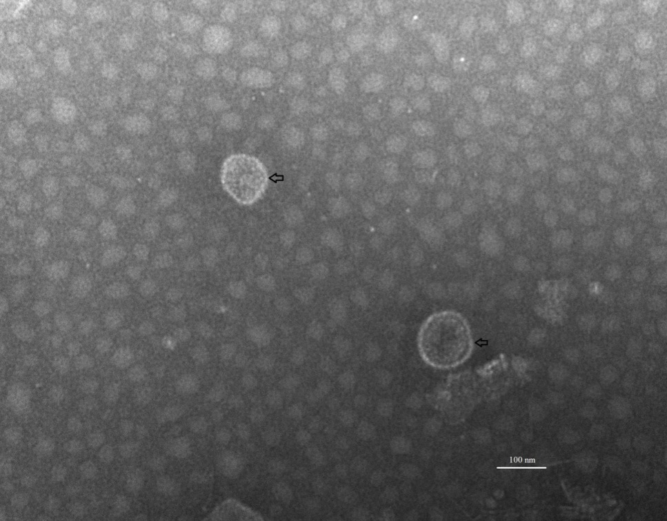
Morphological characterization of extracellular vesicles isolated from salivary glands of tick, Haemaphysalis longicornis. Image was taken by using AMT XR51 CCD cameras of Transmission Electron Microscope.
with nuclear regulation, protein synthesis machinery, enzymes and cytoskeletal proteins. Among the identified proteins, 28% belonged to nuclear regulation, 24% to protein synthesis machinery, 16% to enzymes and 12% to cytoskeletal proteins. This is followed by lowly abundant protein classes that account for structural proteins (8%) and translation regulation (6%) (Fig. 2). In previous studies, these protein groups were also identified within vesicles derived from excretory-secretory products of Fasciola hepatica, Schistosoma mansoni, Leishmania, Echinostoma caproni and Heligmosomoides polygyrus . In addition to this, these protein do exist in vesicles isolated from urine, serum, plasma and milk of other organisms ; ; ; . Keeping this in mind, a comparative analysis was generated between proteins identified from vesicles of tick and other organisms. It was observed that majority of proteins identified in salivary glands secreted vesicles (actin, histones, ribosomal proteins, elongation factor) were also identified in vesicles isolated from other systems ; ; ; ; ; ; (Table I).
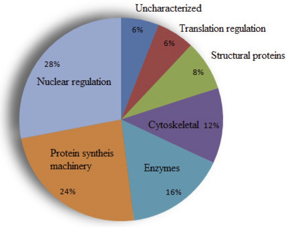
Distribution patterns of proteins identified in tick-derived EVs according to their functions. The EVs were isolated from tick salivary glands using ultracentrifugation technique while proteomics of vesicles was carried out using Liquid chromatography mass spectrometry (LC-MS/MS).
Table I. Common protein among tick-derived vesicles and other parasites.
|
Proteins |
Schistosoma mansoni |
Leishmania (L. donovani and major) |
Echinococcus granulosus |
Aedes aegypti (BBMV) |
Drosophila (Kc167 and S2 MVs) |
|
Actin |
✔ |
✔ |
✔ |
✔ |
✔ |
|
Elongation factor 1-Alpha |
✔ |
✔ |
✔ |
🗷 |
✔ |
|
Histone H2B |
✔ |
✔ |
✔ |
🗷 |
✔ |
|
H3 |
✔ |
🗷 |
✔ |
🗷 |
✔ |
|
Alpha tubulin |
✔ |
✔ |
✔ |
✔ |
|
|
Ubiquitin/polyubiquitin |
✔ |
🗷 |
✔ |
🗷 |
✔ |
|
Acetyl-CoA synthetase |
🗷 |
✔ |
🗷 |
🗷 |
🗷 |
|
Vacuolar ATP synthase subunit e |
🗷 |
🗷 |
🗷 |
✔ |
🗷 |
|
ATP synthase alpha subunit |
🗷 |
🗷 |
🗷 |
✔ |
🗷 |
|
chaperonin-60kD |
🗷 |
🗷 |
🗷 |
✔ |
🗷 |
|
40S ribosomal protein S27a |
🗷 |
🗷 |
🗷 |
🗷 |
✔ |
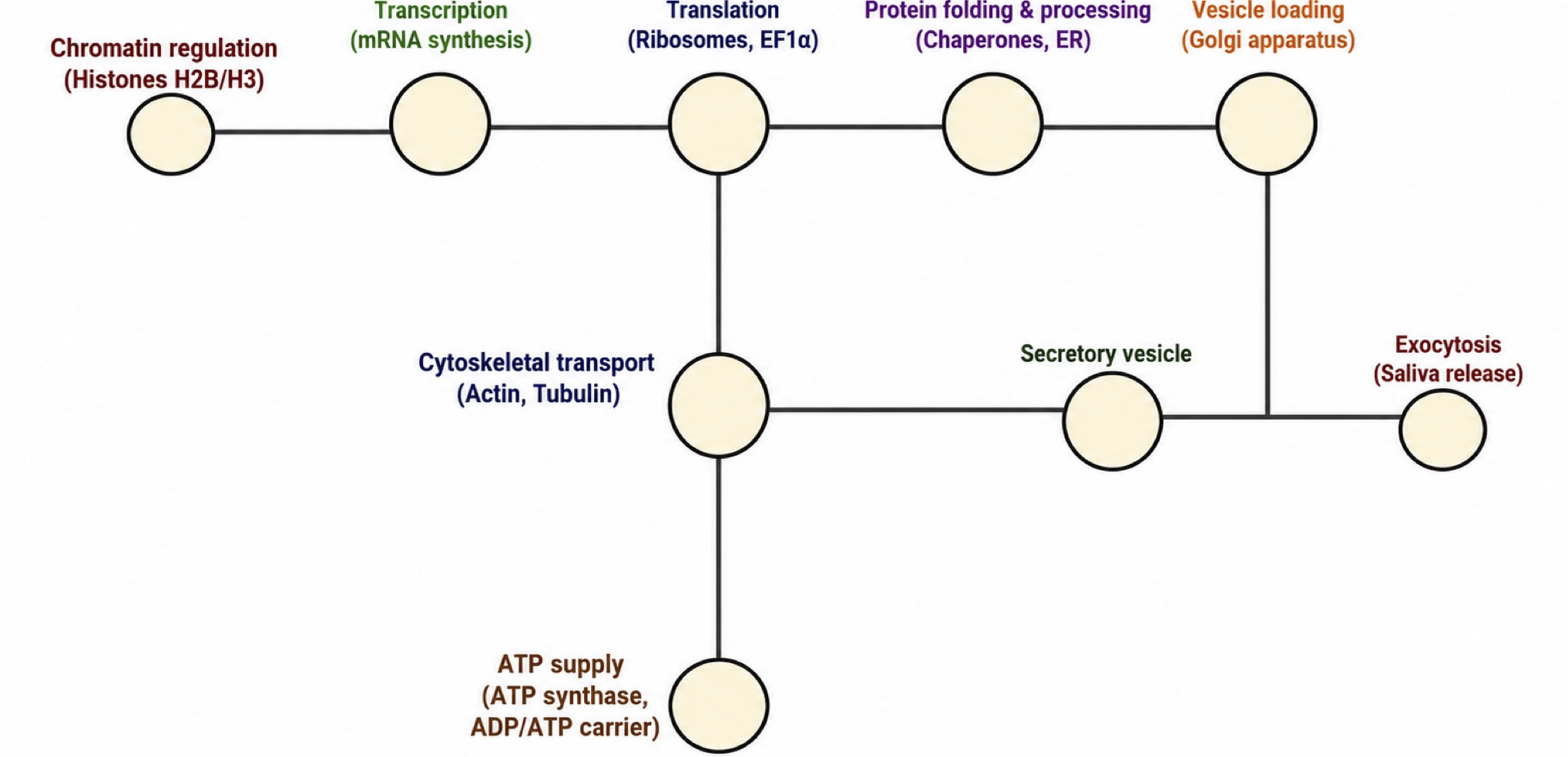
Mechanistic pathway of protein secretion of tick salivary glands proteome.
Mechanistic pathway diagram
The mechanistic schematic illustrates the stepwise biological flow of protein synthesis and secretion in tick salivary glands as shown in Figure 3. It begins with chromatin regulation by histones (H2B, H3), enabling transcriptional activation of salivary protein genes. The synthesized mRNAs are translated by ribosomes and elongation factor-1α, followed by protein folding and processing in the endoplasmic reticulum (ER) with the aid of chaperones. Processed proteins are then packaged into vesicles through the Golgi apparatus, which are transported along the actin–tubulin cytoskeleton towards the salivary ducts. The process is energetically supported by mitochondrial ATP synthase and ADP/ATP translocases, providing the ATP required for active transport and exocytosis. Finally, secretory vesicles fuse with the plasma membrane, releasing functional saliva components that modulate host immune and inflammatory responses.
Functional cluster diagram
The cluster-based view groups identified proteins into six major functional modules: Chromatin regulation (nuclear histones), translation machinery (ribosomal proteins, elongation factors), cytoskeletal dynamics (actin, tubulin), mitochondrial energy metabolism (ATP synthase, translocase), protein processing (chaperonins, acyl-CoA synthetase), and the secretory system (vesicle and lipocalin proteins) as shown in Figure 4. This organization highlights the coordinated roles of nuclear, cytoplasmic, and secretory compartments in maintaining an efficient secretion machinery critical for tick feeding and host manipulation.
Protein–protein interaction (PPI) network
The inferred PPI network integrates the proteomic components into an interaction map, emphasizing central hub proteins such as actin, EF1-α, and ATP synthase, which bridge transcriptional, translational, and secretory functions. The network topology suggests strong functional connectivity among cytoskeletal, mitochondrial, and translational proteins, ensuring synchronized control over energy generation, vesicle trafficking, and secretion as shown in Figure 5. These interactions collectively represent the molecular backbone of tick saliva synthesis and vesicle-mediated exocytosis.
Discussion
The present study provides direct evidence that the salivary glands of H. longicornis are capable of secreting extracellular vesicles (EVs) into the surrounding environment. Transmission electron microscopy confirmed the release of vesicle-like structures with an average size of approximately 100 nm, which falls within the typical range of exosomes ; ; . This finding demonstrates that EV secretion is an active process within tick salivary glands rather than a phenomenon restricted to saliva.
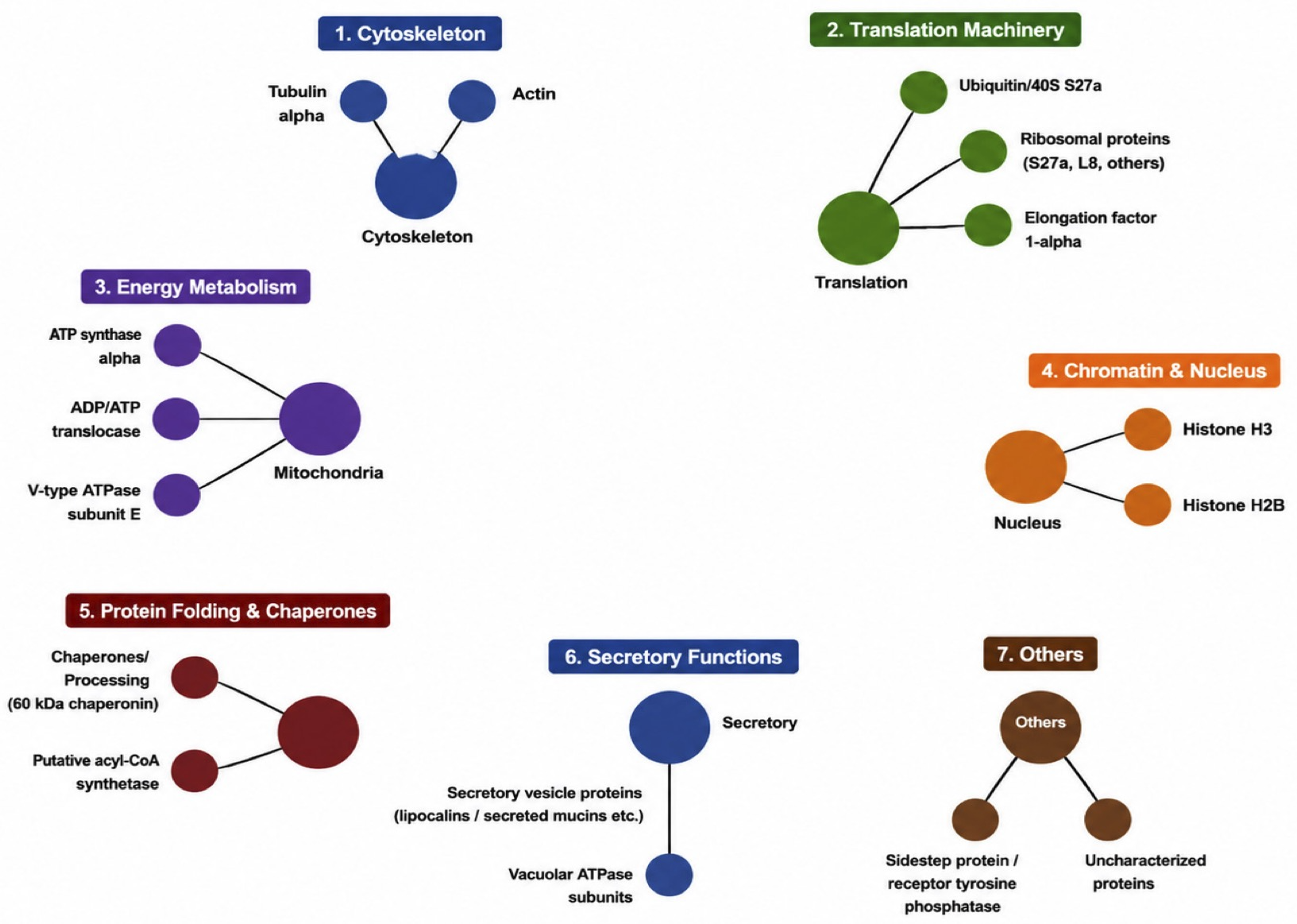
Functional protein clusters identified from salivary glands of tick, Haemaphysalis longicornis.
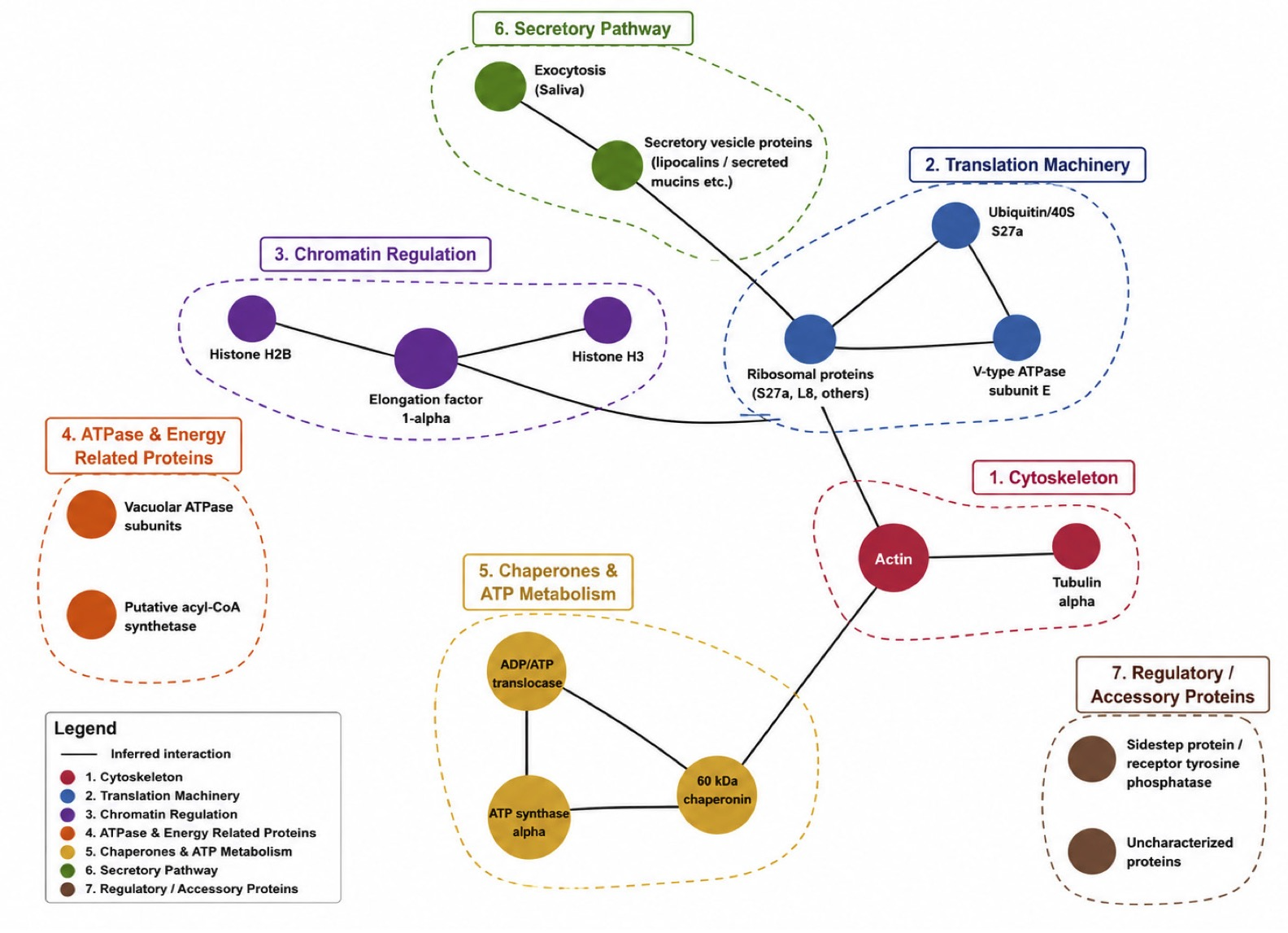
Inferred protein–protein interaction (PPI) network of salivary gland proteins from Haemaphysalis longicornis.
Proteomic analysis of the salivary gland derived EVs revealed a total of 287 proteins belonging to major functional categories, including nuclear regulation, protein synthesis machinery, enzymes, cytoskeletal and structural proteins. The predominance of nuclear and protein synthesis related components suggests active intracellular transport and regulatory processes in the salivary glands. The enrichment of cytoskeletal proteins such as actin and tubulin indicates their likely role in vesicle formation, trafficking, and release.
Among the identified proteins, actin constituted about 10% of the total vesicular proteins. Actin is known to play a key role in maintaining cell structure and enabling vesicle budding ; , implying that similar mechanisms may contribute to EV secretion in ticks. The high representation of nuclear regulatory proteins (28%) reflects a potentially strong link between vesicle formation and gene expression activity in the salivary glands. Furthermore, detection of elongation factor 1 alpha (EF-1α) and ubiquitin points to possible roles of these molecules in protein synthesis and turnover within the tick EV system.
A comparative analysis with previously identified saliva-derived vesicles from the same tick species revealed notable differences in protein composition . Only 9.6% of proteins were shared between the two EV sources, indicating that the vesicles released from the salivary glands and those found in saliva are compositionally distinct. Salivary gland EVs were enriched in nuclear regulation, enzymatic, and cytoskeletal proteins, whereas saliva-derived vesicles were dominated by structural and metabolic proteins, including vitellogenin, alpha-2-macroglobulin, and various antioxidant enzymes . These differences suggest that EVs produced within salivary glands may represent early or immature vesicles, while those secreted into saliva could undergo further modification or enrichment before delivery to the host during feeding.
The presence of SNARE-related proteins in our dataset supports the hypothesis that vesicle secretion from salivary glands is a regulated process mediated by conserved vesicular transport pathways. Such molecular machinery likely facilitates the targeted release of EVs into the extracellular milieu, contributing to tick feeding and possibly to pathogen transmission.
Overall, the data generated in this study extend the current understanding of tick biology by demonstrating that H. longicornis salivary glands actively secrete extracellular vesicles containing a diverse range of functional proteins. These vesicles likely play a role in glandular activity and may participate in modulating host–parasite interactions during blood feeding. Future studies should investigate the functional roles of these gland-derived vesicles, including their potential involvement in immune modulation and pathogen delivery to the host.
Declarations
Acknowledgement
Authors would like to express their sincere gratitude to the members of Animal House, Shanghai Veterinary Research Institute, GSCAAS, China for providing the rabbits.
Funding
This work was supported by a grant (2018YFC0840403) from The National Key Research and Development Programme of China. The funders had no role in study design, data collection and analysis, decision to publish, or preparation of the manuscript.
Ethics approval
The experiments to be performed were approved by the Institutional Animal Care and Use Committee of Shanghai Veterinary Research Institute (IACUC No: SHVRI-SOP-1104- 003). Experimental animals (rabbits) were kept in cages in compliance with the guidelines on the Humane Treatment of Laboratory Animals (Ministry of Science and Technology of the People’s Republic of China).
Data availability
The mass spectrometry proteomics data have been deposited to the ProteomeXchange Consortium (http://proteomecentral.proteomexchange.org) via the iProX partner repository ; with the dataset identifier PXD039607.
Supplementary material
There is supplementary material (Supplementary Table I) associated with this article. Access the material online at: https://dx.doi.org/10.17582/journal.pjz/20200207160258
Generative AI and AI assisted technology statement
The authors declare that no generative AI and AI assisted technology was used in the creation of this manuscript.
Statement of conflict of interest
The authors have declared no conflict of interest.
References
- Akers, J.C., Gonda, D., Kim, R., Carter, B.S. and Chen, C.C., 2013. Biogenesis of extracellular vesicles (EV): Exosomes, microvesicles, retrovirus-like vesicles, and apoptotic bodies. J. Neuro-Oncol., 113: 1-11. https://doi.org/10.1007/s11060-013-1084-8 [CrossRef]
- Al-Kaabi, A., Traupe, T., Stutz, M., Buchs, N. and Heller, M., 2012. Cause or effect of arteriogenesis: Compositional alterations of microparticles from CAD patients undergoing external counterpulsation therapy. https://doi.org/10.1371/journal.pone.0046822 [CrossRef]
- Blans, K., Hansen, M.S., Sørensen, L.V., Hvam, M.L., Howard, K.A., Möller, A., Wiking, L., Larsen, L.B. and Rasmussen, J.T., 2017. Pellet-free isolation of human and bovine milk extracellular vesicles by size-exclusion chromatography. J. Extracell. Vesicles, 6: 1294340. https://doi.org/10.1080/20013078.2017.1294340 [CrossRef]
- Chen, T., Ma, J., Liu, Y., Chen, Z., Xiao, N., Lu, Y., Fu, Y., Yang, C., Li, M. and Wu, S., 2022. iProX in 2021: Connecting proteomics data sharing with big data. Nucl. Acids Res., 50: D1522-D1527. https://doi.org/10.1093/nar/gkab1081 [CrossRef]
- Cox, J. and Mann, M., 2008. MaxQuant enables high peptide identification rates, individualized ppb-range mass accuracies and proteome-wide protein quantification. Nat. Biotechnol., 26: 1367-1372. https://doi.org/10.1038/nbt.1511 [CrossRef]
- Cwiklinski, K., 2015. The extracellular vesicles of the helminth pathogen, Fasciola hepatica: Biogenesis pathways and cargo molecules involved in parasite pathogenesis (eds. E. la Torre-Escudero de, M. Trelis, D. Bernal, P.J. Dufresne, G.P. Brennan, et al.). Mol. Cell Proteom., 14: 3258-3273. https://doi.org/10.1074/mcp.M115.053934 [CrossRef]
- Farooq, R., Hafeez, M.A., Oneeb, M., Rafique, A., Ashraf, K., Aslam, F., Rauf, N., Khalid, K., Bilal, F. and Mahmood, S., 2020. Molecular characterization and phylogenetic analysis of Babesia species isolated from domestic cattle. Pak. Vet. J., 40: 224-228. https://doi.org/10.29261/pakvetj/2019.003 [CrossRef]
- Fu, J., Momčilović, I. and Prasad, P.V., 2012. Roles of protein synthesis elongation factor EF-Tu in heat tolerance in plants. J. Bot., 2012: 835836. https://doi.org/10.1155/2012/835836 [CrossRef]
- Gonzalez-Begne, M., Lu, B., Han, X., Hagen, F.K., Hand, A.R., Melvin, J.E. and Yates III, J.R., 2009. Proteomic analysis of human parotid gland exosomes by multidimensional protein identification technology (MudPIT). J. Proteome Res., 8: 1304-1314. https://doi.org/10.1021/pr800658c [CrossRef]
- Grant, R., Ansa-Addo, E., Stratton, D., Antwi-Baffour, S., Jorfi, S., Kholia, S., Krige, L., Lange, S. and Inal, J., 2011. A filtration-based protocol to isolate human plasma membrane-derived vesicles and exosomes from blood plasma. J. Immunol. Methods, 371: 143-151. https://doi.org/10.1016/j.jim.2011.06.024 [CrossRef]
- Gupta, C.M. and Thiyagarajan, S., 2025. Structure and functions of actin and actin-binding proteins in leishmania. Pathogens, 14: 948. https://doi.org/10.3390/pathogens14090948 [CrossRef]
- Hussain, S., Saqib, M. and Ashfaq, K., 2021. First molecular evidence of Coxiella burnetii in ticks collected from dromedary camels in Punjab, Pakistan. Pak. Vet. J., http://dx.doi.org/10.29261/pakvetj/2021 [CrossRef]
- Jahn, R. and Südhof, T.C., 1999. Membrane fusion and exocytosis. Annu. Rev. Biochem., 68: 863-911. https://doi.org/10.1146/annurev.biochem.68.1.863 [CrossRef]
- Karim, S., Essenberg, R.C., Dillwith, J.W., Tucker, J., Bowman, A.S. and Sauer, J., 2002. Identification of SNARE and cell trafficking regulatory proteins in the salivary glands of the lone star tick, Amblyomma americanum (L.). Insect Biochem. Mol. Biol., 32: 1711-1721. https://doi.org/10.1016/S0965-1748(02)00111-X [CrossRef]
- Kornilov, R., Puhka, M., Mannerström, B., Hiidenmaa, H., Peltoniemi, H., Siljander, P., Seppänen-Kaijansinkko, R. and Kaur, S., 2018. Efficient ultrafiltration-based protocol to deplete extracellular vesicles from fetal bovine serum. J. Extracell. Vesicles, 7: 1422674. https://doi.org/10.1080/20013078.2017.1422674 [CrossRef]
- Kowal, J., Arras, G., Colombo, M., Jouve, M., Morath, J.P., Primdal-Bengtson, B., Dingli, F., Loew, D., Tkach, M. and Théry, C., 2016. Proteomic comparison defines novel markers to characterize heterogeneous populations of extracellular vesicle subtypes. Proc. natl. Acad. Sci., 113: E968-E977. https://doi.org/10.1073/pnas.1521230113 [CrossRef]
- Lejal, E., Moutailler, S., Šimo, L., Vayssier-Taussat, M. and Pollet, T., 2019. Tick-borne pathogen detection in midgut and salivary glands of adult Ixodes ricinus. Parasit. Vectors, 12: 1-8. https://doi.org/10.1186/s13071-019-3418-7 [CrossRef]
- Li, Y., Liu, Y., Xiu, F., Wang, J., Cong, H., He, S., Shi, Y., Wang, X., Li, X. and Zhou, H., 2018. Characterization of exosomes derived from Toxoplasma gondii and their functions in modulating immune responses. Int. J. Nanomed., 13: 467. https://doi.org/10.2147/IJN.S151110 [CrossRef]
- Liu, Z., Cauvi, D.M., Bernardino, E.M., Lara, B., Lizardo, R.E., Hawisher, D., Bickler, S. and De Maio, A., 2018. Isolation and characterization of human urine extracellular vesicles. Cell Stress Chaperones, 23: 943-953. https://doi.org/10.1007/s12192-018-0902-5 [CrossRef]
- Ma, J., Chen, T., Wu, S., Yang, C., Bai, M., Shu, K., Li, K., Zhang, G., Jin, Z. and He, F., 2019. iProX: An integrated proteome resource. Nucl. Acids Res., 47: D1211-D1217. https://doi.org/10.1093/nar/gky869 [CrossRef]
- Margiotta, A., 2022. Membrane fusion and snares: Interaction with ras proteins. Int. J. mol. Sci., 23: 8067. https://doi.org/10.3390/ijms23158067 [CrossRef]
- Mathieu, M., Martin-Jaular, L., Lavieu, G. and Théry, C., 2019. Specificities of secretion and uptake of exosomes and other extracellular vesicles for cell-to-cell communication. Nat. Cell Biol., 21: 9-17. https://doi.org/10.1038/s41556-018-0250-9 [CrossRef]
- Nawaz, M., Malik, M.I., Hameed, M. and Zhou, J., 2019. Research progress on the composition and function of parasite-derived exosomes. Acta Trop., 196: 30-36. https://doi.org/10.1016/j.actatropica.2019.05.004 [CrossRef]
- Nawaz, M., Malik, M.I., Zhang, H., Hassan, I.A., Cao, J., Zhou, Y., Hameed, M., Hussain, K.Z. and Zhou, J., 2020a. Proteomic analysis of exosome-like vesicles isolated from saliva of the tick Haemaphysalis longicornis. Front. Cell. Infect. Microbiol., 10: 542319. https://doi.org/10.3389/fcimb.2020.542319 [CrossRef]
- Nawaz, M., Malik, M.I., Zhang, H., Hassan, I.A., Cao, J., Zhou, Y., Hameed, M., Kuthu, Z.H. and Zhou, J., 2020b. Proteomic analysis of exosome-like vesicles isolated from saliva of the tick Haemaphysalis longicornis. Front. Cell. Infect. Microbiol., 10. https://doi.org/10.3389/fcimb.2020.542319 [CrossRef]
- Oliva-Chávez, A.S., Wang, X., Marnin, L., Archer, N.K., Hammond, H.L., Carroll, E.E.M., Shaw, D.K., Tully, B.G., Buskirk, A.D., Ford, S.L., Butler, L.R., Shahi, P., Morozova, K., Clement, C.C., Lawres, L., Neal, A.J.O., Mamoun, C.B., Mason, K.L., Hobbs, B.E., Scoles, G.A., Barry, E.M., Sonenshine, D.E., Pal, U., Valenzuela, J.G., Sztein, M.B., Pasetti, M.F., Levin, M.L., Kotsyfakis, M., Jay, S.M., Huntley, J.F., Miller, L.S., Santambrogio, L. and Pedra, J.H.F., 2021. Tick extracellular vesicles enable arthropod feeding and promote distinct outcomes of bacterial infection. Nat. Commun., 12: 3696. https://doi.org/10.1038/s41467-021-23900-8 [CrossRef]
- Olsen, J.V., de Godoy, L.M., Li, G., Macek, B., Mortensen, P., Pesch, R., Makarov, A., Lange, O., Horning, S. and Mann, M., 2005. Parts per million mass accuracy on an Orbitrap mass spectrometer via lock mass injection into a C-trap. Mol. Cell. Proteom., 4: 2010-2021. https://doi.org/10.1074/mcp.T500030-MCP200 [CrossRef]
- Olsen, J.V., Ong, S.-E. and Mann, M., 2004. Trypsin cleaves exclusively C-terminal to arginine and lysine residues. Mol. Cell. Proteom., 3: 608-614. https://doi.org/10.1074/mcp.T400003-MCP200 [CrossRef]
- Perkins, D.N., Pappin, D.J., Creasy, D.M. and Cottrell, J.S., 1999. Probability-based protein identification by searching sequence databases using mass spectrometry data. Electrophoresis: Int. J., 20: 3551-3567. https://doi.org/10.1002/(SICI)1522-2683(19991201)20:18<3551::AID-ELPS3551>3.0.CO;2-2 [CrossRef]
- Raimondo, F., Morosi, L., Corbetta, S., Chinello, C., Brambilla, P., Della Mina, P., Villa, A., Albo, G., Battaglia, C. and Bosari, S., 2013. Differential protein profiling of renal cell carcinoma urinary exosomes. Mol. BioSyst., 9: 1220-1233. https://doi.org/10.1039/C3MB25582D [CrossRef]
- Ryu, K.J., Lee, J.Y., Park, C., Cho, D. and Kim, S.J., 2020. Isolation of small extracellular vesicles from human serum using a combination of ultracentrifugation with polymer-based precipitation. Annls Lab. Med., 40: 253-258. https://doi.org/10.3343/alm.2020.40.3.253 [CrossRef]
- Samoil, V., Dagenais, M., Ganapathy, V., Aldridge, J., Glebov, A., Jardim, A. and Ribeiro, P., 2018. Vesicle-based secretion in schistosomes: Analysis of protein and microRNA (miRNA) content of exosome-like vesicles derived from Schistosoma mansoni. Sci. Rep., 8: 1-16. https://doi.org/10.1038/s41598-018-21587-4 [CrossRef]
- Samuel, M., Chisanga, D., Liem, M., Keerthikumar, S., Anand, S., Ang, C.-S., Adda, C.G., Versteegen, E., Jois, M. and Mathivanan, S., 2017. Bovine milk-derived exosomes from colostrum are enriched with proteins implicated in immune response and growth. Sci. Rep., 7: 1-10. https://doi.org/10.1038/s41598-017-06288-8 [CrossRef]
- Silverman, J.M., Clos, J., de’Oliveira, C.C., Shirvani, O., Fang, Y., Wang, C., Foster, L.J. and Reiner, N.E., 2010. An exosome-based secretion pathway is responsible for protein export from Leishmania and communication with macrophages. J. Cell Sci., 123: 842-852. https://doi.org/10.1242/jcs.056465 [CrossRef]
- Šimo, L., Kazimirova, M., Richardson, J. and Bonnet, S.I., 2017. The essential role of tick salivary glands and saliva in tick feeding and pathogen transmission. Front. Cell. Infect. Microbiol., 7: 281. https://doi.org/10.3389/fcimb.2017.00281 [CrossRef]
- Simons, M. and Raposo, G., 2009. Exosomes–vesicular carriers for intercellular communication. Curr. Opin. Cell Biol., 21: 575-581. https://doi.org/10.1016/j.ceb.2009.03.007 [CrossRef]
- Sotillo, J., Valero, L., Sanchez Del Pino, M., Fried, B., Esteban, J., Marcilla, A. and Toledo, R., 2008. Identification of antigenic proteins from Echinostoma caproni (Trematoda) recognized by mouse immunoglobulins M, A and G using an immunoproteomic approach. Parasit. Immunol., 30: 271-279. https://doi.org/10.1111/j.1365-3024.2007.01019.x [CrossRef]
- Sotillo, J., Valero, M.L., Del Pino, M.M.S., Fried, B., Esteban, J.G., Marcilla, A. and Toledo, R., 2010. Excretory/secretory proteome of the adult stage of Echinostoma caproni. Parasitol. Res., 107: 691-697. https://doi.org/10.1007/s00436-010-1923-x [CrossRef]
- Surpin, M., Zheng, H., Morita, M.T., Saito, C., Avila, E., Blakeslee, J.J., Bandyopadhyay, A., Kovaleva, V., Carter, D. and Murphy, A., 2003. The VTI family of SNARE proteins is necessary for plant viability and mediates different protein transport pathways. Plant Cell, 15: 2885-2899. https://doi.org/10.1105/tpc.016121 [CrossRef]
- Tancini, B., Buratta, S., Sagini, K., Costanzi, E., Delo, F., Urbanelli, L. and Emiliani, C., 2019. Insight into the role of extracellular vesicles in lysosomal storage disorders. Genes, 10: 510. https://doi.org/10.3390/genes10070510 [CrossRef]
- Théry, C., Amigorena, S., Raposo, G. and Clayton, A., 2006. Isolation and characterization of exosomes from cell culture supernatants and biological fluids. Curr. Protoc. Cell Biol., 30: 3.22. 21-23.22. 29. https://doi.org/10.1002/0471143030.cb0322s30 [CrossRef]
- Théry, C., Zitvogel, L. and Amigorena, S., 2002. Exosomes: Composition, biogenesis and function. Nat. Rev. Immunol., 2: 569-579. https://doi.org/10.1038/nri855 [CrossRef]
- Urbanelli, L., Buratta, S., Sagini, K., Ferrara, G., Lanni, M. and Emiliani, C., 2015. Exosome-based strategies for diagnosis and therapy. Recent Patents CNS Drug Discovery (Discontinued), 10: 10-27. https://doi.org/10.2174/1574889810666150702124059 [CrossRef]
- Urbanelli, L., Magini, A., Buratta, S., Brozzi, A., Sagini, K., Polchi, A., Tancini, B. and Emiliani, C., 2013. Signaling pathways in exosomes biogenesis, secretion and fate. Genes, 4: 152-170. https://doi.org/10.3390/genes4020152 [CrossRef]
- Van Troys, M., Vandekerckhove, J. and Ampe, C., 1999. Structural modules in actin-binding proteins: towards a new classification. Biochim. biophys. Acta (BBA)-Mol. Cell Res., 1448: 323-348. https://doi.org/10.1016/S0167-4889(98)00152-9 [CrossRef]
- Villarreal, A.M., Adamson, S.W., Browning, R.E., Budachetri, K., Sajid, M.S. and Karim, S., 2013. Molecular characterization and functional significance of the Vti family of SNARE proteins in tick salivary glands. Insect Biochem. mol. Biol., 43: 483-493. https://doi.org/10.1016/j.ibmb.2013.03.003 [CrossRef]
- Wisniewski, J.R., Zougman, A. and Mann, M., 2009. Combination of FASP and StageTip-based fractionation allows in-depth analysis of the hippocampal membrane proteome. J. Proteome Res., 8: 5674-5678. https://doi.org/10.1021/pr900748n [CrossRef]
- Yáñez-Mó, M., Siljander, P.R.M., Andreu, Z., Bedina, Z.A., Borràs, F.E., Buzas, E.I., Buzas, K., Casal, E., Cappello, F. and Carvalho, J., 2015. Biological properties of extracellular vesicles and their physiological functions. J. Extracell. Vesicles, 4: 27066. https://doi.org/10.3402/jev.v4.27066 [CrossRef]
- Zaborowski, M.P., Balaj, L., Breakefield, X.O. and Lai, C.P., 2015. Extracellular vesicles: Composition, biological relevance, and methods of study. Bioscience, 65: 783-797. https://doi.org/10.1093/biosci/biv084 [CrossRef]
- Zamanian, M., Fraser, L.M., Agbedanu, P.N., Harischandra, H., Moorhead, A.R., Day, T.A., Bartholomay, L.C. and Kimber, M.J., 2015. Release of small RNA-containing exosome-like vesicles from the human filarial parasite Brugia malayi. PLoS Neglected Trop. Dis., 9: e0004069. https://doi.org/10.1371/journal.pntd.0004069 [CrossRef]
- Zhang, Y., Bi, J., Huang, J., Tang, Y., Du, S. and Li, P., 2020. Exosome: A review of its classification, isolation techniques, storage, diagnostic and targeted therapy applications. Int. J. Nanomed., pp. 6917-6934. https://doi.org/10.2147/IJN.S264498 [CrossRef]
- Zhu, L., Liu, J., Dao, J., Lu, K., Li, H. and Gu, H., 2016. Molecular characterization of S. japonicum exosome-like vesicles reveals their regulatory roles in parasite-host interactions. Sci. Rep., 6: 25885. https://doi.org/10.1038/srep25885 [CrossRef]
- Zubiri, I., Posada-Ayala, M., Sanz-Maroto, A., Calvo, E., Martin-Lorenzo, M., Gonzalez-Calero, L., de la Cuesta, F., Lopez, J.A., Fernandez-Fernandez, B. and Ortiz, A., 2014. Diabetic nephropathy induces changes in the proteome of human urinary exosomes as revealed by label-free comparative analysis. J. Proteom., 96: 92-102. https://doi.org/10.1016/j.jprot.2013.10.037 [CrossRef]

Services
Article usage statistics
Loading…
Related articles
- The Effects of Parity on Protective Protein Components and MMP Proteolytic Activity of Mammary Secretion during Involution in Tropical Dairy CowsAdvances in Animal and Veterinary Sciences · 2019
- Immunohistochemical Expression of Transforming Growth Factor-Alpha between Rat Submandibular and Sublingual Salivary Glands during Post-Natal DevelopmentPakistan Journal of Zoology · 2019
- Regulation of Transforming Growth Factor-Alpha Expression by Thyroid Hormone during the Post-Natal Development of Rat Submandibular and Sublingual Salivary GlandsPakistan Journal of Zoology · 2019
- Proteomic Analysis by Shotgun LC-MS/MS of Cyst Wall Protein Components From the Protozoan Parasite Cryptocaryon irritans, the Causative Agent of White Spot Disease in Marine FishPakistan Journal of Zoology · 2020
- Increased Gliricidia sepium in Ration Containing Rice Straw on Rumen Fermentation and Microbial Protein Synthesis of Indigenous Bali CattleAdvances in Animal and Veterinary Sciences · 2018
- Comparative Effects of β-Tricalcium Phosphate and Mesenchymal Stem Cell–Derived Extracellular Vesicles on Ilial Fracture Healing in a Mongrel Dog ModelAdvances in Animal and Veterinary Sciences · 2026