
Gastrointestinal parasites (GIP) are among the most common health constraints affecting cattle (Bos indicus) production. It can lead to reduced productivity and economic losses. This study was conducted to determine the prevalence and infection intensity of GIP in cattle from selected barangays of General Santos City, Philippines. Fecal samples were collected from 149 extensively grazed cattle (≥1 year old) with no reported deworming within the previous six months from nine agricultural barangays of General Santos City and examined using the McMaster egg counting technique. Out of the 149 fecal samples, 92 tested positive for GIP, resulting in an overall prevalence of 61.74%. Prevalence ranged from 50.00% to 83.33% among barangays; however, the differences were not statistically significant (χ² = 4.784, df = 8, p = 0.780). Four parasite taxa were identified, namely Eimeria spp., Trichostrongylidae, Fasciola spp., and Toxocara spp. Among these, Eimeria spp. was the most frequently detected parasite, followed by Trichostrongylidae, Fasciola spp., and Toxocara spp. Low-intensity infections predominated across parasite–barangay combinations and were observed in 27 out of 28 combinations. Moderate- and high-intensity infections were each observed in 4 out of 28 combinations. These findings indicate high occurrence of GIP infection among the sampled cattle and highlight the importance of regular parasite monitoring and strategic parasite control programs. The results provide baseline information that can assist cattle producers in improving herd health management and support veterinary services in developing evidence-based parasite surveillance and deworming strategies.
Research Article · Volume 15, Issue 1, Pages 39–45, 14 November 2026 · Open Access
Prevalence and Identification of Gastrointestinal Parasites in Cattle Fecal Samples in General Santos City, Philippines
, , , , , , ,
Affiliations & Notes ⌄
- 1Department of Animal Science, Mindanao State University - General Santos, 9500, Philippines
- 2Mindanao State University - General for Animal Research and Ethics in SOCCSKSARGEN (MSU-CARES), General Santos City, 9500, Philippines
- *Correspondence | Alexis N. Miole; Email: alexis.miole@msugensan.edu.ph
Article Info ⌄
- Journal:
- Journal of Animal Health and Production
- Submitted Date:
- 15 June 2026
- Accepted Date:
- 9 September 2026
- Published Date:
- 14 November 2026
Abstract
Introduction
Cattle (Bos indicus) production is an important component of the agricultural sector, contributing to food security, rural livelihoods, and household income. In the Philippines, the cattle population was estimated at 2.54 million heads as of September 2025, with approximately 86.3% raised in smallholder farms (PSA, 2025). As a result, cattle production continues to play a significant role in supporting the livelihood and economic stability of rural communities.
Despite its importance, cattle production is affected by several health problems, one of the most common being gastrointestinal parasites (GIP). These parasites can reduce animal performance by affecting feed utilization, growth, reproduction, and milk production. Infected cattle may also be more vulnerable to other diseases, resulting in economic losses for farmers . Although infected animals do not always show obvious signs of illness, gastrointestinal parasitism remains a major challenge to cattle production worldwide .
The occurrence of gastrointestinal parasites is influenced by several factors, including management practices, grazing systems, environmental conditions, and host-related factors . Numerous studies have reported the prevalence of gastrointestinal parasites in cattle from different regions; however, information regarding their occurrence in General Santos City remains limited despite the city’s importance in livestock production. The lack of local data may hinder the development of effective parasite monitoring and control programs.
Several studies conducted in the Philippines have reported gastrointestinal parasitism in cattle. In Bukidnon, Van Aken et al. (2000) documented strongyle infections among backyard cattle, while Balbin et al. (2025) reported widespread gastrointestinal parasitic infections in backyard cattle in Southern Isabela. Despite these findings, information on gastrointestinal parasites in cattle from General Santos City remains limited, highlighting the need for locally generated baseline data.
The need for local information is particularly relevant in the Soccsksargen Region, which had an estimated cattle population of 190,450 heads as of September 2025 (PSA, 2025). Understanding the occurrence of gastrointestinal parasites in cattle within the area can provide baseline information to support future parasite control strategies and livestock health programs.
In addition to determining prevalence, this study identifies the gastrointestinal parasite taxa present and evaluates infection intensity among cattle from selected barangays, thereby providing local epidemiological information that can help identify the extent and distribution of gastrointestinal parasitism and areas where parasite-control interventions may be prioritized. The findings may also support veterinary program planning and evidence-based allocation of livestock health resources at the city and regional levels, particularly in designing parasite surveillance and control strategies appropriate to local conditions. Therefore, this study was conducted to determine the prevalence and infection intensity of gastrointestinal parasites in cattle from selected barangays of General Santos City, Philippines.
Materials and Methods
Sampling design and sample collection
Fecal samples were collected from nine (9) agricultural barangays within General Santos City, namely: Conel, Katangawan, Ligaya, Mabuhay, Olympog, San Jose, Siguel, Sinawal, and Upper Labay (Figure 1). Samples were collected from cattle aging 1 year and above which were raised under extensive grazing systems. Samples were collected from the middle portion of freshly voided feces. No handling or restraint of animals was performed.
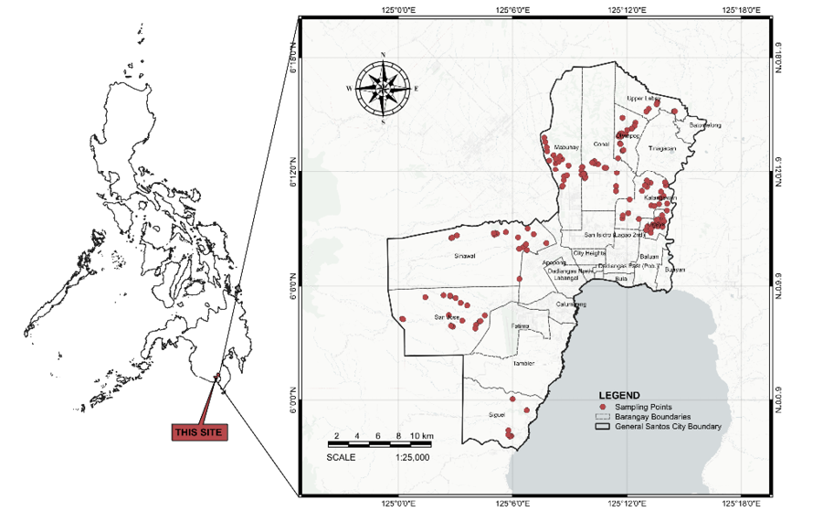
Location of the sampling sites in selected barangays of General Santos City, Philippines.
The distance between sampling locations was considered during sample collection. Where feasible, a minimum distance of approximately 100 meters between cattle owners or grazing locations was maintained. It was done to reduce the likelihood of repeatedly sampling animals exposed to highly similar environmental conditions. Distance was assessed using Google Maps during field sampling. Only cattle with no reported history of deworming within the previous six months, based on owner information, were included in the study. No formal sample size calculation was performed. Instead, the final sample size (n= 149) was determined by the number of cattle that met the inclusion criteria and whose owners consented to participate during the study period. Variation in sample size among barangays was primarily due to the application of the study’s inclusion criteria. Consequently, the number of samples collected from each barangay was not intended to proportionally represent the cattle population of that barangay. In some barangays, a limited number of cattle met the eligibility requirements for sampling. Some tethered cattle could not be included because their owners could not be located during field visits to obtain the necessary information and consent for sample collection. Cattle herd sizes among sampled owners varied and were categorized as follows: 1–5 heads (n= 110 owners), 6–10 heads (n= 20), 11–15 heads (n= 8), 16–20 heads (n= 5), and 21 heads and above (n= 6). One cattle was sampled per owner/herd; therefore, each sampled animal represented a separate owner/herd, and no multiple animals from the same owner or herd were included in the analysis.
Sample storage and preservation
Collected fecal samples were placed in sealed containers containing 10% formaldehyde solution and stored at 4–8 °C to preserve parasite eggs and oocysts . To minimize potential distortion of parasitic structures during preservation and storage, all samples were processed and examined within 32 hours after collection.
Flotation solution (Sheather’s Sugar) preparation
The flotation solution was prepared by dissolving 454 g of granulated sugar in 355 mL of water under low heat. After complete dissolution, the solution was allowed to cool before the addition of 6 mL of formaldehyde to inhibit microbial growth. The flotation solution was maintained at a specific gravity of 1.27, which was verified using a hydrometer . To ensure consistency throughout the study, the specific gravity of the flotation solution was checked prior to each sample processing session.
Fecal sample preparation
For each sample, 4 g of feces was mixed with 56 mL of Sheather’s flotation solution in a beaker. The mixture was then filtered through cheesecloth to remove large debris. The mixture was then transferred into a test tube where it was allowed to stand for at least 10 minutes to allow the parasite eggs and oocysts to float. Using a pipette, mixture from the top part was collected to fill the chambers of the McMaster slide . The loaded slide was allowed to stand for approximately 3–5 minutes prior to microscopic examination to permit parasite eggs and oocysts to rise into the chamber grid for examination .
Identification of parasites
Fecal samples were examined under a microscope using the McMaster technique . Parasite identification was based on the morphological characteristics of eggs and oocysts using published photo images by Zajac and Conboy (2012), Dewair and Bessat (2020), Loginova et al. (2024), and Haryadi et al. (2025). Multiple individuals performed the identification by cross-referencing from the published sources.
EPG and OPG counting and infection intensity
Eggs per gram (EPG) and oocysts per gram (OPG) were determined by counting all eggs and oocysts within the grid areas of both McMaster chambers, excluding those located outside the counting grids. The combined count from the two chambers was then multiplied by 50 to obtain the final EPG or OPG value .
Infection intensity was categorized as low, moderate, or high based on established EPG/OPG threshold values. Infections were considered low at 50–799 EPG/OPG, moderate at 800–1,200 EPG/OPG, and high at >1,200 EPG/OPG . In the absence of locally established threshold values, these published criteria were used as reference guides for interpreting parasite burden. The classifications were intended for comparative assessment of egg and oocyst counts and do not necessarily indicate equivalent pathogenic effects among different parasite taxa. Infection intensity was evaluated separately for each parasite taxon and the potential cumulative effects of co-infections were not assessed.
Statistical analysis
Prevalence was expressed as percentages with corresponding 95% confidence intervals (Wilson score method). Differences in parasite prevalence among barangays were evaluated using a chi-square test based on the observed frequencies of positive and negative samples. Infection intensity was classified as low, moderate, or high to indicate the level of parasite burden. Since sample sizes varied among barangays and purposive sampling was employed, the results should be interpreted with caution and are representative only of the sampled cattle rather than the entire cattle population of each barangay or General Santos City.
Results and Discussion
Prevalence of gastrointestinal parasites
The prevalence of GIP differed across the 9 barangays in General Santos City. Siguel exhibited the highest prevalence at 83.33%, followed by Mabuhay (75%), Sinawal (72.22%), Conel (70%), Ligaya (65%), and San Jose (63.16%). Lower prevalence rates were observed in Katangawan and Olympog, both at 55%, while Upper Labay recorded the lowest prevalence at 50%. Overall, 92 out of the 149 fecal samples examined were positive for GIP. The overall prevalence of 61.74% was recorded. Even with the observed variation in prevalence among barangays, no significant difference was detected between barangays (χ²= 4.784, df= 8, p= 0.780). The relatively wide confidence intervals observed in some barangays indicate limited precision in the estimated prevalence values and should therefore be interpreted cautiously. Table 1 shows these results.
Table 1: Prevalence of gastrointestinal parasites in cattle across selected barangays in General Santos City, Philippines.
| Barangay | Number of samples | Positive samples | Prevalence (%) | Confidence interval (95%) |
| Conel | 20 | 14 | 70 | 48.10–85.45 |
| Katangawan | 20 | 11 | 55 | 34.21–74.18 |
| Ligaya | 20 | 13 | 65 | 43.29–81.88 |
| Mabuhay | 20 | 15 | 75 | 53.13–88.81 |
| Olympog | 20 | 11 | 55 | 34.21–74.18 |
| San Jose | 19 | 12 | 63.16 | 41.04–80.85 |
| Siguel | 6 | 5 | 83.33 | 43.65–96.99 |
| Sinawal | 18 | 13 | 72.22 | 49.13–87.50 |
| Upper Labay | 6 | 3 | 50 | 18.76–81.24 |
| Total | 149 | 92 | 61.74 | 53.74–69.16 |
Confidence intervals were calculated using the Wilson score method. No significant difference was observed among barangays (χ²= 4.784, df = 8, p= 0.780). Total prevalence was calculated using the overall number of positive samples (92/149 × 100) rather than the average of barangay prevalence values. Prevalence estimates for barangays with small sample sizes (e.g., n = 6) should be interpreted with caution, as sample numbers were determined by the study inclusion criteria and owner participation during the sampling period.
Out of the 149 fecal samples examined, 92 were positive for GIP. Four (4) taxa were identified. Among the identified, Eimeria spp. exhibited the highest prevalence (36.91%), followed by Trichostrongylidae (30.20%), Fasciola spp. (8.72%), and Toxocara spp. (7.38%). The relatively low occurrence of Fasciola spp. and Toxocara spp. should be interpreted with caution because of the small number of positive samples that may affect precision. A summary of the prevalence of the detected GIP is presented in Table 2.
Table 2: Prevalence of Identified Gastrointestinal Parasites in Cattle from General Santos City, Philippines (n = 149).
| Parasite | Number of positive samples | Prevalence (%) | Confidence interval (95%) |
| Eimeria spp. | 55 | 36.91 | 29.58–44.90 |
| Trichostrongylidae | 45 | 30.20 | 23.40–37.99 |
| Fasciola spp. | 13 | 8.72 | 5.17–14.35 |
| Toxocara spp. | 11 | 7.38 | 4.17–12.74 |
Confidence intervals were calculated using the Wilson score method.
The predominance of Eimeria spp. observed in the present study is consistent with previous reports identifying coccidia among the most common gastrointestinal parasites of cattle. According to Ninditya et al. (2024), Eimeria spp. infections are often associated with environmental contamination, particularly under warm and humid conditions that favor oocyst survival and transmission.
The occurrence of Trichostrongylidae is consistent with the study of Dereje et al. (2023) where strongyle-type nematodes are among the predominant gastrointestinal parasites of cattle. They emphasized that the widespread distribution may be attributed to the direct life cycle of the parasite and the continuous exposure of grazing animals to infective larvae present in contaminated pastures.
Fasciola spp. showed lower prevalence compared to Eimeria spp. and Trichostrongylidae. It may be due to the environmental conditions that can affect parasite transmission . According to Ogwal et al. (2025), low prevalence can be attributed to the complex life cycle of Fasciola spp. that requires Lymnaeid snail to complete its life cycle.
In this study, Toxocara spp. showed the lowest prevalence. Tiele et al. (2023) reported a similar finding where Toxocara spp. had a lower prevalence than Trichostrongylidae. The low prevalence of Toxocara spp. may be related to the age of the sampled cattle. Only cattle aged one year and above were included. According to Suwito et al. (2025), Toxocara spp. infections are more commonly observed in younger calves. As animals grow older, they become less susceptible to infection due to the development of acquired immunity.
Infection intensity
Infection intensity varied among parasite–barangay combinations (Figure 2). The distribution of infection intensity is presented using a color-coded scheme. The color green, yellow, and red indicate low, moderate, and high infection levels, respectively. The EPG/OPG values may vary within each intensity category. Therefore, the categories shown in Figure 2 represent general levels of infection rather than the exact EPG/OPG values recorded for each parasite–barangay combination.
Low-intensity infections predominated across the parasite–barangay combinations and were observed in 27 out of 28 combinations. Moderate- and high-intensity infections were less frequent, each occurring in 4 out of 28 combinations. Several combinations exhibited a mixture of intensity categories; which means that the frequencies of low, moderate, and high infections are not mutually exclusive.
Low-intensity infections predominated in Eimeria spp. infections across all positive barangays. Conel, Katangawan, Ligaya, Mabuhay, Olympog, Upper Labay, and Siguel exhibited exclusively low-intensity infections. In contrast, San Jose showed a combination of low- and moderate-intensity infections, while Sinawal exhibited the widest range of infection intensities, with low-, moderate-, and high-intensity infections occurring simultaneously.
For Trichostrongylidae, low-intensity infections predominated across all positive barangays. Conel, Katangawan, Ligaya, Mabuhay, Olympog, Siguel and Upper Labay exhibited exclusively low-intensity infections. In contrast, San Jose displayed a mixture of low-, moderate-, and high-intensity infections, while Sinawal showed both low- and high-intensity infections.
Fasciola spp., infection exhibited exclusively low-intensity infections in Katangawan, Ligaya, and Mabuhay. For Toxocara spp., low-intensity infections were observed in Conel, Katangawan, Ligaya, Mabuhay, and San Jose. Olympog exhibited both low- and moderate-intensity infections, while Sinawal recorded high-intensity infections. Overall, low-intensity infections predominated among the positive barangays.
The predominance of low-intensity infections observed in the present study suggests that, although gastrointestinal parasites were widespread among the sampled cattle, most infected animals carried relatively low parasite burdens. Similar findings were reported by Thapa et al. (2022), who documented low infection intensities despite the occurrence of gastrointestinal parasites in cattle populations. As noted by Teixeira et al. (2021), low egg and oocyst counts may indicate limited parasite burdens and are generally associated with less severe clinical effects.
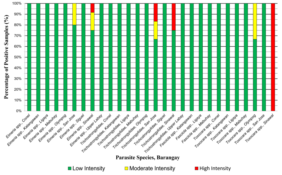
Distribution of infection intensity levels of gastrointestinal parasites in cattle across selected barangays in General Santos City, Philippines. Infection intensity was classified based on EPG/OPG values as low (50–799 EPG/OPG), moderate (800–1,200 EPG/OPG), and high (>1,200 EPG/OPG) according to Thapa et al. (2022).
Moderate- and high-intensity infections were observed in only a few parasite–barangay combinations, suggesting that heavier parasite burdens were relatively uncommon within the study area. As observed by Ninditya et al. (2024), higher infection intensities may occur under conditions that favor parasite survival and transmission, particularly in environments with greater levels of contamination.
The occurrence of Fasciola spp. in the present study was limited to barangays with irrigation systems used for rice farming. This observation may be associated with the presence of water-filled environments that facilitate parasite transmission. Shahzad et al. (2025) explained that irrigation and drainage canals can promote the spread of Lymnaeid snails, the intermediate hosts of Fasciola spp., thereby increasing the risk of Fascioliasis.
Representative photomicrographs of the gastrointestinal parasites identified in this study are presented in Figure 3, namely: (A) Eimeria spp.; (B) Trichostrongylidae; (C) Fasciola spp.; and (D) Toxocara spp. Due to the thickness of the McMaster slide chambers, parasite eggs and oocysts were primarily examined at a total magnification of 100×. Images were digitally enlarged to facilitate clearer visualization of the morphological characteristics used for parasite identification.
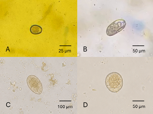
Representative photomicrographs of identified gastrointestinal parasite oocyst and eggs in cattle: (A) Eimeria spp.; (B) Trichostrongylidae; (C) Fasciola spp.; and (D)Toxocara spp. Images were obtained using light microscopy at 100× total magnification with a McMaster slide. Photomicrographs were digitally enlarged to facilitate clearer observation of morphological characteristics.
This study focused on parasite prevalence, identification, and infection intensity. Host- and management-related factors (e.g., age, sex, breed, body condition score, and herd management practices) were not evaluated and warrant investigation in future studies.
Conclusion and Recommendation
This study revealed an overall GIP prevalence of 61.74% among the cattle sampled from selected barangays of General Santos City. Four (4) parasite taxa were identified, namely: Eimeria spp., Trichostrongylidae, Fasciola spp., and Toxocara spp. The Infection intensity was predominantly low across most parasite–barangay combinations, indicating generally low levels of parasite burden within the sampled cattle population. Nevertheless, moderate- and high-intensity infections were observed in a limited number of parasite–barangay combinations, suggesting localized areas of increased parasite transmission.
Regular parasite monitoring and strategic deworming are recommended to reduce gastrointestinal parasite infections in cattle. Further studies on co-infections, seasonal variation, and parasite-associated risk factors are also recommended.
Acknowledgement
The authors gratefully acknowledge the General Santos City Veterinary Office and the local government units of the selected barangays for their cooperation and assistance during the conduct of this study. The authors also thank Vince Louies P. Macul and Jenifer P. Pepito for their valuable assistance, and Joren M. Sumapal for logistical support. Special thanks are extended to Dr. Jose Alexander Abella for his mentorship and instruction in veterinary parasitology, which contributed to the corresponding author’s foundational knowledge relevant to this study. The authors also acknowledge Engr. Harry Oliveros for preparing the map used in this study.
Novelty Statement
This study provides baseline information on the prevalence and infection intensity of gastrointestinal parasites in cattle from selected barangays of General Santos City, Philippines, using the McMaster egg counting technique. The findings contribute to the limited local data on cattle gastrointestinal parasites and may serve as a reference for future epidemiological studies and parasite control programs in the area.
Author’s Contribution
WFR, SGQL, DDFD: Contributed to laboratory works, analysis of the data, and manuscript writing. ARLA, MGMC, JERO, FAMS and ANM: Contributed to conceptualization, reviewed protocols, supervised the study, data analysis and revised the manuscript.
Ethical approval
Only freshly voided fecal samples were collected for this study. No cattle were handled, restrained, or subjected to invasive procedures during sample collection. The study complied with the Animal Welfare Act of the Philippines (Republic Act No. 8485, as amended by Republic Act No. 10631). Human ethics clearance was obtained from Mindanao State University - General Santos, and informed consent was secured from cattle owners before sample collection.
Generative AI and AI-assisted technology statement
The authors declare that the AI-assisted tool (ChatGPT) was used solely to improve English language quality and grammar.
Conflict of interest
The authors have declared no conflict of interest.
References
- Abdisa T (2018). Examination of feces. Int. J. Vet. Sci. Res., S1: 45-50. https://doi.org/10.17352/ijvsr.s1.106 [CrossRef]
- Balbin AJM, Nayga JN, Bacton J, Bautista J, Catalonia HMD, Dela Cruz K, Fajardo MAD, Ignacio AD, Quiming L, Sapaden J (2025). Gastrointestinal parasitic infections of ruminants in backyard farms of Southern Isabela, Philippines. BIO Web. Conf., 191: 00015. https://doi.org/10.1051/bioconf/202519100015 [CrossRef]
- Bricarello PA, Longo C, Da Rocha RA, Hötzel MJ (2023). Understanding animal-plant-parasite interactions to improve the management of gastrointestinal nematodes in grazing ruminants. Pathogens, 12(4): 531. https://doi.org/10.3390/pathogens12040531 [CrossRef]
- Dereje T, Bantayehu M, Beshir H, Zerihun A (2023). Prevalence of gastrointestinal parasites in cattle kept under pastoral management system in selected districts of Borana Zone, Ethiopia. Int. J. Livest. Prod., 14(2): 31-36. https://doi.org/10.5897/IJLP2022.0800 [CrossRef]
- Dewair A, Bessat M (2020). Molecular and microscopic detection of natural and experimental infections of Toxocara vitulorum in bovine milk. PLoS One, 15(5): e0233453. https://doi.org/10.1371/journal.pone.0233453 [CrossRef]
- Haryadi F, Nurcahyo R, Priyowidodo D, Indarjulianto S, Ekawasti F, Ninditya VI (2025). Determination of the morphology of Eimeria spp. in beef cattle in Bandung Regency, West Java Province, Indonesia. Open Vet. J., 15(1): 85. https://doi.org/10.5455/OVJ.2025.v15.i1.7 [CrossRef]
- Loginova O, Efeykin B, Krutikova A, Mizin I, Spiridonov S (2024). Fasciola hepatica: Updates on egg morphology, host range, and distribution. Food Waterborne Parasitol., 36: e00237. https://doi.org/10.1016/j.fawpar.2024.e00237 [CrossRef]
- Ninditya VI, Ekawasti F, Prastowo J, Widiyono I, Nurcahyo W (2024). Prevalence of gastrointestinal parasites in cattle in Indonesia: A meta-analysis and systematic review. Vet. World, 17: 2675-2687. https://doi.org/10.14202/vetworld.2024.2675-2687 [CrossRef]
- Ogwal A, Okello DM, Aliro T, Owiny DO, Ndyomugyenyi EK (2025). Prevalence of bovine fasciolosis, financial losses and risk factors associated with the disease in Lira and Gulu Districts, Northern Uganda. Vet. Med. Int., 2025: 7757654. https://doi.org/10.1155/vmi/7757654 [CrossRef]
- PSA (2025). Livestock and poultry quarterly bulletin. Philippine Statistics Authority, Philippines.
- Shahzad R, Rasheed J, Irum S (2025). Discuss the transmission dynamics of Fasciola hepatica, including the role of intermediate hosts, water contamination, and human behavior. J. Adv. Parasitol., 12: 29-33. https://doi.org/10.17582/journal.jap/2025/12.29.33 [CrossRef]
- Stice B, Bennett LH, Kirby CL, Williams AL, Diehl BN (2024). Guide to performing McMaster’s fecal egg count for small ruminants: VM266, 12/2024. EDIS, 2024(6). https://doi.org/10.32473/edis-vm266-2024 [CrossRef]
- Suwito W, Andriani A, Kusumaningtyas E, Primatika R, Heryanto R (2025). Prevalence and risk factors of toxocariasis in beef calves in Pandak, Indonesia: A cross-sectional study. Open Vet. J., 15(4): 1-10. https://doi.org/10.5455/OVJ.2025.v15.i4.15 [CrossRef]
- Teixeira WFP, Gomes LVC, Felippelli G, Buzzulini C, Zapa DMB, Cavalcante ASA, Borges DGL, Ferreira LL, Santos TR, Soares VE, Arnhold E, Costa AJ, Borges FA, Lopes WDZ (2021). Investigation of fecal egg counts versus worm burden and helminth fauna in cattle treated or not with macrocyclic lactones in a tropical region. Vet. Parasitol., 300: 109618. https://doi.org/10.1016/j.vetpar.2021.109618 [CrossRef]
- Thapa B, Parajuli RP, Dhakal P (2022). Prevalence and burden of gastrointestinal parasites in stray cattle of the Kathmandu Valley. J. Parasit. Dis., 46(3): 845-853. https://doi.org/10.1007/s12639-022-01499-x [CrossRef]
- Tiele D, Sebro E, H/Meskel D, Mathewos M (2023). Epidemiology of gastrointestinal parasites of cattle in and around Hosanna Town, Southern Ethiopia. Vet. Med. Res. Rep., 14: 1-9. https://doi.org/10.2147/VMRR.S389787 [CrossRef]
- Van Aken D, Dargantes A, Valdez L, Flores A, Dorny P, Vercruysse J (2000). Comparative study of strongyle infections of cattle and buffaloes in Mindanao, the Philippines. Vet. Parasitol., 89(1–2): 133–137. https://doi.org/10.1016/S0304-4017(00)00190-4 [CrossRef]
- Ybañez RHD, Resuelo KJG, Kintanar APM, Ybañez AP (2018). Detection of gastrointestinal parasites in small-scale poultry layer farms in Leyte, Philippines. Vet. World, 11(11): 1587-1591. https://doi.org/10.14202/vetworld.2018.1587-1591 [CrossRef]
- Zajac AM, Conboy GA (2012). Veterinary Clinical Parasitology. 8th Ed. Wiley-Blackwell, Ames, Iowa, USA.

Services
Article usage statistics
Loading…
Related articles
- Prevalence and Identification of Gastrointestinal Parasites in Chicken Fecal Samples in General Santos City, PhilippinesAdvances in Animal and Veterinary Sciences · 2026
- Prevalence and Associated Risk Factors of Gastrointestinal Parasites in Cattle in Sirajganj, BangladeshThe Journal of Advances in Parasitology · 2026
- Prevalence of Gastrointestinal Parasite of Cattle in Ullapara Upazila, BangladeshThe Journal of Advances in Parasitology · 2023
- Identification of Zoonotic Intestinal Parasites in Madura Cattle in Bangkalan, Madura, IndonesiaAdvances in Animal and Veterinary Sciences · 2026
- Detection and Antimicrobial Susceptibility of Escherichia coli in Retail Chicken Breast Meat in General Santos City, PhilippinesAdvances in Animal and Veterinary Sciences · 2026
- Prevalence of Gastrointestinal Parasites in Free Range Cattle; a Case Study in Haa District, BhutanJournal of Animal Health and Production · 2013