
Cystic Urolithiasis in Dogs: A Case Report and Review of the Literature


Cystic Urolithiasis in Dogs: A Case Report and Review of the Literature
Chandrakala Rana1*, Deepak Subedi1*, Shanti Kunwar1, Rajesh Neupane1, Birendra Shrestha2, Khan Sharun3, Dinesh Kumar Singh4 and Krishna Kaphle2
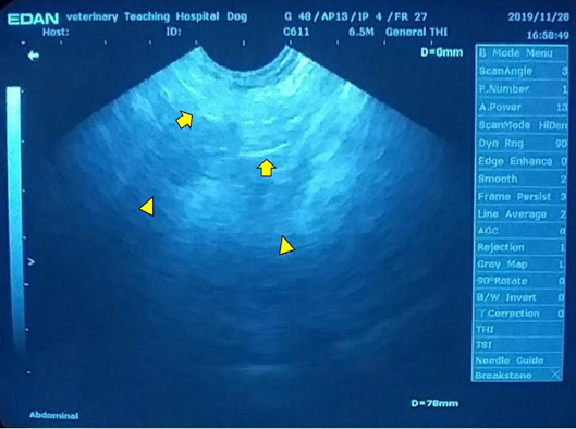
Hyperechoic focal echogenicity (arrows) creating distal acoustic shadow (arrowheads) in the dependent portion of the bladder, confirming the presence of cystic calculus.
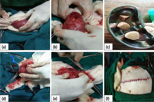
Image showing the steps of cystotomy in a dog. Distended bladder incised at the base of the bladder (a), Bladder distended with calculi (b), Removed three bladder calculi (c), Inserting catheter from bladder to urethral opening (d), Double-layer suturing of the bladder (e), Skin closure by simple interrupted pattern (f).
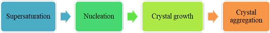
Steps involved in lithogenesis (supersaturation, nucleation, crystal growth, and aggregation irrespective of crystal nature) (Espinosa-Ortiz et al., 2018).


