
Effects of Nano-Cerium Oxide and Alpha Lipoic Acid on Sciatic Nerve Regeneration in Rats


Effects of Nano-Cerium Oxide and Alpha Lipoic Acid on Sciatic Nerve Regeneration in Rats
Dhuha Adel Yasser*, Ammar M. Hashim, Rafid Majeed Naeem
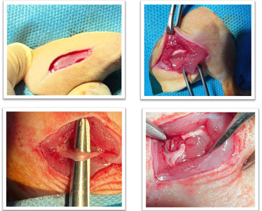
showed the surgical site, showed exposure of sciatic nerve, sciatic nerve a complete transect.
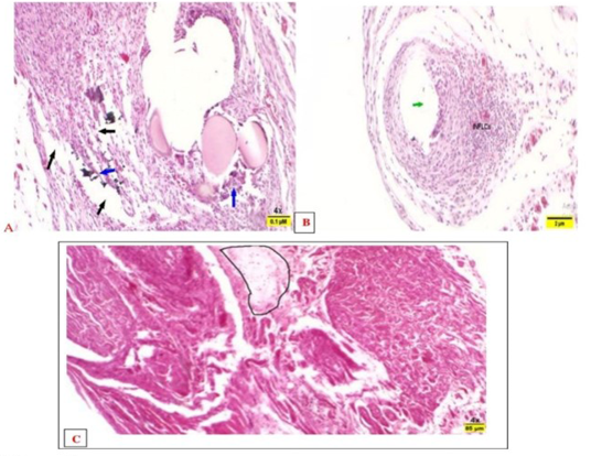
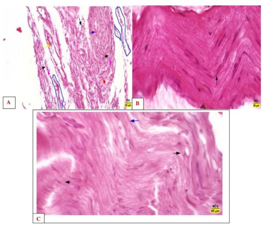
A-control-4 weeks. Photomicrographs of the sciatic nerve the positive control showed distributed of the arrangement of fibers, degenerated nerve fibers (blue arrows), and they had swollen or missing axons with various degrees of edema (black arrows). H and E, 4x, B-control-8 weeks. Photomicrographs showed edema (ED), and infiltration of Inflammatory cells; H and E, 4X. C-control-12 weeks. Photomicrographs showed severe disarrangement of fibers, notable degeneration of axon sheaths, edema, and infiltration of inflammatory cells (black circle). H and E, 4x.
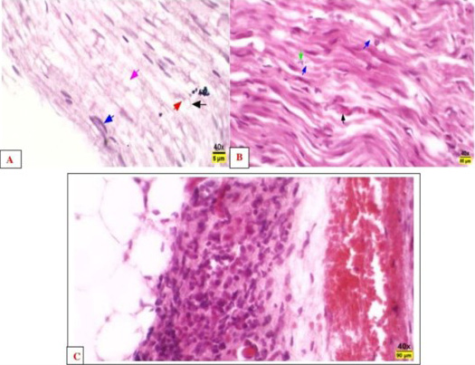
A-4 week. Photomicrographs of the sciatic nerve in the ALA group showed a significant decrease in the thickness of perineural fibrosis compared to the control group. There is significant large vacuolation (pink arrow), degenerated axon (red arrow), prominent perineural cells (blue arrow), and Schwann cells (black arrows). H and E, 40x. B-8 weeks. Sections of the Sciatic nerve of the ALA group showed mild degeneration, dilated axon sheaths (blue arrows) containing debris (fragmented axons and/or myelin) (green arrow), and macrophages (black arrow) that engulf this debris. H and E, 40x. C-12 weeks. Sections of the Sciatic nerve of the ALA group showed thickening of the perineurium layer, infiltration of the inflammatory cells (black arrow), and congestion (yellow arrows). H and E, 40x.
A-4 weeks. Photomicrographs of the sciatic nerve of the mix-treated group showed almost normal myelinated nerve fibers (blue arrow), but slightly disarranged, atrophied, and myelin sheath loss (blue circled area) were also detected, normal Schwann cells (pink arrows), normal axon (black arrows), congestion and increase blood vessels (vellow arrow), and edema also evident. H and E, 40x. B-8 weeks. Photomicrographs of the sciatic nerve of the mix-treated group showed epineurium (black arrows), and normal nerve fiber arrangements (black arrows) in the endoneurium. H and E, 40x. C-12 weeks. Sections from the Sciatic nerves of the mix-treated group revealed almost normally arranged nerve fibers (blue arrow), normal Schwan cells and axons (black arrows). H and E, 10x.
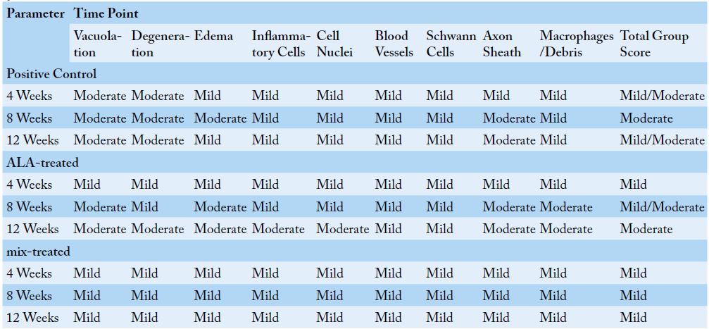
Comparison of histopathological changes across different groups and time points using the descriptive scoring system.


