Expression of Somatostatin Type-2 Receptors in Mouse Dorsal Root Ganglion at Early Stage of Pain Models: Evidence for the Inhibitory Role of Somatostatin in Pain
Expression of Somatostatin Type-2 Receptors in Mouse Dorsal Root Ganglion at Early Stage of Pain Models: Evidence for the Inhibitory Role of Somatostatin in Pain
Qiong Xiang1, Jing-Jing Li1, Qian Zhang1,3, Rong-Bo Tian1 and Xian-Hui Li1,2*
Procedures for establishing pain-related mouse model and behavioral tests. Following the Pinch-nerve injury, mice in the Pinched group, and sham group were housed and subjected to thermal hyperalgesia behavior and cold allodynia tests consecutively within 7days and prolonged subsequently for 14days for Von-Frey tests. n = 5 per group. After Carrageenan injection (Carrageenan-treated group), and sham group were respectively injected with saline, mice were sacrificed after 15min , 90min, 1day and 4days and prolonged on the 7th day. After Dorsal Rhizotomy (DR) was completed, mice were housed for 7 days and subjected to DRGs dissection. n = 5 per group. The mice were performed Spared nerve injury (SNI) surgery; kept for 21days (3 weeks) and also subjected to DRGs dissection. n = 5 per group.
Mechanical hyperalgesia induced after Pinch nerve injury. Time course of mechanical pain withdrawal threshold measured by the von Frey filaments after unilateral Pinch (n=5). Ipsilateral hind paw displays a strongly painful behavior measured by pain withdrawal threshold compared with sham group animals (n=5); This mechanical hyperalgesia is not seen in the contralateral paw compared with sham group animals (n=5). Data are expressed as mean±S.EM. *, P <0.05 compared with the sham group.
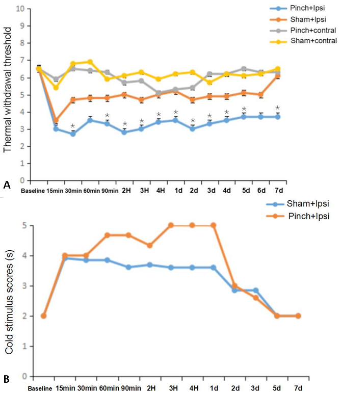
Thermal hyperalgesia and cold allodynia induced after Pinch nerve injury. (A)Time course of thermal hyperalgesia tests after Pinch. The paw withdrawal latency (Seconds) after nociceptive hot stimulation significantly decreased at 30min time point in Pinch group (n=5) compared with sham group (n=5); This thermal hyperalgesia sustained for 7days. The contralateral paw has not shown this mechanical hyperalgesia compared with sham group animals (n=5). (B)cold allodynia tests after Pinch.Time course of cold stimulus scores measured by the Actone after Pinch-nerve injury (n=5). Ipsilateral hind paw displays a strongly but not significantly painful behavior measured by pain scores compared with sham group animals (n=5); This cold allodynia is not done in the contralateral paw compared with sham group animals (n=5). Data are expressed as mean±S.EM. *, P <0.05 compared with the sham group.
The expression of SSTR2 protein in DRGs by western bolt. (A) SSTR2 expression in DRGs after Carrageenan injection. Mice sacrificed at 15min, 90 min, 1day and 4 day after Carrageenan injected in left- hind paw(L), compared with right-side untreated hind paw, showed up-regulated SSTR2 expression in DRG neurons at 15min and 90 min these two time points (no significant differences existed, respectively); However, 1day after injection of Carrageenan, SSTR2 protein showed down-regulated in left-side DRGs(L), after 4 day, The SSTR2 protein sharply reduced in left-side DRGs(L) compared to right-side DRGs(R). Error bars represent standard error of the mean±SEM. Significant differences are indicated by *, P<0.05; **. P<0.01 (n=5). (B) The expression of SSTR2 in DRGs on the basis of DR and SNI models. Tissues from ipsilateral hind paw DRGs (L) are compared with tissues from contralateral hind paw DRGs(R) after DR 1 week and SNI 3weeks surgery. SSTR2 expression are not showed significant variations in neither of these two models (n=5).


