Iron Excess Impact on Pancreatic Beta Cell Structure and Fasting Blood Glucose Levels in Male Wistar Rats (Rattus norvegicus)
Anisa Muthia Fakhira1, Madihah Madihah1, Susi Susanah2, Erick Khristian3, Ratu Safitri1*
1Department of Biology, Faculty of Mathematics and Natural Sciences, Universitas Padjadjaran, Bandung, West Java, Indonesia; 2Department of Child Health, Faculty of Medicine, Universitas Padjadjaran, Bandung, West Java, Indonesia; 3Program Study of Medical Laboratory Technology, Faculty of Health and Science Technology Universitas Jenderal Achmad Yani, Cimahi, West Java, Indonesia.
*Correspondence | Ratu Safitri, Department of Biology, Faculty of Mathematics and Natural Sciences, Universitas Padjadjaran, Bandung, West Java, Indonesia; Email:
[email protected]
Figure 1:
Histopathological Representation of Islets of Langerhans in Rat Pancreas Post-Treatment with PAS Staining at 1000× Magnification.
Note: (A) Control; (B) ID 10 mg/kg BW; (C) ID 20 mg/kg BW; (D) ID 30 mg/kg BW; (E) ID 40 mg/kg BW; (F) ID 50 mg/kg BW; (G) ID 60 mg/kg BW; (↑) necrotic beta cells; (▲) normal beta cells; (∆) alpha cells.
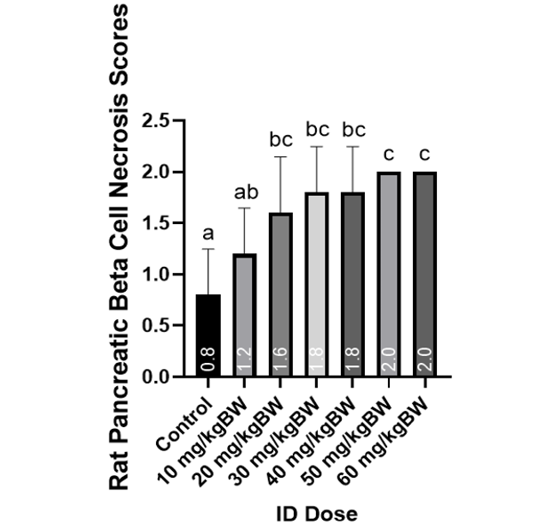
Figure 2:
Graph of rat pancreatic beta cell necrosis score with iron excess conditions.
Note: Different letters indicate the results of Mann-Whitney U analysis, indicating significant differences between treatments (P<0.05).
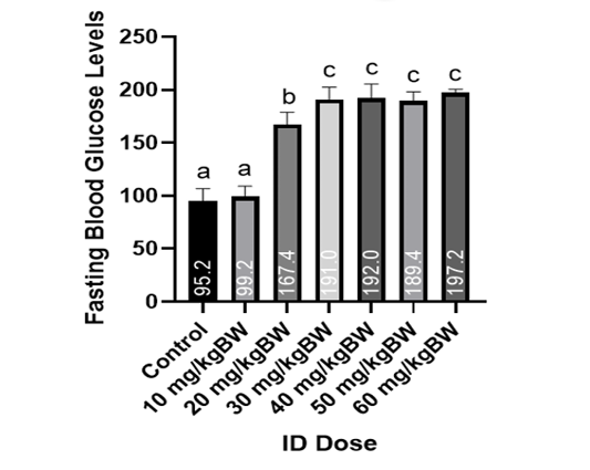
Figure 3:
Graph of rat fasting blood glucose levels from rat blood serum in iron excess conditions.
Note: different letters indicate significant differences between Duncan’s advanced test treatments (P<0.05).


