Isolation and Identification of Mycoplasma mycoides Subspecies mycoides from the Ear Canal of Cattle in Plateau State, North-Central Nigeria
Isolation and Identification of Mycoplasma mycoides Subspecies mycoides from the Ear Canal of Cattle in Plateau State, North-Central Nigeria
Paul I. Ankeli, Mashood A. Raji, Haruna M. Kazeem, Moses O. Odugbo, Nendir J. Umaru, Idowu O. Fagbamila, Livinus T. Ikpa, Obinna O. Nwankiti, Issa A. Muraina, Pam D. Luka and Nicholas D. Nwankpa
Map of Plateau State showing Local Government Areas sampled (Source: National Bureau of Statistics)
Photomicrograph of Mycoplasma mycoides subspecies mycoides (Mmm) colonies isolated from ear swab samples from cattle on mycoplasma agar. Note: raised dense centres and lighter peripheries (arrows), x35
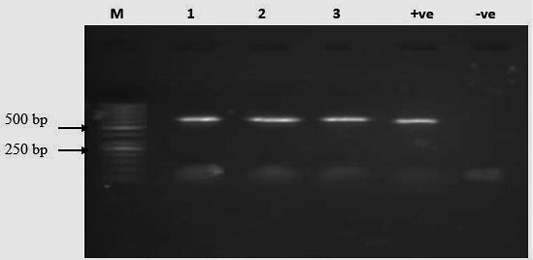
Mmm-specific PCR showing 574 bp: Lane M is the molecular marker (50 bp ladder), Lane 1, 2, 3, for the three positive Mmm, respectively, Lane +ve, -ve for negative and positive (T1/44 vaccine strain) controls
Mmm-specific PCR showing 1.1 kb amplicon sizes ran in duplicates: Lane M is the molecular marker (1kp plus ladder), Lane 1A-1B, 2A-2B, 3A-3B, for the three positives, respectively, Lane –ve, +ve for negative and positive (T1/44 vaccine strain) controls


