
Management of Spinal Osteochondroma in Young Golden Retriever Dog


Management of Spinal Osteochondroma in Young Golden Retriever Dog
Neeranoot Detcharoenyos1, Nakrob Pattanapon1, Soontaree Petchdee2*
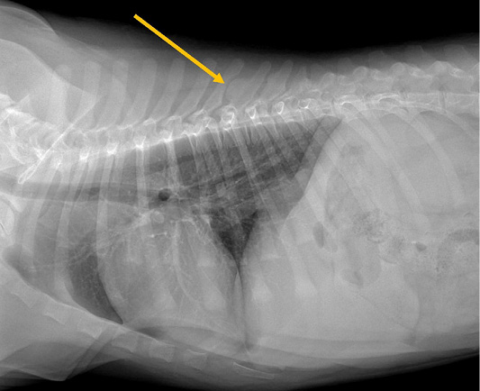
Figure 1:
Radiograph of thoracic (lateral view) with exostoses at T8-T9 dorsal spinous processes (yellow arrow).
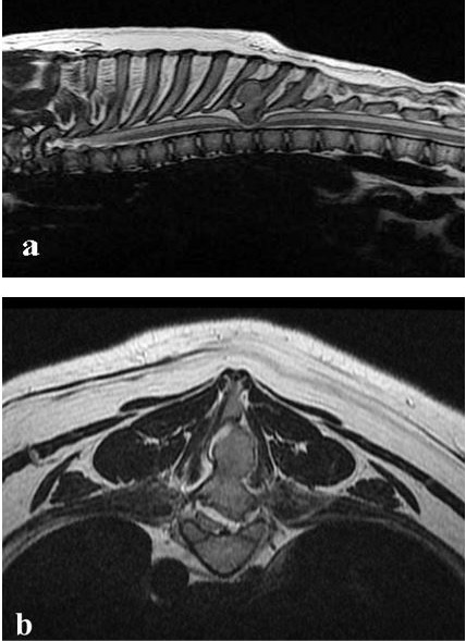
Figure 2:
T2-weighted sagittal (a) and transverse (b, c) MRI showing bony mass at T8 with severely compressed the spinal cord.
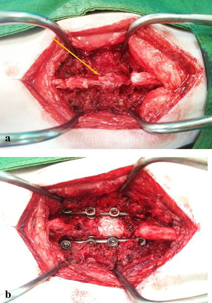
Figure 3:
Intraoperative view of pathological exostoses located at T8 dorsal spinous process (a, yellow arrow). The modified pedicle screw-rod fixation was placed to T7-T10 for stabilization after exostoses were excised and the defect was covered with an autologous fat graft (b).
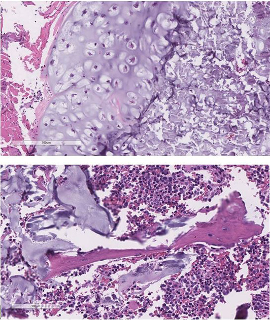
Figure 4:
The microscopic finding presented a nodule of the cartilaginous cap with well-differentiated chondrocytes interspersed with bone spicules surrounded by marrow cells.
July 2022
Vol. 10, Iss. 7, Pages 1423-1658
