

Morphological and Immunohistochemical Study on the Distribution of Endocrine Cells in the Gastrointestinal Tract of Partridge, Alectoris chukar


Morphological and Immunohistochemical Study on the Distribution of Endocrine Cells in the Gastrointestinal Tract of Partridge, Alectoris chukar
Zekeriya Özüdoğru1, Hülya Kara2*, Adem Kara3, Derviş Özdemir3 and Hülya Balkaya2

Histological structure of gastrointestinal tissues of red legged partridge stained with anti-Motilin antibodies; (a) proventriculus; (b) ventriculus; (c) duodenum; (d) jejenum and (e) ileum. Arrows show immunoperoxidase staining positive stained cells.
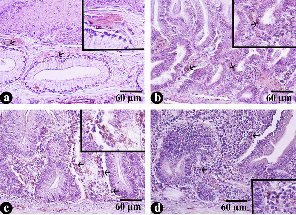
Histological structure of gastrointestinal tissues of red legged partridge stained with anti-VIP antibodies; (a) proventriculus; (b) ventriculus; (c) duodenum and (d) jejenum. Arrows show immunoperoxidase positive stained cells.

Histological structure of gastrointestinal tissues of red legged partridge stained with anti-GIP antibodies; (a) proventriculus; (b) ventriculus; (c) duodenum; (d) jejenum and (e) ileum. Arrows show immunoperoxidase positive stained cells.

Histological structure of gastrointestinal tissues of red legged partridge stained with anti-Grelin antibodies; (a) proventriculus; (b) ventriculus; (c) duodenum; (d) jejenum and (e) ileum. Arrows show immunoperoxidase positive stained cells.

Histological structure of gastrointestinal tissues of red legged partridge stained with anti-CCK antibodies; (a) proventriculus; (b) ventriculus; (c) duodenum; (d) jejenum and (e) ileum. Arrows show immunoperoxidase positive stained cells.


