
Optimizing Estrus Detection Techniques in Tropical Saanen Does (Capra aegagrus hircus): A Comparative Study


Optimizing Estrus Detection Techniques in Tropical Saanen Does (Capra aegagrus hircus): A Comparative Study
Muhammad Evan Magistrama1, Dio Fico Felsidan Diatmono1, Mira Tsurayya Masruroh1, Fransisca Gani Padmawati1, Pradita Iustitia Sitaresmi2, Yustina Yuni Suranindyah3 and Diah Tri Widayati1*
Research location, in Center for Livestock Development (PPT), Faculty of Animal Science, Universitas Gadjah Mada, Sleman Regency, Daerah Istimewa Yogyakarta Province (in tropical climate).
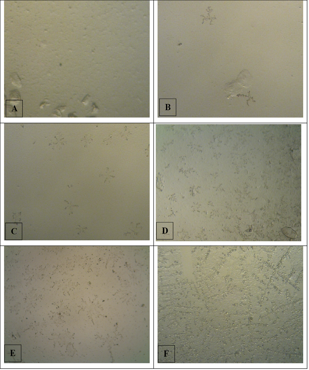
Photomicrograph of salivary fern pattern’s scoring with magnification of 10x10; score of 1 (A), score of 2 (B), score of 3 (C), score of 4 (D), score of 5 (E), and score of 6 (F).
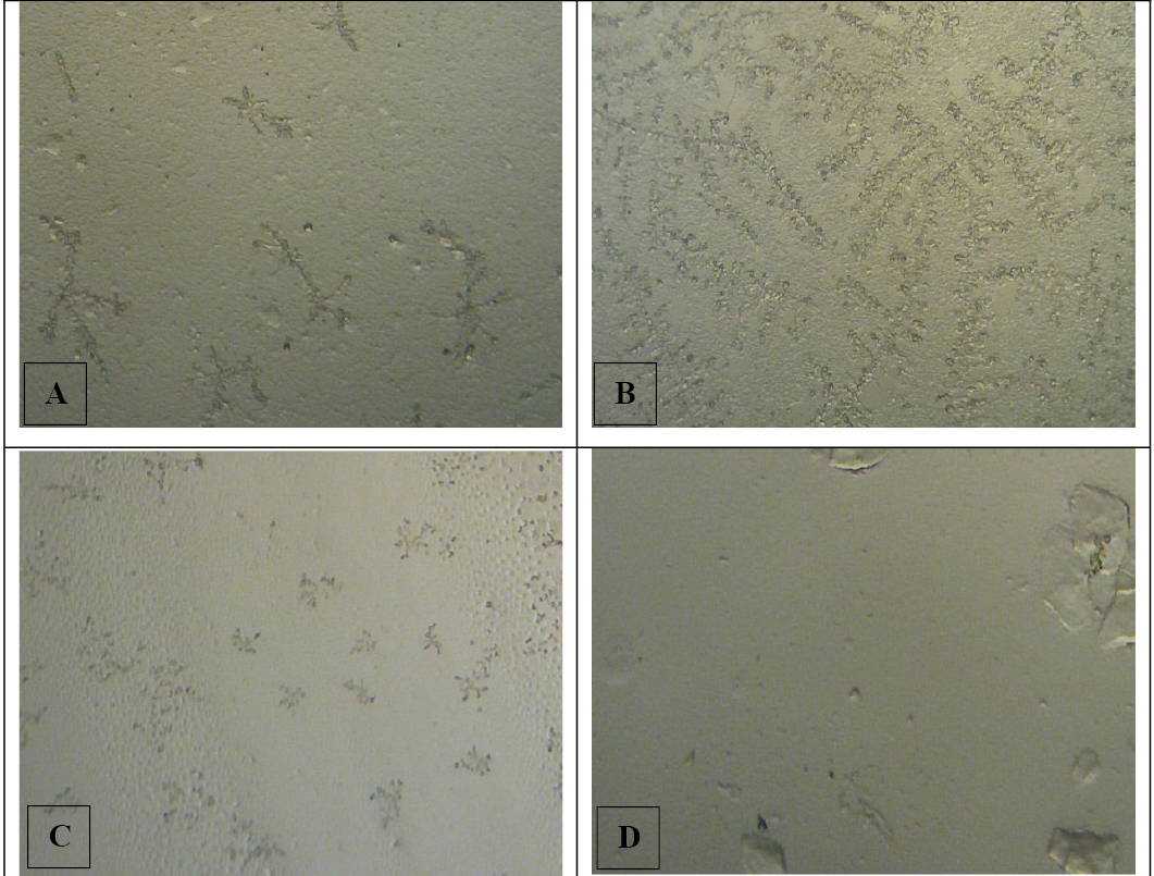
Photomicrograph of salivary fern pattern on each estrus phases with magnification of 10x10; proestrus (A), estrus (B), metestrus (C), and diestrus (D).
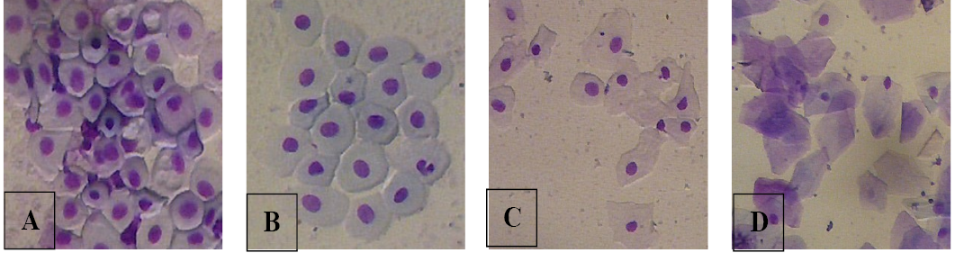
Photomicrograph of vaginal epithelial cells with magnification of 10x10; parabasal (a), intermediate (b), superficial (c), and superficial with keratinization (d).
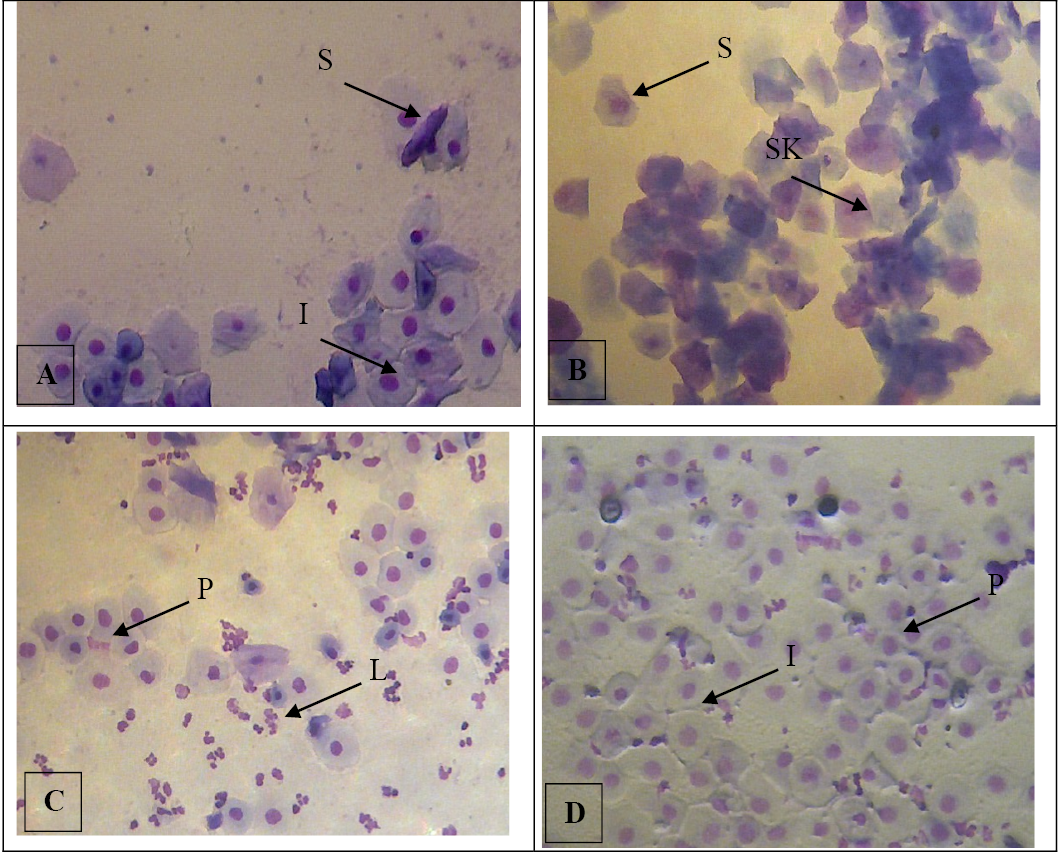
Photomicrographs of vaginal smear on each estrus phases with magnification of 10x10; proestrus (A), estrus (B), metestrus (C), diestrus (D), also various of epithelial cells in vaginal cytology; parabasal cell (P), intermediete cell (I), superficial cell (S), superficial with keratinization (SK), and leucocyte (L).

