Prevention of Rhabdomyolysis-Induced Acute Kidney Injury via Attenuation of Oxidative Injury and Inflammation by Cinnamic Acid Coated Gold Nanoparticles in Mice
Prevention of Rhabdomyolysis-Induced Acute Kidney Injury via Attenuation of Oxidative Injury and Inflammation by Cinnamic Acid Coated Gold Nanoparticles in Mice
Rehan Ahmed Siddiqui1,2*, Shabana Usman Simjee2,3, Nurul Kabir4, Muhammad Ateeq3,5, Kevin Joseph Jerome Borges6, Muhammad Raza Shah3 and Rahman M. Hafizur2
(A) UV-visible spectrum and (B) AFM analysis of CA coated AuNPs.
Calculation of Damaged areas in proximal convoluted tubules in different animal groups. The graph demonstrating no proximal tubular damage in normal control. Whereas, there is a marked increase in damaged area in glycerol treated AKI group compared to the normal control (*p <0.001). A significant decrease in the damaged areas was observed in animals treated with CA as compared with the glycerol treated AKI group (*p <0.001) and nearly complete protection can be seen in CA-AuNPs treated groups at a relatively low dose (*p <0.001).
Serum urea and creatinine levels. Serum urea and creatinine levels were significantly elevated in the glycerol treated AKI group as compared to the normal control (*p <0.001). The levels were significantly decreased in CA and CA-AuNPs treated animals as compared to the glycerol treated AKI group (*p <0.001).
PAS stained microscopic images demonstrating brush borders of kidney tubules in the cortex and medulla. Images A and E shows normal tubular brush borders. Images B and F are of glycerol treated AKI group showing damaged brush borders of loop of Henle and proximal tubules. Images C and G are taken from CA treated animals, a remarkable reduction in the damage of brush borders can be observed while images D and H displays protection with CA-AuNPs. (Magnification; 600x).
CA and CA-AuNPs prevent damage in actin cytoskeleton and reduces expression of COX-2. Normal distribution of actic can be seen in figure A while figure B shows disrupted actin cytoskeleton. The protection of actin cytoskeleton can be observed in figure C and D which were treated with CA and CA-AuNPs respectively. (Magnification; 200x). There is no expression of COX-2 in normal control (E) whereas higher expression level can be observed in glycerol treated AKI group (F). A decrease expression of COX-2 can be noticed in CA (G) and CA-AuNPs (H) treated groups. Red is actin and COX-2, green is DAPI and gray is DIC image. (Magnification; 600x).
Effects of CA and CA-AuNPs on iNOS, NF-κB, HO-1 and Kim-1 mRNA expressions. CA and CA-AuNPs attenuates mRNA expression of iNOS (A) and NF-κB (B) while both increases the mRNA expression of HO-1 (c) and Kim-1 (d) (***p < 0.001, **p < 0.01, * p < 0.05).
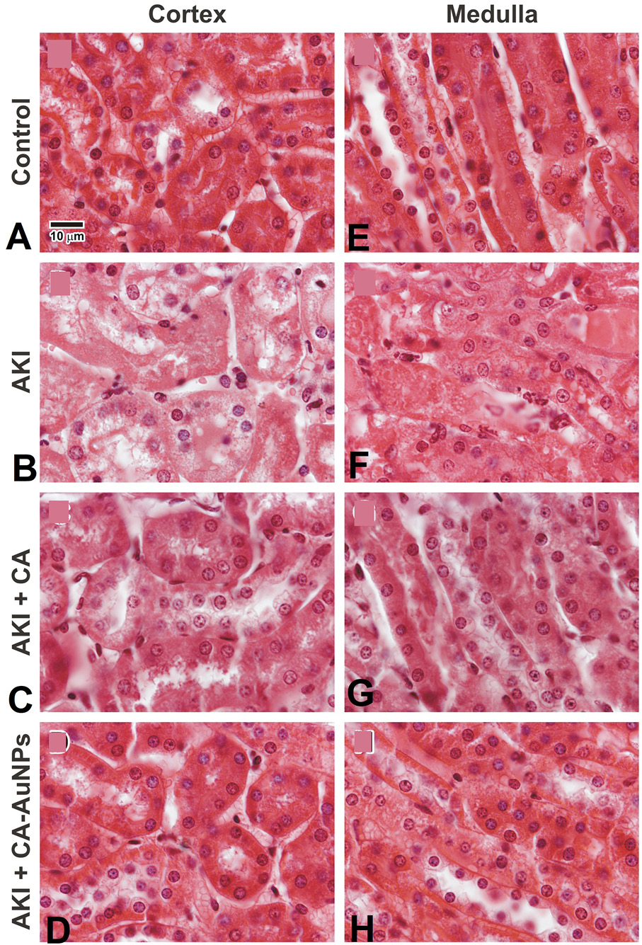
H and E stained microscopic images demonstrating kidney cortex and medulla. Images A and E are showing normal kidney architectures. Images B and F are of glycerol treated AKI sections showing damaged proximal tubules and cast deposition in tubules. Images C and g are CA treated group presenting notable decline in tubular cast deposition and damage. While images D and H are CA-AuNPs treated kidney sections displaying more or less complete protection. (Magnification; 600x).


