
Some Epidemiological Studies on Theileria annulata Infection in Egypt


Some Epidemiological Studies on Theileria annulata Infection in Egypt
Ahmed Abdel-Rady3*, Mohamed Karmi2, Menna_allah Youssef1, Aml M. Abdel-Ra’ouf1, Bahaaa Madkour1

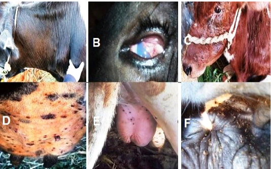
A) Enlargement of prescapular L.N. in Theileria annulata infected cattle. B) Corneal opacity in Theileria annulata infected cattle. C) Severe eye and skin affections around the eye. D) Heavy infestation of udder with ticks.
E) Ticks infestation on scrotum and perineal region. F)Heavy infestation with ticks around the anus.
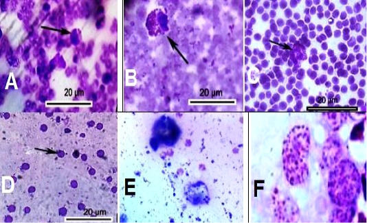
Blood and lymph smears of Theileria annulata infected cattle. A) arrow refers to macro-schizont inside lymphocyte (Koch’s blue bodies).B) arrow shows micro-schizont inside lymphocyte. C) arrow shows raptured schizont. D) arrow shows Theileria annulata piroplasm inside RBCs. E and F) schizont of Theileria annulata inside lymphocytes (koch’s blue bodies) in lymph smears.
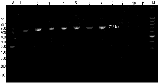
PCR findings of blood of Theileria annulata suspected infected cattle. M: Ladder of 100 base pair. Lane 1: control +ve T. annulata (Dept. of parasitology, Beni-Suef University) showed the specific amplicon size of 768 bP. Lanes 2, 3, 4, 5, 6 and 7: T. annulata +ve samples showed the specific 768 bp amplicon size. Lanes 8, 9, and 10: -ve samples. Lane 11: a control -ve.

