
Treatment of Severe Canine Parvoviral Enteritis Associated with Coccidia


Treatment of Severe Canine Parvoviral Enteritis Associated with Coccidia
Mohammed Mijbas Mohammed Alomari1, Nawar Jasim Alsalih2*, Samir Sabaa Raheem2, Mohenned Alsaadawi2, Ali Mosa Rashid Al-Yasari3
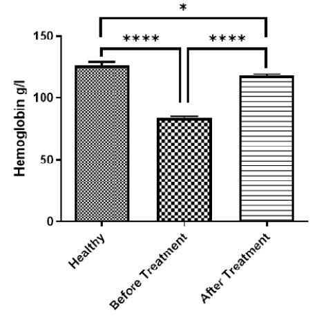
Figure 1:
Concentration of Hemoglobin g/l.
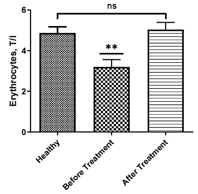
Figure 2:
Total number of Erythrocytes, T/l.
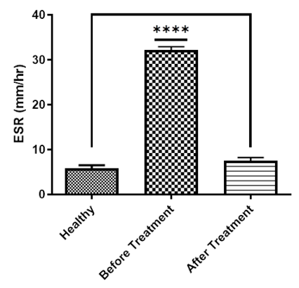
Figure 3:
ESR, mm/h.
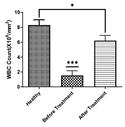
Figure 4:
Total number of leucocyte.
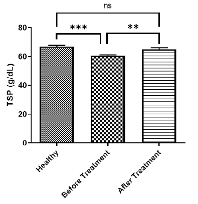
Figure 5:
Concentration of Total serum protein (g/dl).
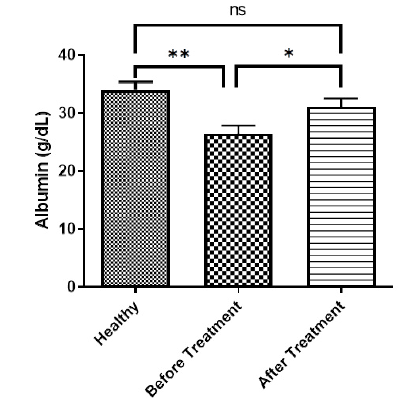
Figure 6:
Concentration of Albumin (g/dl).
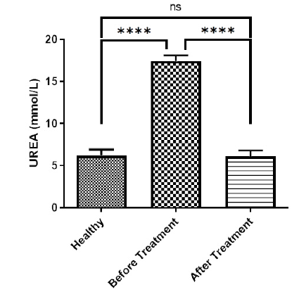
Figure 7:
Concentration of UREA (mmol/L).
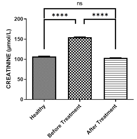
Figure 8:
Concentration of Creatinine (µmol/l).
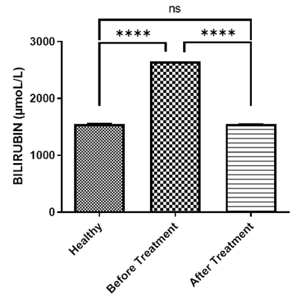
Figure 9:
Concentration of bilirubin (µmol/l).
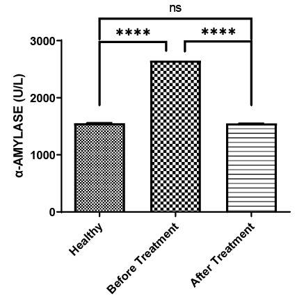
Figure 10:
Concentration of α-amylase (U/L), CEC, the activity of ALT, AST as in Figures 11,12 and 13.
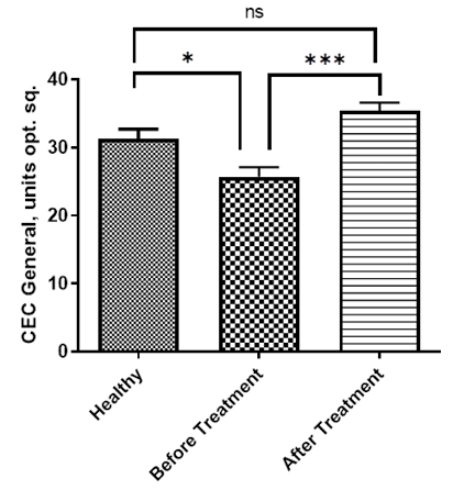
Figure 11:
CEC general, units opt. sq.
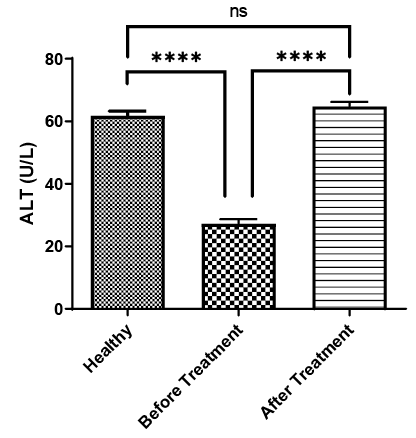
Figure 12:
Concentration of alanine aminotransferases (U/L).
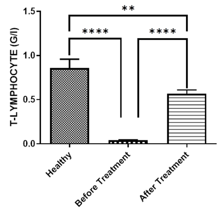
Figure 14:
Total number of T. Lymphocyte (G/L).
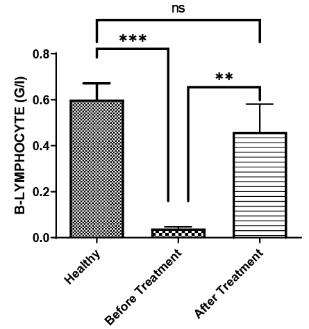
Figure 15:
Total number of B. Lymphocyte (G/L).
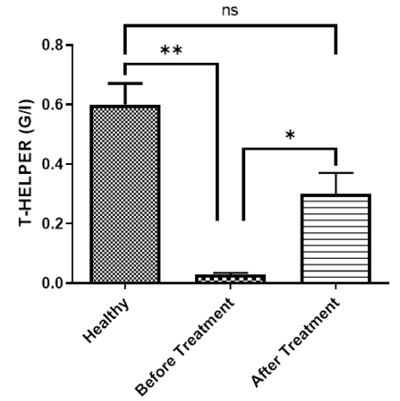
Figure 16:
Total number of T. Helper (G/L).
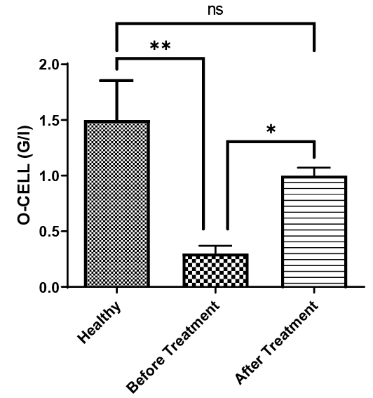
Figure 17:
Total number of O-cells (G/L).
Note: * – р≤0.05; ** – р≤0.01; *** – р≤0.001 (Reliability of the difference between the indicators of sick dogs before and after treatment).
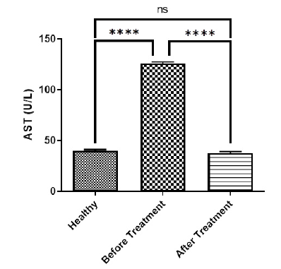
Figure 13:
Concentration of Aspartic i aminotransferases (U/L).
August 2024
Vol. 12, Iss. 8, pp. 1410-1621
