Antiproliferative-Effect-of-Garlic-Extract-on-Cancer-Cell-Lines


Research Article
Antiproliferative Effect of Garlic Extract on Cancer Cell Lines
Nasr Tarq Mohammed
Department of Microbiology, College of Veterinary Medicine, University of Baghdad, Iraq.
Abstract | Breast cancer is one of the most common neoplasms globally and is even more prevalent in some animal species, such as dogs, than in humans. Garlic organosulfur compounds possess anti-carcinogenic, antibacterial, antifungal, antiviral, and immune-boosting properties. This study aimed to evaluate the anticancer potential of aqueous garlic extract (AGE) against breast cancer cell lines. The use of a simple aqueous extract allows for easy and cost-effective preparation while retaining bioactivity. In this research, the effectiveness of AGE in inhibiting cancer cell viability was assessed using cancer cell lines CAL51 and AMN3. The cell lines were treated with various concentrations of AGE (100, 200, 300, 400, and 500 µL) in vitro. The findings indicate that at a concentration of 500 µL, AGE caused 95% destruction of CAL51 cells, while lower concentrations resulted in cell death rates of approximately 83%, 78%, 54%, and 27% at 400, 300, 200, and 100 µL, respectively. For the AMN3 cell line, the destruction rate was 93% at 500 µL, and 90%, 79%, 65%, and 36% at 400, 300, 200, and 100 µL, respectively. In conclusion, this study demonstrates the significant, dose-dependent anticancer effects of aqueous garlic extract on breast cancer cell lines, highlighting its potential as a promising and accessible therapeutic agent in cancer treatment strategies.
Keywords | Antiproliferative, Apoptosis, Garlic extract, Breast cancer, Cytotoxicity, Cell lines.
Received | May 07, 2024; Accepted | August 24, 2024; Published | September 05, 2024
*Correspondence | Nasr Tarq Mohammed, Department of Microbiology, College of Veterinary Medicine, University of Baghdad, Iraq; Email: [email protected]
Citation | Mohammed NT (2024). Antiproliferative effect of garlic extract on cancer cell lines. J. Anim. Health Prod. 12(3): 444-449.
DOI | http://dx.doi.org/10.17582/journal.jahp/2024/12.3.444.449
ISSN (Online) | 2308-2801

Copyright: 2024 by the authors. Licensee ResearchersLinks Ltd, England, UK.
This article is an open access article distributed under the terms and conditions of the Creative Commons Attribution (CC BY) license (https://creativecommons.org/licenses/by/4.0/).
Introduction
Breast cancer is the major type of neoplasms globally, affecting both industrialized and developing nations. The risk is even higher in some animal species, such as dogs, compared to humans (Schiffman and Breen, 2015). Over the past 20 years, the significance of this issue has increased, underscoring the need for innovative treatments to reduce cancer incidence (Ferlay et al., 2013). While conventional treatments include radiation, hormone, and chemotherapy, and surgery have reduced the invasiveness and proliferation of breast cancer, there is ongoing research into more effective treatments. Many conventional chemotherapeutic agents are associated with drug resistance, cancer relapse, and adverse side effects. Researchers are exploring natural chemicals and plant extracts as alternative therapies for breast cancer. These natural molecules, derived from living organisms, can stimulate apoptosis and suppress metastasis, thereby inhibit cancer progression. Natural bioactive substances have been demonstrated to have a major impact on the advanced stages of several types of malignancies, such as breast carcinoma, increasing the rate of survival and lowering the fatalities (Noel et al., 2020; Sung et al., 2021).
Allium sativum, or garlic, is a vegetable in the Allium genus. Traditionally utilized as a medicinal plant, it became well-known for its ability to treat cancer, microbial infections, heart disease, high cholesterol, obesity, and hypertension. Additionally, its bioactive components, Organosulfur compounds (Allicin, diallyl trisulfide (DATS), 2- vinyldithiines, allylmethyl (AMS), Diallyl disulfide (DADS), and ajoene (E, Z) Influence on signaling processes for arrest of cell cycle in breast cancer, including lipid peroxidation, nitric oxide biosynthesis, epidermal growth factor receptor (EGFR), nuclear factor kappa B (NF-κB), and protein kinase C (Bhandari, 2012: Pandey et al., 2023).
Garlic is widely recognized for its phenolic compounds, which have been shown in animal models to potentially slow neoplasm progression and decrease carcinogenesis. In traditional medicine, A. sativum L. is utilized in the Elimination and curing of several carcinomas, such as those of the blood, breast, prostate, ovary, and gastrointestinal tract malignancies (Matysiak et al., 2015).
Numerous natural antioxidants, high flavonoids and phenolic acid compounds, and a wide range of pharmacological substances can all directly or indirectly increase the expression of normal cells antioxidant enzymes (Chen et al., 2013; Piątkowska et al., 2015). Therefore, the goal of this study is to estimate the antiproliferative action of garlic extract against different human as well animal cancer cell lines.
Material and methods
Experimental Design
Two types of breast cancer cells (human (CAL-51) and mice mammary adenocarcinoma (AMN-3)) were provided from Iraqi Center for Cancer and Medical Genetic Research (ICCMGR) to assess the cytotoxicity in the cell line that used in this study.
Preparation of aqueous garlic extract
Garlic, also known as Allium sativum, was bought from a native market in Baghdad. Cloves were sliced, ground, and made into a fine paste. Next, 200 grams of garlic paste were weighed and soaked in 250 milliliters of distilled water. The ingredients was magnetically stirred for three hours. The aqueous garlic extract (AGE) was then filtered over the course of a day, resulting in a final concentration of 150 mg/200 mL. The AGE was then set aside for use in cancer cell studies.
Experimental Groups
The breast cancer cell lines were handled with multiple concentrations of AGE (100, 200, 300, 400, and 500 µL) to evaluate its dose-dependent effects on cell viability. A range of concentrations was used to determine the optimal dose for inducing apoptosis, causing cell cycle arrest, and inhibiting cell division.
Experimental Assays
To determine the impact of the garlic extract on cancer cell lines, a number of tests were carried out, including assessments of cell viability such MTT and crystal violet staining.
Reagents
Following material including chemicals/reagents were purchased: RPMI-1640 Medium, MEM Medium (HiMedia India), Antibiotics (Penicillin/Streptomycin), Antifungal (Amphotericin –B), Fetal bovine serum (CAPRICOR Germany), Sodium bicarbonate (BDH chemicals Ltd. England), 2-[4-(2-hydroxyethyl) piperazin-1-yl] ethanesulfonic acid (HEPES) (Sigma Germany), trypsin –versene powder (US biological USA), Trypan blue dye (Pharmacia Sweden), Phosphate buffer saline (BDH England), and Dimethyl Sulphoxide (DMSO) (Bio World USA).
Garlic extract treatment and cell cultivation
The cell culture process was conducted in a vertical laminar flow bench that had been disinfected with 70% ethanol and UV light exposure. Every piece of equipment and solution used autoclaved, sterile. Using two different types of tumor cell lines (CAL-51 and AMN3) and varying doses of garlic extract, the in vitro approach involves exposing the cells for 72 hours before measuring the results using the MTT assay.
All the cell lines were sub-cultured using the following procedure: After discarding the growth medium and rinsing the cells two times by sterile phosphate buffer saline (PBS), 2 - 3 milliliters of trypsin/versene solution were added to the side of the flask opposite the cells (Freshney, 2010).
After completely flooding the monolayer in the flask and gently rocking it for a few minutes, the growth medium (0.1–0.2 ml/cm2) was added. The cells were then pipetted into the growth medium, dispersing them across the monolayer’s surface. After that, the cells were transferred to cell cultivation flasks at the required concentration and incubated at 37˚C. The media was initially orange, but as the cells grew, it turned yellow, necessitating the replacement of the reducing media. Turbidity indicates that the culture is probably contaminated and needs to be thrown away. Following the falcon’s incubation period, the cells formed a monolayer, and the confluent monolayer was handled similarly to the previously mentioned subculture. Twenty milliliters of growth medium were added after trypsinization, and the cells were pipetted into the growth medium. Using CAL-51 AMN3 cell lines.To assess the cytotoxicity and anticancer action of the samples, the (3-(4, 5-dimethyl thiazol-2yl)-2, 5-diphenyl tetrazolium bromide) MTT technique was done.
Detection of cytotoxicity by MTT assay
The (MTT) solution was prepared by dissolving 0.2gm MTT in 100ml of PBS (Benov, 2021). The solution was filtered through Millipore filter (0.2 µm) to get rid of any purple formazan bodies. then kept at 4°C. in sterile, opaque, screw-capped vials. After preparation, the solution was utilized for no more than two weeks (Freshney, 2010).
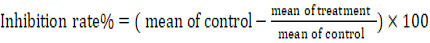
The colorimetric MTT technique was used to assess the proliferation of cancer cells (CAL-51, AMN3). Cells were cultured at a density of 1 × 10⁴ cells/well in tissue culture plates (96-well) with flat bottoms then incubated at 37°C for 24 hours to form a monolayer, which was monitored using an inverted microscope. After 72 hours, the CAL-51 and AMN3 the confluent monolayer treated with different concentrations of AGE (100, 200, 300, 400, or 500 µL). Following treatment, 100 µL of yellow-colored tetrazolium MTT solution was added to each well, and the microplate was incubated at 37°C for 4 hours. The medium was then replaced with 50 µL of DMSO in each well, and the plate was re-incubated for 40 minutes at 37°C. The intracellular purple formazan crystals were solubilized by the DMSO and evaluated spectrophotometrically. Optical density OD of each well was determined using a micro-ELISA reader at 570 nm.. The inhibitory rate of cell proliferation was calculated using the following equation (Benov, 2021).
Detecting viability of cultured cells by Crystal Violate assay
A 96-well plate was used for cell seeding. The medium was added to three wells without any cells. The culture media was adjusted to a volume of ≥100 μL/well in order to prevent evaporation effects. Cells were incubated at 37°C for 24 hours in order to facilitate cell adherence to wells. Following the aspiration of the medium from each well, ≥100 μL of fresh medium enriched with garlic extract was applied to each well. The cells were incubated under the proper conditions for the intended duration. After aspirating the medium, the cells underwent two gentle tap water washes. As soon as the wells filled with water, the water was promptly sucked out of them. To get rid of any leftover liquid, the plate was lightly taped after being cleaned and inverted onto filter paper. After adding 50 μL of 0.5% crystal violet staining solution to each well, the plates were incubated at room temperature for 20 minutes using a bench rocker set to oscillate at a frequency of 20 times per minute. Four times, a stream of tap water was used to wash the plate. To get rid of any leftover liquid, the plate was lightly taped after being cleansed and inverted onto filter paper. At room temperature, let the plate air dry without a lid for at least two hours. To prepare a crystal violate stain, five grams of crystal violate powder were dissolved in 200 milliliters of methanol. The resulting mixture was then filtered through Whatman No. 1 filter paper. After adding distilled water, 50 ml of a formaldehyde solution with a 37% concentration was added to bring the liquid’s volume to 1000 ml. Whatman No. 1 filter paper was used to remove the extra solid residue, and it was then incubated at room temperature and away from light sources in a black container. Using a plate reader, the optical density (at OD570) of each well was determined. The average OD570 of unstimulated cells was adjusted to 100%. Then, compared the average OD570 values of stimulated cells to those of non-stimulated cells to calculate the percentage of viable (attached) cells. The mean and the standard error of the mean for at least three independent experiments were calculated (Freshney, 2010; Feoktistova et al., 2016).
Statistical analysis
In order to assess group differences for multiple comparisons, the one-way analysis of variance (ANOVA) was utilized. Statistical significance was defined as a p-value of below 0.05 (p < 0.05).
Results
Cytotoxicity by MTT assay
The viability of CAL-51cells examined by MTT assay after 72 hrs of exposure to different concentrations (500,400, 300, 200, or 100 µL) of AGE, showed significant (P<0.05) suppression of the cells’ growth, however, the reduction was concentration-dependent manner. The cancer cells treated with a concentration of 500 µg/mL showed a significant inhibition rate of 95%, compared to the 27% inhibition rate observed at the 100 µg/mL concentration. This significant variation is illustrated in Figure 1.
The viability of AMN3 cells examined by MTT assay after 72 hrs of exposure to different concentrations (500,400, 300, 200, or 100 µL) of AGE, showed significant (P<0.05) inhibition of the growth of the cells, however, the reduction was concentration-dependent manner. The cancer cells treated with a concentration of 500 µg/mL exhibited a significant inhibition rate of 93%, compared to the 36% inhibition rate observed at the 100 µg/mL concentration. This significant variation is illustrated in Figure 2.
Microscopic findings
A crystal violet method was employed to evaluate the anti-tumor potentials of AGE toward breast carcinoma cell lines CAL-51 and AMN3. The viability of cancerous cells significantly declined when reviewed microscopically after 72 hrs. The cells that had been exposed to AGE in a concentration of 500 µL per a 5-mL MEM, and RPMI-1640 medium showed morphological alterations and apoptosis. These alterations included a reduction in size and a loss of the typical fusiform shape. In addition, cells began to separate from the surfaces they were attached to, as well as from one another as shown in (Figure 3, 4).
Discussion
In current research, we investigate the efficacy of aqueous AGE on the breast cancer cell lines AMN3 and CAL-51 to ascertain their anticancer and antiproliferative properties. Our findings showed that AGE showed growth inhibition in dose-dependent cytotoxicity against both cancer cell lines. At 500 μg/ml, the maximum AGE concentration, 95% of CAL-51 cells and 93% of AMN3 cells were eliminated. Garlic has strong anticancer effects, as evidenced by the substantial reduction of proliferation observed even at the lowest dose of 100 μg/ml AGE. These results are agree with earlier research by (Bagul et al., 2015), which showed that crude garlic extract, in a concentration-dependent manner, inhibited growth and produced a decreased cell death in cancer cell line.
Several studies suggest that immunotherapy, combined with allium’s immunomodulatory properties and its cytotoxic effects on cancer cells, might make it a promising candidate for cancer therapy development. However, since different cell lines may respond differently to cytotoxic compounds, it is essential to test garlic extract on various cell lines to evaluate its effectiveness in reducing cell viability (Ghazanfari et al., 2011).
Organosulfur compounds found in garlic, such as diallyl trisulfide and ajoene, are primarily responsible for its anticancer effects. These substances can influence signaling pathways related to apoptosis, cell cycle progression, and proliferation. Garlic’s organosulfur compounds have been associated with tubulin polymerization and microtubule damage. The mechanism of action includes downregulating CDK1 expression in cancer cells, leading to cell cycle arrest in the G1/G0 and G2/M phases. Additionally, it can result in G2/M arrest by increasing cyclin E expression and decreasing CDK2 gene expression, as well as by temporarily lowering intracellular glutathione levels, which is linked to growth inhibition (Yi and Su, 2013).
Both intrinsic mitochondrial mechanisms and extrinsic death receptor pathways are involved in the apoptosis caused by garlic organosulfur compounds (Chmelikova et al., 2018). To inhibit proliferation, the sulfur compounds in garlic, allicin and DATS, can also control the pathways that lead to phosphatidylinositol 3-kinase (PI3K)/Akt, c-Jun N-terminal kinases (JNK), and extracellular signal-regulated kinases (ERK) (Almatroodi et al., 2017).
Certain studies indicate that oxidative stress and reactive oxygen species (ROS) play crucial roles in DADS-induced apoptosis by increasing ROS production in cancer cells. Endogenously produced ROS may also be significant in the apoptotic process triggered by various stimuli, including compounds found in garlic (Chen et al., 2011).
Garlic organosulfur compounds can inhibit cancer growth by modulating various cell signaling pathways, inducing cell cycle arrest, increasing tumor suppressor gene expression, inhibiting angiogenesis, and promoting apoptosis. Garlic derivatives can regulate DNA damage, thereby slowing oncogenic changes. They also have the potential to activate caspases, which can prevent or delay p53 gene mutations, thus limiting the development of various types of cancer (Upadhyay, 2017).
The study’s findings showed that the AGE can prevent breast cancer by acting as an anti-proliferative agent. To determine the precise signaling pathways and the most effective therapeutic doses in vivo, further research is necessary. Organosulfur chemicals and polyphenols derived from garlic could be a promising natural cancer treatment with low toxicity.
Conclusion
Our findings showed that AGE (aqueous garlic extract) exhibited strong antiproliferative and anticancer properties against CAL51 and AMN3 breast cancer cell lines. AGE demonstrated dose-dependent cytotoxicity and growth inhibition, resulting in 95% and 93% cancer cell death for CAL51 and AMN3 cells, respectively, at the maximum dosage of 500 μg/ml. It significantly decreases the cancer cells proliferation even at the lowest AGE concentration examined.
This study underscores the need for further investigation into the therapeutic uses of garlic extract in the treatment of breast cancer. More research is needed to determine the optimal doses required for in vivo anticancer activity in animal models. Garlic holds potential as a natural source of anticancer agents that could eventually provide breast cancer patients with therapeutic benefits and low toxicity. Future studies should employ molecular methods to elucidate the effects of garlic extract on cancer cell lines, conduct in vivo studies to assess the pharmacokinetics of garlic extract, and investigate histopathological changes in animal tissues.
Acknowledgements
My deep thanks to the Iraqi center for Cancer and medical genetic research (ICCMGR). For providing all facilities required in this research.
Conflict of Interest
The author has declared no conflict of interest.
Novelty Statement
Our study is the first to illustrate the antiproliferative and cytotoxicity of AGE specifically on the breast cancer cell lines CAL-51 and AMN3. We determined the optimal concentrations required for growth inhibition and cancer cell death. The dose-dependent response and IC50 values established in this study provide original quantitative data on AGE potency. Furthermore, we visually confirmed the cell toxicity of AGE by crystal violet staining. The images of morphological changes and loss of adhesion in AGE-treated breast cancer cells provide a novel morphologic study and analysis.
authors contribution
The Author Nasr Tariq Mohammed conducted all the article contain because she is the only author in this article.
References
Almatroodi S. A., Alsahli M. A., Almatroudi A., Rahmani A. H. (2019). Garlic and its active compounds: a potential candidate in the prevention of cancer by modulating various cell signalling pathways. Anti-Cancer Agents Med. Chem. (Formerly Current Medicinal Chemistry-Anti-Cancer Agents), 19(11), 1314-1324. https://doi.org/10.2174/1871520619666190409100955
Bagul M., Kakumanu S., Wilson T. A. (2015). Crude garlic extract inhibits cell proliferation and induces cell cycle arrest and apoptosis of cancer cells in vitro. J. Med. Food., 18(7): 731-737. https://doi.org/10.1089/jmf.2014.0064
Beato V.M., Orgaz F., Mansilla F., Montan˜o A. (2011). Changes in phenolic compounds in garlic (Allium sativum L.) owing to the cultivar and location of growth. Plant Food Hum. Nutr. 66: 218– 223. https://doi.org/10.1007/s11130-011-0236-2
Benov L. (2021). Improved Formazan Dissolution for Bacterial MTT Assay. Microbiol. Spectr. 2021 Dec 22;9(3):e0163721. doi: 10.1128/spectrum.01637-21. Epub 2021 Dec 22. PMID: 34937171; PMCID: PMC8694201. https://doi.org/10.1128/spectrum.01637-21
Bhandari P. (2012). Garlic (Allium sativum L.): a review of potential therapeutic applications. Int. J. Green Pharm. 6: 118–129. http://dx.doi.org/10.4103/0973-8258.102826.
Chen W.C., Hsu S.S., Chou C.T., Kuo C.C., Huang J.K., Fang Y.C., Chang H.T., Tsai J.Y., Liao W.C., Wang B.W., Shieh P., Kuo D.H., Jan C.R. (2011). Effect of diallyl disulfide on Ca2+ movement and viability in PC3 human prostate cancer cells. Toxicol. In Vitro 25: 636–643. https://doi.org/10.1016/j.tiv.2010.12.015
Chmelikova E., Nemecek D., Dvorakova M., Heroutova I., Sedmikova M. (2018). Organo-sulphur garlic compounds influence viability of mammalian cells: A review. Scient. Agricult. Bohemica, 49(1): 9-16. https://doi.org/10.2478/sab-2018-0002
Feoktistova M., Geserick P., Leverkus M. (2016). Crystal violet assay for determining viability of cultured cells. Cold Spring Harbor Protocols, 2016(4): 343–346. https://doi.org/10.1101/pdb.prot087379
Ferlay J., Steliarova F., Lortet T.E., Rosso J., Coebergh S., Comber J. W., Bray F. (2013). Cancer incidence and mortality patterns in Europe: estimates for 40 countries in 2012. Euro. J. Cancer., 49(6): 1374-1403. https://doi.org/10.1016/j.ejca.2012.12.027
Freshney R.I. (2010). Culture of animal cell: a manual for basic technique (sixth edition). Wiley-Liss: Hoboken, NJ, Canada. ISBN 9780470-52812-9. https://doi.org/10.1002/9780470649367
Ghazanfari T., Yaraee R., Rahmati B., Hakimzadeh H., Shams J., Jalali-Nadoushan M. R. (2011). In vitro cytotoxic effect of garlic extract on malignant and nonmalignant cell lines. Immunopharmacol. Immunotoxicol., 33(4): 603–608. https://doi.org/10.3109/08923973.2011.551832
Hong Y. S., Ham Y., Choi J. H., Kim J. (2000). Effects of allyl sulfur compounds and garlic extract on the expression of Bcl-2, Bax, and p53 in non small cell lung cancer cell lines. Experimen. Molecule. Med., 32(3): 127-134. https://doi.org/10.1038/emm.2000.22
Matysiak M., Gawel-Beben K., Rybczynska K., Gminski J., Surma S. (2015). Porównanie wybranych właściwości biologicznych czosnku (Allium sativum L.) pochodzącego z Polski i Chin. Żywność Nauka Technolog. Jakość, 22(2).
Noel B., Singh S. K., Lillard J. W., Singh R. (2020). Role of natural compounds in preventing and treating breast cancer. Front. Biosci. Sch. Ed. 3 (12): 137–160. https://doi.org/10.2741/s544
Pandey P., Khan F., Alshammari N., Saeed A., Aqil F., Saeed M. (2023). Updates on the anticancer potential of garlic organosulfur compounds and their nanoformulations: Plant therapeutics in cancer management. Front. Pharmacol., 14: 1154034. https://doi.org/10.3389/fphar.2023.1154034
Piątkowska E., Kopec´ A., Leszczyn´ska T. (2015). Basic chemical composition, content of micro- and macroelements and antioxidant activity of different varieties of garlic’s leaves polish origin. Zywnosc.Nauka.Technologia.Jakosc/Food Sci. Technol. Qual. 1: 181–192. http://dx.doi.org/10.15193/zntj/2015/98/014.
Schiffman J. D., Breen M. (2015). Comparative oncology: what dogs and other species can teach us about humans with cancer. Philosoph. Transact. Royal Society B: Biolog. Sci., 370(1673): 20140231. https://doi.org/10.1098/rstb.2014.0231
Sung H., Ferlay J., Siegel R. L., Laversanne M., Soerjomataram I., Jemal A., et al. (2021). Global cancer statistics 2020: GLOBOCAN estimates of incidence and mortality worldwide for 36 cancers in 185 countries. CA: a cancer J. Clin. 71 (3): 209–249. https://doi.org/10.3322/caac.21660
Upadhyay R. K. (2017). Garlic induced apoptosis, cell cycle check points and inhibition of cancer cell proliferation. J. Cancer Res., 5(2): 35-54.
Yi Lan, Su Qi. (2013). Molecular mechanisms for the anti-cancer effects of diallyl disulfide. Food Chem. Toxicol., (57): 362–370. https://doi.org/10.1016/j.fct.2013.04.001
To share on other social networks, click on any share button. What are these?