
Biochemical and Histological Effects of Long-Term Administration of Estrogen on Female Mice


Biochemical and Histological Effects of Long-Term Administration of Estrogen on Female Mice
Talal Jabal Hussen1, Sattar J.J. Al-Shaeli1*, Baidaa H.R. Al-Mahna2, Hasanain A.J. Gharban3

Estrogen administration increases the concentration of ALT enzyme.
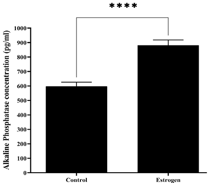
Estrogen administration increases the concentration of ALP enzyme.
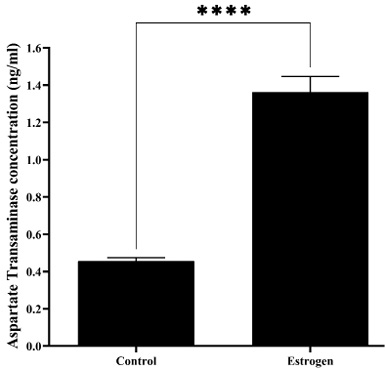
Estrogen administration increases the concentration AST enzyme.

Estrogen administration increases the concentration BUN.

Estrogen administration increases the concentration Cr.
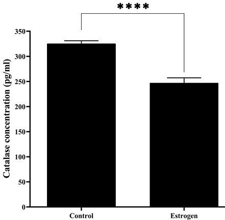
Estrogen administration decreases the concentration of CAT.
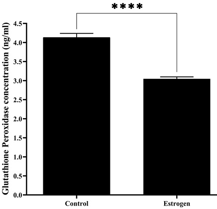
Estrogen administration decreases the concentration of GSH-Px.

Estrogen administration decreases the concentration of SOD.
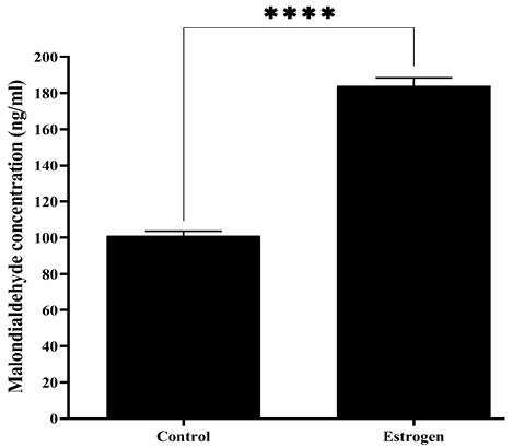
Estrogen administration increases the concentration MDA.

Normal histological structure of liver in control group mice. The prepared section was stained with Hematoxylin and Eosin stain and examined at 10X. The image is represented experimental mice.
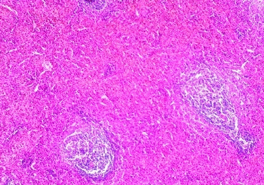
Normal histological structure of spleen in control group mice. The prepared section was stained with Hematoxylin and Eosin stain and examined at 10X. The image is represented experimental mice.

Normal histological structure of kidney in control group mice. The prepared section was stained with Hematoxylin and Eosin stain and examined at 10X. The image is represented experimental mice.

Histopathological changes of liver in mice received estrogen. (A): Sinusoidal dilation (Black arrow) and infiltration of mononuclear cells (Red arrow); (B): Droplets of cytoplasm (Black arrow); (C): Congestion of veins (Black arrow), Amyloidosis (Blue arrow), Sinusoidal space (Red arrow), Degeneration of parenchyma (Yellow arrow); (D): Thrombus (Black arrow), Deposition of fibrin (Blue arrow), Perivascular cuff (Red arrow), Degeneration of parenchyma (Yellow arrow). The sections were stained with Hematoxylin and Eosin stain, examined at 10X, and the images are represented experimental mice.

Histopathological changes of spleen in mice received estrogen. (A): Amyloidosis (Black arrow); (B): Hyperplasia (Black arrow); (C): Hyperplasia (Black arrow), Congestion of blood vessels (Yellow arrow). The sections were stained with Hematoxylin and Eosin stain, examined at 10X, and the images are represented experimental mice.

Histopathological changes of kidney in mice received estrogen. (A): Damaging of proximal tubules (Black arrow) and Inflammatory cells infiltration (Blue arrow); (B): Damaging of venous wall (Black arrow), Venous congestion (Blue arrow), Narrowing of ureter’s lumen (Red arrow); (C): Amyloid degeneration (Black arrow), Amyloid infiltration in veins (Blue arrow), Amyloid infiltration in proximal tubules (Red arrow); (D): Congestion of veins (Black arrow), Atrophy of glomerular tufts (Red arrow), Hypertrophy of proximal tubules (Yellow arrow); (E): Congestion of veins (Black arrow), Parenchymal fibrosis (Blue arrow), Atrophy of glomerular tufts (Red arrow), Hypertrophy of tubular lumen (Yellow arrow); (F): Congestion of veins (Black arrow), Hypertrophy of glomerular space (Red arrow), Hypertrophy of tubular lumen (Yellow arrow). The sections were stained with Hematoxylin and Eosin stain, examined at 10X, and the images are represented experimental mice.
