
Clinical and Macroscopical Evaluation the Effects of Acellular Tunica vaginalis and Acellular Dermal Matrix on Reconstruction of Abdominal Wall Hernia in Bucks


Clinical and Macroscopical Evaluation the Effects of Acellular Tunica vaginalis and Acellular Dermal Matrix on Reconstruction of Abdominal Wall Hernia in Bucks
Anas M. S. Al-Haj Afandi*, Ahmed H. F. AL-Bayati

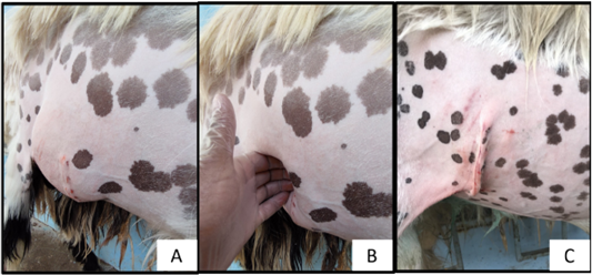
Shows; the detection of ventro-lateral abdominal wall hernia, 30 days post-inducing by the presence of hernial sac (A) and hernial ring (B) and the disappearance of hernia sac directly post-treatment (C).

Macroscopic appearance of implantation sites, 2 weeks post-treatment of BTV treated hernias (A), and caprine ADM treated hernias (B), shows: numerous blood vessels (yellow arrows), deposition of white fibrous connective tissue in BTV more than caprine ADM (black arrows), incorporation of mesh with host tissue (blue arrows).

Macroscopic appearance of implantation site 8 weeks post-treatment of BTV (A), and caprine ADM (B) shows; deposition of white fibrous connective tissue at the center of implantation (black arrows), sprouting blood vessels (yellow arrows), while, good incorporation between sheet and surrounding tissue (blue arrows)(C) and (D).

Macroscopic appearance of implantation site 8 weeks post-treatment of BTV (A), and caprine ADM (B) shows; deposition of white fibrous connective tissue at the center of implantation (black arrows), sprouting blood vessels (yellow arrows)
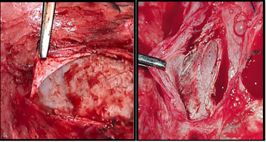
Macroscopic appearance of implantation site 16 week post-treatment of BTV (A) and caprine ADM(B) shows; decrease in thickness of meshes due to degradation (yellow arrows).

