Differential DNA Methylation of Growth Factors in Antlers of Sika Deer and Reindeer
Differential DNA Methylation of Growth Factors in Antlers of Sika Deer and Reindeer
Jian-Cheng Zhai1,2, Sheng-Nan Wang1,3, Qiang-Hui Wang1, Yan-Ling Xia1, Wei-Shi Liu1, Ya-Jie Yin4 and He-Ping Li1*
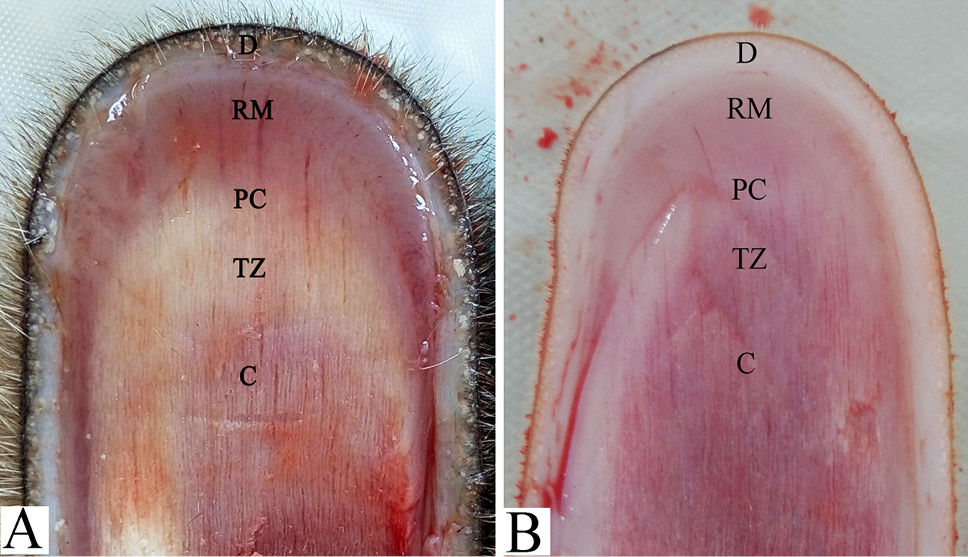
A schematic diagram of the apical structure of reindeer antler (A) and sika deer antler (B). D, dermis; RM, reserve mesenchyme; P, precartilage; TZ, transition zone; C, cartilage.
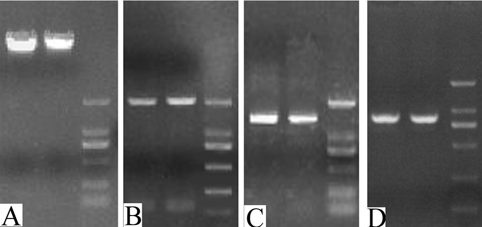
Electrophoresis of genomic DNA and IGF1, KGF and NGF gene promoters’ amplification. A: Genomic DNA of reindeer and sika deer antler reserve mesenchyme. B, C, and D, represent the amplification of reindeer and sika deer IGF1, KGF and NGF gene promoters, respectively.
Methylation status of IGF1 gene in antler mesenchyme of female, male reindeer and sika deer. A: A schematic represents the distribution of the CpG site in the IGF1 gene and the analyzed sequence represents a 666 base pair fragment (positions −50 ~ +615) in the promoter region of IGF1 gene. B: Electrophoresis of BSP products of IGF1. C: IGF1 methylation levels in antler mesenchyme of female, male reindeer and sika deer. TSS, transcription start sites; vertical line, CpG sites; RF, female reindeer antler mesenchyme; RM, male reindeer antler mesenchyme; SM, male sika deer antler mesenchyme; *0.01<P<0.05, **P<0.01.
Methylation status of KGF gene in antler mesenchyme of female, male reindeer and sika deer. A: A schematic represents the distribution of the CpG site in the KGF gene and the analyzed sequence represents a 493 base pair fragment (positions −285 ~ +207) in the promoter region of KGF gene. B: Electrophoresis of BSP products of KGF. C: KGF methylation levels in antler mesenchyme of female, male reindeer and sika deer. For abbreviations and statistical detail, see Figure 3.
Methylation status of NGF gene in antler mesenchyme of female, male reindeer and sika deer. A: A schematic represents the distribution of the CpG site in the NGF gene and the analyzed sequence represents a 468 base pair fragment (positions −388 ~ +109) in the promoter region of NGF gene. B: Electrophoresis of BSP products of NGF. C: NGF methylation levels in antler mesenchyme of female, male reindeer and sika deer. For abbreviations and statistical detail, see Figure 3.


