Distribution and Morphology of Ghrelin-Immunopositive Cells in the Pancreas of the African Ostrich
Distribution and Morphology of Ghrelin-Immunopositive Cells in the Pancreas of the African Ostrich
Jia-xiang Wang1,2,* and Peng Li1,2
Histological structure of the African Ostrich pancreas (HE staining). A, the pancreas; B, the pancreatic parenchyma; C, the pancreatic exocrine portion; D, the pancreatic parenchyma. EV, envelope; PA, pancreatic acinus; PI, islet; BV, blood vessels; ILD, intralobular duct; ICD, intercalary duct; GC, pancreatic gland cells; AC, centroacinar cell; PD, islet cells. (A) Scale bar: 100 μm; (B) scale bar: 50 μm; and (C and D) scale bar: 20 μm.
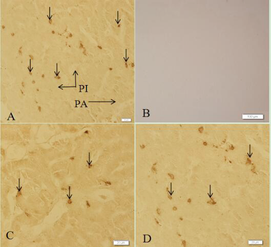
Histological structure of pancreas showing ghrelin-immunopositive cells in the pancreas of the African ostrich (SABC staining). A, a large number of ghrelin cells (arrows) were found within the pancreas; B, microphotograph of the absorption testing in the pancreas; C, ghrelin-ip cells (arrows) amound the Pancreatic acinar cells; D, ghrelin-ip cells (arrows) within the islets. PA, pancreatic acinus; PI, islet. (A, C and D) Scale bar: 20 μm; (B) scale bar: 100 μm.
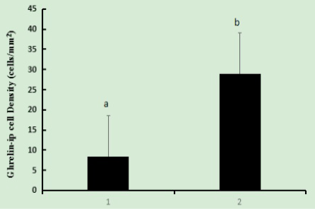
Histogram showing the densities of ghrelin-immunoposive cells (cells/mm2) in the 334 day-old African ostrich pancreas. The number of ghrelin-immunopositive cells were a steady decrease from pancreatic islet to pancreatic acinus. Ghrelin-immunoposive were present throughout the pancreas. a-b, Different letters within the same column indicate significant differences among segments according to Duncan’s multiple range (P≤0.05). 1, pancreatic acinar; 2, islet.


