
Experimental Induction and Control of Cellulitis in Broiler Chickens


Experimental Induction and Control of Cellulitis in Broiler Chickens
Mohamed Mahrous Amer1*, Hanaa Sayed Fedawy2, Hoda Mohamed Mekky2, Khaled Mohamed Elbayoumi2, Ahmed Ali El-Shemy3, Mohamed Abd El-Rahman Bosila2
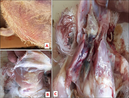
Lesion in broiler chicken s.c infected with S. aureus and /or E. coli showing. A: Thick rough dark colored skin. B: S.c lesion. C: S.c lesion, pericarditis and prehepatitis with subscapular hemorrhage in liver.
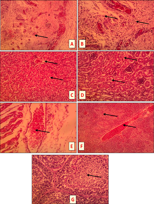
Tissue sections of S. aureus and /or E. coli infected birds at 3 DPI (HandEX, 200) showing the following lesions. A: Subcutis of E. coli infected bird: Severe suppurative inflammation characterized by heterophils and mononuclear cells infiltration in s.c tissues (head of arrow). B: Subcutis of S. aureus and E. coli infected bird: severe suppurative inflammation with marked accumulation of heterophils and mononuclear cells in s.c fatty tissue (head of arrow). C: Liver of S. aureus infected bird: hydropic degeneration of the hepatocytes (head of arrow). D: Kidney of S. aureus + E. coli infected bird: severe hydropic degeneration (head of arrow). E: Subcutis of S. aureus infected bird: subcutaneous hemorrhages (head of arrow). F: Spleen of S. aureus infected bird: congested red bulb and blood vessels with necrotic area (head of arrow). G: Bursa of E. coli and/or S. aureus infected bird: depletion of the lymphoid follicle (head of arrow).
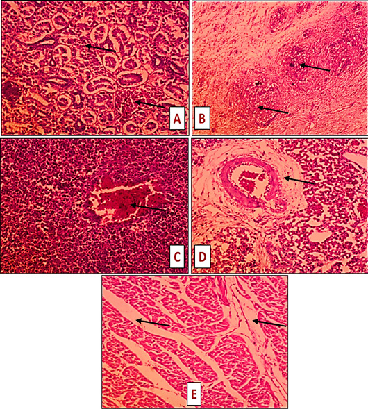
Tissue sections of S. aureus and /or E. coli infected non treated birds (7 DPT) (HandE, X 200) showing. A: E. coli infected bird: kidney hemorrhages accompanied with coagulative necrosis of some tubules (head of arrow). B: E. coli infected bird: s.c severe suppurative inflammation with marked accumulation of heterophils and mononuclear cells in s.c fatty tissue (head of arrow). C: E. coli infected bird: spleen hemorrhages (head of arrow). D: S. aureus infected bird: perivascular edema of the pulmonary artery (head of arrow). E: S. aureus + E. coli infected bird: muscle edema (head of arrow).
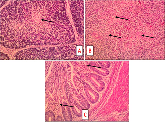
Tissue sections of S. aureus and /or E. coli infected treated birds at 7 DPT (HandE, X 200) showing the following lesions. A: S. aureus + E. coli infected treated bird: bursa slight depletion of the lymphoid follicle (head of arrow). B: S. aureus + E. coli infected treated bird: spleen slight depletion of the lymphoid follicle (head of arrow). C: S. aureus infected treated bird: intestine light inflammation characterized by lymphocytic infiltration of the mucosa (head of arrow).

