Exploring the Protective Efficacy of Lactobacillus acidophilus Against Interleukin-1beta (IL-1β) Expression in Mice Induced with Canine Coronavirus Vaccine
Exploring the Protective Efficacy of Lactobacillus acidophilus Against Interleukin-1beta (IL-1β) Expression in Mice Induced with Canine Coronavirus Vaccine
Rondius Solfaine1, Faisal Fikri2, Salipudin Tasil Maslamama3, Muhammad Thohawi Elziyad Purnama4, Iwan Sahrial Hamid5*
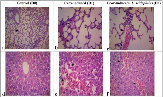
The histological examination of the control group (a, d) demonstrates typical cell structure in the lung tissues and liver. In the D1 group (b), lung tissues exhibit histomorphological feature such as diffuse alveolar cell damages, hyaline tissues deposition, septa alveolar hemorrhages, leukocyte cells infiltration, and alveolar septa proliferation. The D2 group (c) shows neutrophil cell infiltration and plasma fibrin accumulation in lung tissues. In the liver tissues of D1 group (d) shows an accumulation of glycogen in hepatocyte cells, hemorrhage with dilated sinusoids, infiltration of lymphocyte cells. The D2 group (e) present sinusoidal congestion and dilatation (HE, 400x, changes are indicated by arrows).
Positive TNF-α expression, shown as brown aggregates in the intestinal mucosa and submucosa duodenal (b), was significantly higher compared to the non-reactive control group D0 (a). in contrast, TNF-α expression in the isntestinal villi showed a moderate decrese (c) with non-specific IL-1β expression in the epithelia and intestinal crypts (d). Strong IL-1β immunoreaction, depicted as brown aggregates in the intestinal villi’s epithelial and lamina propria area (e), contrasted with the weak IL-1β immunoreaction in the lamina propria area (immunohistochemistry, hematoxylin counterstained, arrows indicate morphological changes).


