
Haematological and Histological Evaluation of Camel Bone as a Potential Orthopaedic Biomaterial


Haematological and Histological Evaluation of Camel Bone as a Potential Orthopaedic Biomaterial
Umar Salisu Ahmad1,2*, Adamu Zoaka Hassan2, Echiobi Gaba Emmanuel2, Munir Ari Sani2, Fatai O. Anafi3, Adamu Abdul Abubakar4,1
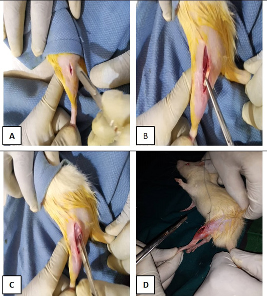
A, incisions site, B and C, positioning of the implants and D muscle and skin losures following implantations.
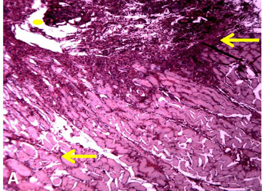
A, Photomicrograph of muscle tissue of rats at 2 weeks post-implantation of K-wire (yellow dote) showing moderate cellular infiltration around the implant (arrows), H and E stain ×100.
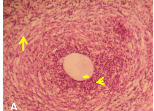
A, Photomicrograph of muscle tissue of rats at 2 weeks post-implantation of camel bone (yellow dote) showing moderate cellular infiltration and capsule formation around the implant (arrow head), H and E stain × 100.

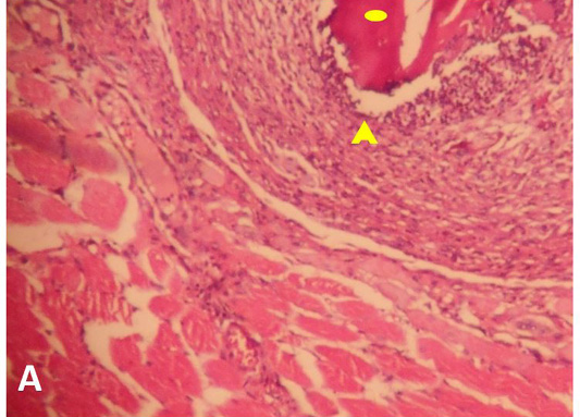
A, Photomicrograph of muscle tissue of rats at 4 weeks post-implantation of camel bone (yellow dote) showing moderate cellular infiltration and capsule formation around the implant (arrow) and mild muscular degeneration (arrow head), H and E stain ×100.
A, Photomicrograph of muscle tissue of rats at 4 weeks post-implantation of K-wire (yellow dote) showing moderate cellular infiltration around the implants (arrow head) and muscular degeneration (arrow), H and E stain ×100.


