
Histological Characterization of Normal Gill Tissue of Oscar Fish and Goldfish


Histological Characterization of Normal Gill Tissue of Oscar Fish and Goldfish
Habeeb M. Alsudani1*, Sura K. Abduljabbar1, Firas A. Alhasson2

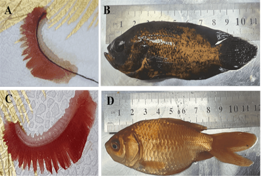
A and B measuring the surface area of gills in Oscar fish. C and D measuring the surface area of gills in Goldfish.
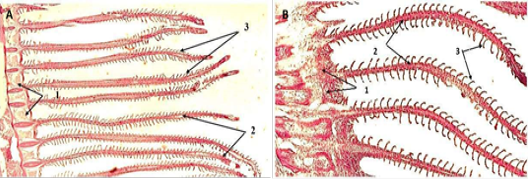
Histological section of Gills in (A) Carassius auratus and (B) Astronotus ocellatus show (1) gill arch, (2) gill filaments, and (3) Lamella (H & E, 10X).
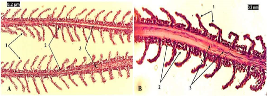
Histological section of gill filaments in (A) Carassius auratus and (B) Astronostus ocellatus show (1) secondary lamellae, (2) filament epithelium, and (3) venous sinus (H & E, 20X).
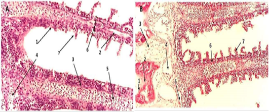
Histological section of Gill arch in (A) Carassius auratus and (B) Astronotus ocellatus show (1) gill raker (2) submucosa, (3) adipose tissue, (4) efferent branchial arterioles, (5) afferent branchial artery, (6) primary lamellae, and (7) secondary lamellae (H & E, 40X).
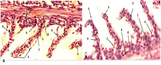
Histological section of Gill arch in (A)Carassius auratus and (B) Astronotus ocellatus show (1) Primary lamella, (2) secondary lamella, (3) epithelial cell, (4) mucous cell, (5) pillar cell, (6) lacuna (capillary lumen), (7) erythrocyte within capillary lumen, (8) chloride cell rodlet cell, (9), and (10) undifferentiated basal cell (H & E, 100X).
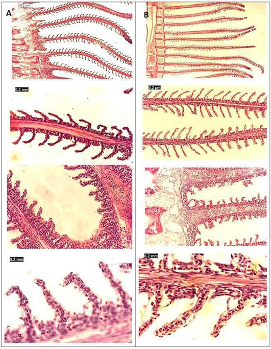
The histological differences between the Astronotus ocellatus (A) and Carassius auratus (B) (H & E).
