
Identification and Antifungal Susceptibility Profile of Aspergillus niger Isolates from Chicken Blood in the Federal Capital Territory, Nigeria


Identification and Antifungal Susceptibility Profile of Aspergillus niger Isolates from Chicken Blood in the Federal Capital Territory, Nigeria
Bridget Maria Jessica Adah1, Samuel Mailafia1, H.O.K Olabode1, James Agbo Ameh1, Martha Echioda Ogbole1, Ebenezer Odey Odey1, Ifeanyi Cajetan Cashmir1, Hakeem Onigbanjo1, Monday Onakpa2, Enid Godwin3, Chinwe Elizabeth Okoli3, Nicodemus Nnabuike Mkpuma4, Oluwa Adikpe Agbonu5, Rabi Rebecca Mairabo6
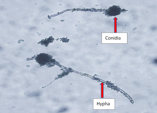
Showing the microscopic appearance of Aspergillus niger using Lacto Phenol Cotton Blue stain on Leica DM 300 binocular compound microscope at x40 Magnification.
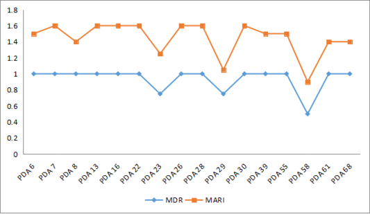
Line graph showing the multiple drug resistance (MDR) and multiple antifungal resistance index (MARI) of the isolates.

Powdery black colonies of Aspergillus niger on PDA.

Reverse white cultural morphology of Aspergillus niger on PDA after 7 days of culture at 250 C with arrow showing the radial fissure.
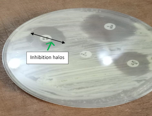
Clear zones of inhibition from antifungal susceptibility testing of Aspergillus niger on PDA using the Disc diffusion method after 20 hours of incubation on PDA.

Antifungal resistance of Aspergillus niger to various antifungal agents on PDA with blue arrows indicating resistant zones while red arrow indicates clear zone of inhibition suggestive of isolate susceptibility to the antifungal agent.

