
Impact of Trichomonas gallinae in the Infected Domestic Pigeon and Antitrichomonal Potentials of Garlic


Impact of Trichomonas gallinae in the Infected Domestic Pigeon and Antitrichomonal Potentials of Garlic
Rand Kamil Abbas, Zahra Sadoon Hadi*, Ahmad Hassan Sahib

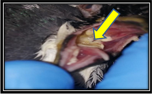
The lesion in buccal cavity of infected pigeon.

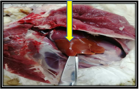
The lesion in liver of infected pigeon.
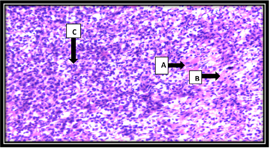
Light photographs of section in the larynx show heavy mixed inflammatory cells infiltration (Lymphocytes, macrophages and neutrophils) (A, B) and vascular proliferation (C).
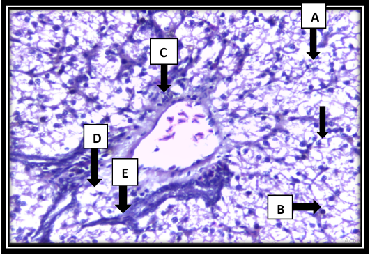
Light photographs of section in the liver shows necrosis of hepatocytes (A, B), bile duct proliferation (C). Mononuclear cell infiltration (mainly lymphocytes) (D), sinusoidal congestion and Kupffer cells hyperplasia.
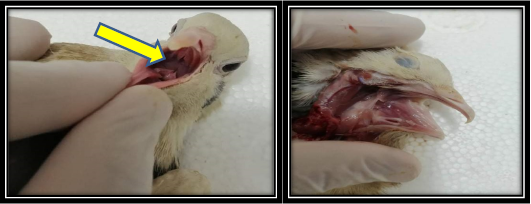
Buccal cavity of pigeon after treatment by garlic showing no caseous mass.

The liver of pigeon after treatment by garlic showing no necrotic foci.
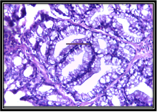
Section in the larynx shows hypertrophy and hyperplasia of mucous secreting gland.
The liver of pigeon after treatment by garlic showing no necrotic foci.
