
Investigation of Cryptosporidium Species Infecting Dogs


Investigation of Cryptosporidium Species Infecting Dogs
Suhad I.J. Al-Asady1*, Mohammad H. Al-Hasnawy2
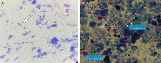
Morphology of Cryptosporidium spp.: A: Oocysts (40X) by using modified Ziehl-Neelsen stain. B: Oocysts (100X) by using modified Ziehl-Neelsen stain with oil immersion and using a digital camera
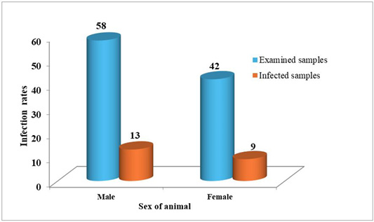
Infection rate of Cryptosporidium spp. in dog according to the sex.

Infection rate of Cryptosporidium spp. in dog according to the age group.
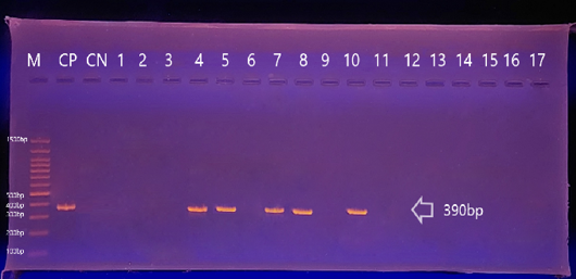
Agarose gel electrophoresis image shows nested PCR product for Cryptosporidium Canis, GP60 gene 390bp in dogs’ samples. Bands were fractionated by electrophoresis on a 1.5% agarose gel, and visualized under U.V. light after staining with red stain. Lane: M (M: 100bp-1500bp ladder). (M=marker) (CP= control positive) (CN= control negative).
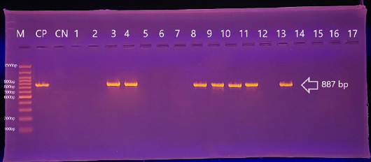
Agarose gel electrophoresis image shows nested PCR product for cryptosporidium parvum, GP60 gene 887bp in dogs samples. Bands were fractionated by electrophoresis on a 1.5% agarose gel, and visualized under U.V. light after staining with red stain. Lane: M (M: 100bp-1500bp ladder). M= marker, CP= control positive, CN= control negative.
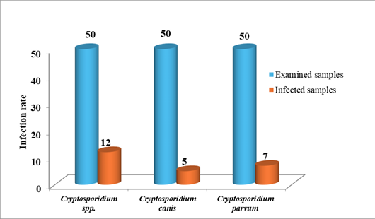
Infections rates of Cryptosporidium species based on the nested PCR.
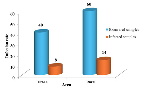
Infection rate of Cryptosporidium spp. in dog according to the area of study.

Phylogenetic tree analysis based on the partial sequence of GP60 gene explains identity between the local isolates of Cryptosporidium canis (A) and Cryptosporidium parvum (B) in dogs and NCBI-BLAST isolates.

