
Investigation of Physiological and Immunohistochemical Changes in Rat Treated with 5-Fluorouracil


Investigation of Physiological and Immunohistochemical Changes in Rat Treated with 5-Fluorouracil
Zainab A. Shehab1*, Assad H. Eissa2, Hind A.A. Alahmed1

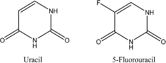
Chemical structure of uracil and 5-fluorouracil.
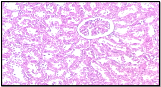
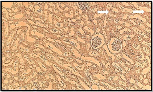
Histological structure of the kidney control group the kidney structure consists of renal tubules that are embedded in the the malpighian corpuscle that is externally surrounded by a connective tissue capsule H and E.
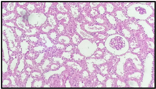
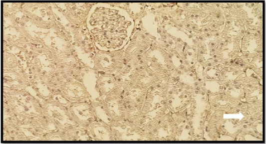
Histological structure of the kidney 5FU treated group showing degeneration, increase in bowman capsular area, hemorrhage, atrophy, necrosis (H and E).
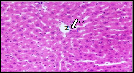
Liver tissues in control rat group stained by (H and E staining) showing the normal architecture of the liver with hepatic lobules around the central vein (CV) and each lobule consisting of hepatic strands of hepatocytes.
Immunohistochemical staining of control group kidney with bcl2, A control group showing moderate immunoreactivity in the cortex region of the kidney.
Immunohistochemical staining of 5FU treated group kidney with Bcl2. A 5FU group showing weak immunoreactivity in the kidney section.
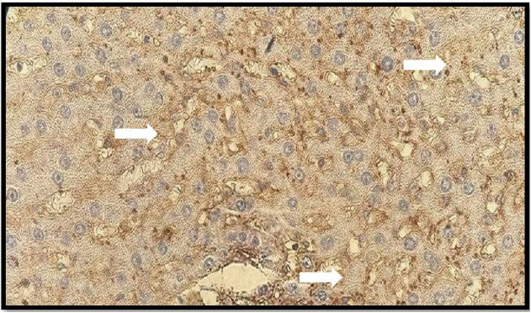
Immunohistochemical staining of liver in control group with Bcl2. A control group showing strong positive reaction.
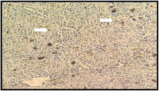
Immunohistochemical staining of liver in 5 FU treated group with Bcl2. A5 FU group showing weak reaction.
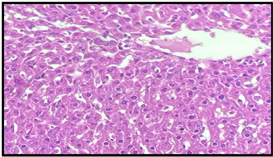
Liver tissues in 5FU treated group stained by (H and E staining). A5FU group showing the leukocytes infiltration, vacuolated cells and severe congestion in central vein (CV).

