
Mareks Disease Virus in Egypt Historical Overview and Current Research Based on the Major MDV-Encoded Oncogene Meq


Mareks Disease Virus in Egypt Historical Overview and Current Research Based on the Major MDV-Encoded Oncogene Meq
Fatma Abdallah1*, Ola Hassnain2, Elsayed Attar3, Haytham Ali3,5, Mohamed Megahed1 and Venugopal Nair4
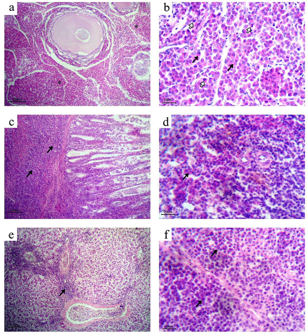
Histopathology of MDV naturally infected birds (HandE stain). a): Ovary. Massive diffuse infiltration with myelocytes (asterisks) with its characteristic eosinophilic cytoplasm; b): Ovary. Myeloid cells proliferation (black arrows) with numerous mitotic figures (white arrows); c): Proventriculus. Diffuse infiltration with neoplastic lymphoid cells both in the lamina propria (arrows) and the proventricular glands; d): Spleen. Mixed population of lymphoid tumour cells and myeloid cells (arrow) adjacent to the central arterioles; e): Liver. Periportal infiltrations with neoplastic lymphoid cells (arrow); f): Bursa of Fabricius. Diffuse infiltrations with pleomorphic neoplastic lymphoid cells (arrows).

Phylogenetic analysis of MDV based on Meq protein amino acid sequences. phylogenetic tree was constructed via multiple alignments of amino acid sequence of Meq protein of 28 reference strains and the eight Egyptian sequences using a distance-based neighbour joining method with bootstrapping (1000) with MEGA6.06 software. Egyptian MDV Sequences of this study and earlier one was indicated using green triangle and red circle, respectively. Clades EUA and NA are abbreviation for Eurasian and North American.

