Molecular Detection of Entamoeba histolytica in Human and Cattle
Molecular Detection of Entamoeba histolytica in Human and Cattle
Mohammed Jawad Kadhim*, Qasim Jawad Amer
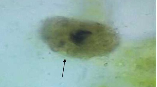
The histolytic cyst stage of E. histolytica, stained with iodine (40X), was isolated from feces.
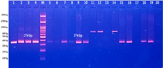
The PCR amplification results of internal transcribed spacer1 gene (ITS1) of Entamoeba histolytica isolated from human stool. Agarose gel picture appears the PCR product bands with molecular weight of 374 bp. (M) refers to (3000 bp) DNA ladder, (1) positive control. (2) Negative control. (3-20) some of PCR results of stool samples.
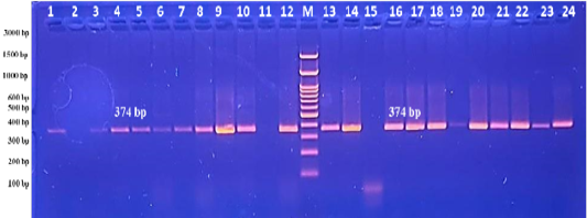
The PCR amplification results of internal transcribed spacer1 gene (ITS1) of Entamoeba histolytica isolated from cattle feces. Agarose gel picture appears the PCR product bands with molecular weight of 374 bp. (M) refers to (3000 bp) DNA ladder, (1) positive control. (2) Negative control. (3-20) some of PCR results of stool samples.


