
Ontogenesis of Rectum in Local Awassi Sheep Fetuses (Ovis aris) During Prenatal Periods


Ontogenesis of Rectum in Local Awassi Sheep Fetuses (Ovis aris) During Prenatal Periods
Ali Mobder Niyf*, Jafar Ghazi Abbas Al-Jebori
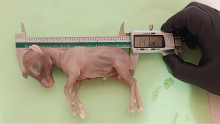
Photograph of sheep fetus using vernier caliper to measure the crown rump length to detect the age of fetus.
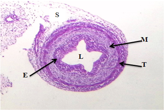
Cross section of rectum at (50-55) days of gestation showing: Epitheliallayer (E), lumen (L), mesenchymallayer (M), tunicamuscularis (T) and serosa (S). (H and E stain, 4X).
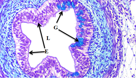
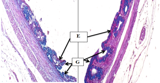
Cross section of rectum at (50-55) days of gestation showing: Epithelial layer (E) stratified columnar cells, Goblet cells (G). (PAS combined Alician blue stain, 10X).
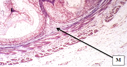
Cross section of rectum at (50-55) days of gestation showing: Undifferentiated mesenchymal layer (M), lumen of rectum (L). (trichrome Masson’s stain, 10X).
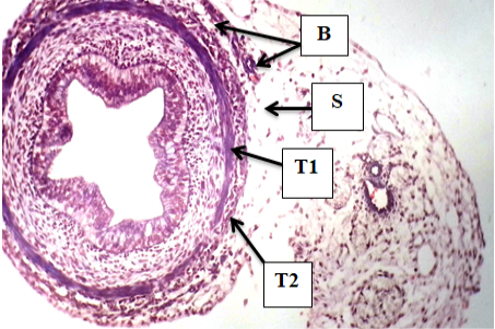
Cross section of rectum at (50-55) days of gestation showing: Inner layer of tunica muscularis (T1), outer layer of tunica muscularis (T2), blood vessels (B).(mason’s trichrome stain, 4X).
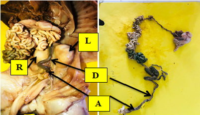
Photograph showing the rectum location in pelvic cavity below the descending colon (D) which extend between left (L) and right (R) kidneys medio- caudally. at 70-75 days of gestation in sheep fetuses.
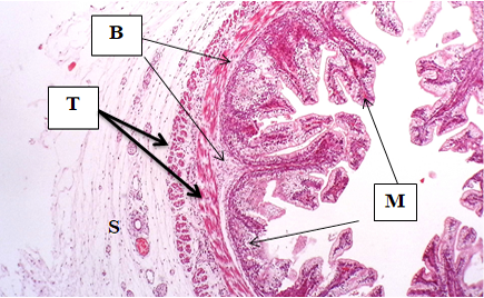
Cross section of rectum at (70-75) days of gestation showing: Tunica mucosa (M), tunica submucosa (B), tunica muscularis (T) and serosa or adventitia (S) (H and E stain, 10X).

Cross section of rectum at (70-75) days of gestation showing: Simple columnar epithelial cells (E) on the apex of villi, stratified epithelial cells on base of villi (E2) and goblet cells (G). (PAS combined Alician blue stain, 10X).
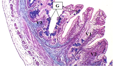
Cross section of rectum at (70-75) days of gestation showing: Rectal villi (V), long villi (V1), middle villi (V2), short villi (V3) and goblet cells (G), (PAS stain, 10X).
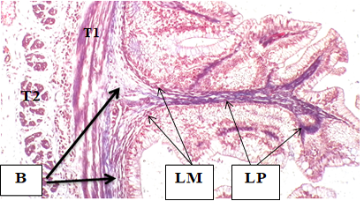
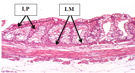
Cross section of rectum at (70-75) days of gestation showing: Lamina propria (LP), lamina muscularis (LM), tunica submucosa, and inner layer (T1), outer layer (T2) of tunica muscularis. (Trichrome massons stain, 10X).
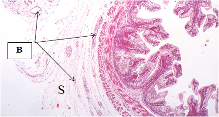
Cross section of rectum at (70-75) days of gestation showing: Tunica serosa (adventitia) (S), blood vessels (B) (H and E satin, 4X).
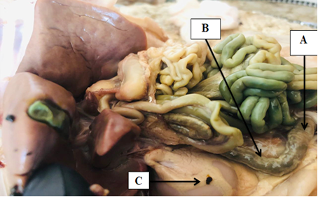
Photograph showing the rectum (A), descending colon (B) and right kidney (C) at 100-105 days of gestation in sheep fetuses.
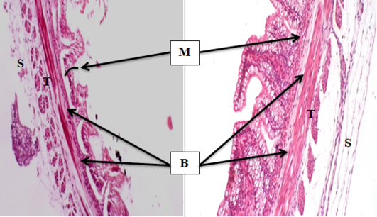
cross section of rectum at (100-105) days (right) and (130-140) days (left) showing: Tunica mucosa (M), tunica submucosa (B), tunica muscularis (T) and tunica serosa (adventitia) (S), (H and E stain,10X).
Cross section of rectum at (100-105) days (left) and (130-140) days (right) showing: Simple columnar epithelial cells (E) and goblet cells(G). (PAS combined Alician blue, 10X).
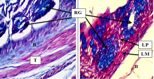
Cross section of rectum at (100-105) days (left) and (130-140) days (right) showing: Rectal glands (RG), lamina propria (LP), lamina muscularis (LM), tunica submucosa (B) and tunica muscularis (T). (PAS combined Alician blue, 40X).
Cross section of rectum at (130-140) days showing: Lamina muscularis (LM) and lamina propria (LP) (H and E stain, 10X).
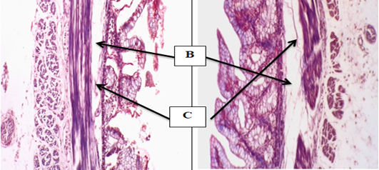
Cross section of rectum at (130-140) days (right) and (100-105) days (left) showing: Tunica submucosa (B) and collagen connectival tissue fibers (C), (Trichrome massons stain, 10X).
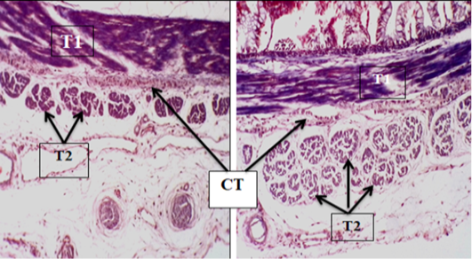
Cross section of rectum at (130-140) days (right) and (100-105) days (left) showing: Inner circular layer of tunica muscularis (T1), outer longitudinal layer of tunica muscularis (T2), separated by layer of connective tissue (CT), (trichrome massons stain, 10X).

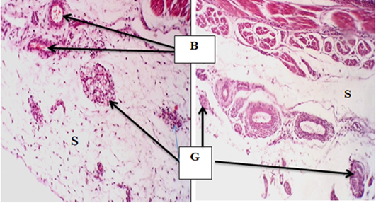
Cross section of rectum at (130-140) days (left) and (100-105) days (right) showing: tunica serosa (adventitia) (S), blood vessels (V) and aggregation of lymphatic tissue (G), (H and E stain, 10X).
