

Protective Effect of Paeoniflorin on Hypoxia Reoxygenation Cardiomyocytes and its Mechanism Based on MAPK Signal Pathway


Protective Effect of Paeoniflorin on Hypoxia Reoxygenation Cardiomyocytes and its Mechanism Based on MAPK Signal Pathway
Shu-Zhi Qin, Wen-Pei Ling, Mei-Fang Yin, Chun-Yu Luo and Cheng-Guo Zhao*
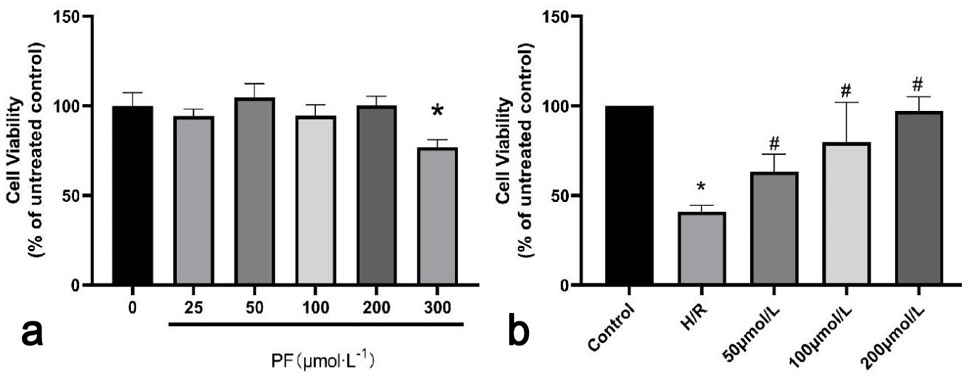
Fig. 1.
Effect of different PF concentrations on H9C2 cell viability (a) under normal oxygen culture and (b) with H/RI treatment.
Notes: x̄ ± s, n = 3; Compared with 0 μmol/L, * P <0.05; Compared with H/R group, # P <0.05.
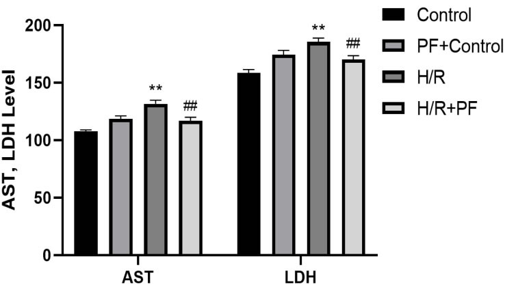
Fig. 2.
Effect of PF on AST and LDH levels in H9C2 cells injured by H/R.
Notes: x̄ ± s, n = 3; ** P <0.01, compared with control group; ## P <0.01, compared with H/R group.
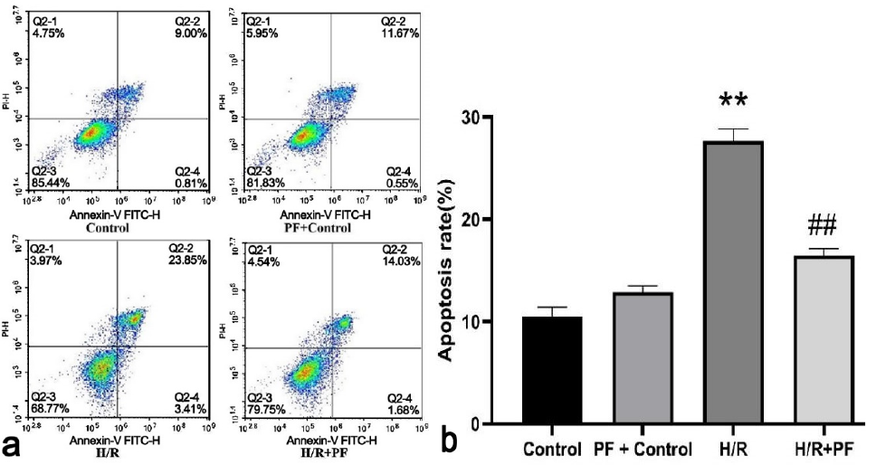
Fig. 3.
Effect of PF on apoptosis rate of H9C2 cells injured by H/R.
Notes: x̄ ± s, n = 3; ** P <0.01, compared with Control group; ## P <0.01, compared with H/R group.
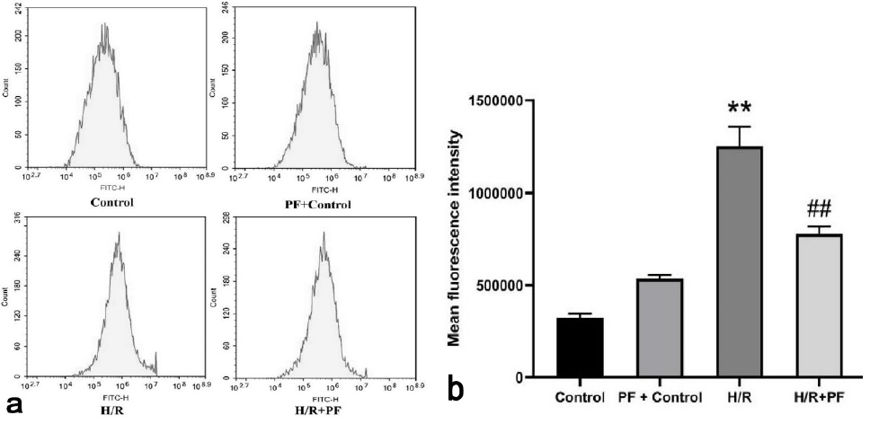
Fig. 4.
Effect of PF on ROS level in H9C2 cells injured by H/R.
Notes: x̄ ± s, n=3; ** P <0.01, compared with Control group; ## P <0.01, compared with H/R group.
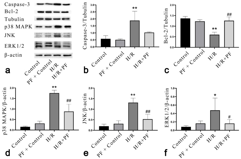
Fig. 5.
Representative picture of Caspase-3, Bcl-2, p38 MAPK, ERK1/2, and JNK in H9C2 cell of each group (a), and Effect of PF on the expression of Caspase-3 (b), Bcl-2 proteins (c), p38 MAPK (d), JNK (e), and ERK1/2 (f) proteins in H9C2 cells.
Notes: x̄ ± s, n=3; * P <0.05, ** P <0.01, compared with control group; # P <0.05, ## P <0.01, compared with H/R group.
February 2024
Pakistan J. Zool., Vol. 56, Iss. 1, pp. 01-501

