Protective Mechanism of Velvet Antler Polypeptide on Myocardial Ischemia-Reperfusion Injury in Rats
Ying Chen1, Chao-Zheng Li2, Zhun Yu3, Jia Zhou4, Yan Xu4, Zhe Lin4, He Lin4* and Xiao-Wei Huang4*
1Department of Cardiology, the Affiliated Hospital of Changchun University of Chinese Medicine, Changchun 130021, China
2College of Chinese Medicine, Changchun University of Chinese Medicine, Changchun 130117, China
3Department of Cardiology, The Third Affiliated Hospital of Changchun University of Chinese Medicine, Changchun 130117, China
4School of Pharmacy, Changchun University of Chinese Medicine, Changchun 130117, China
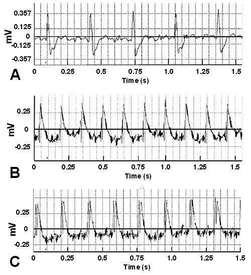
Fig. 1.
ECG changes of rats under treatment in each group. A, normal rats; B, ligation ischemia; C, reperfusion 30 min after ligation ischemia.
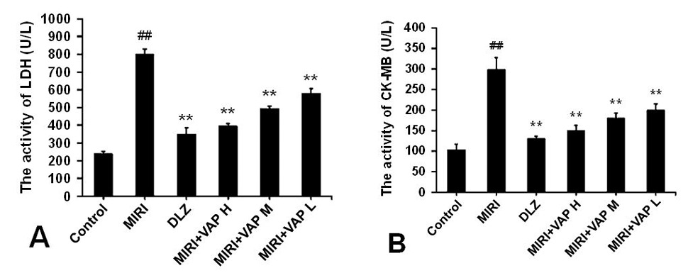
Fig. 2.
LDH (A) and CK-MB (B) activities in serum of each group.
Note: ## p<0.01, compared with control group; ** p<0.01, compared with MIRI group.
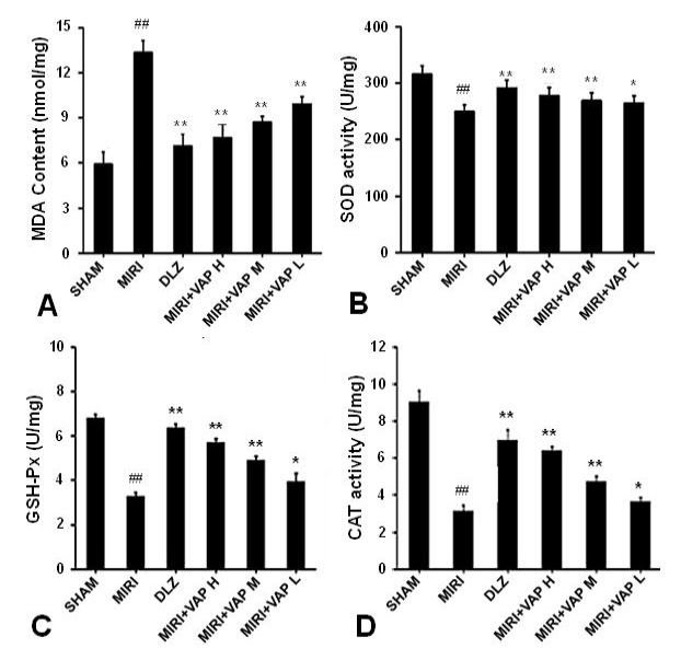
Fig. 3.
MDA (A), SOD (B), GSH-Px (C) and CAT (D) activities in myocardial tissue.
Note: Compared with the control group, # p<0.05, ## p<0.01; Compared with MIRI group,* p<0.05, ** p<0.01.
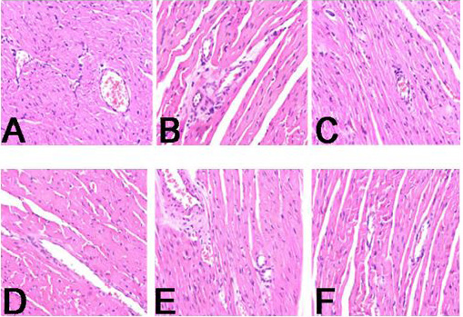
Fig. 4.
H and E staining of myocardial tissue in each group (×300). A, Control group; B, MIRI group; C, DLZ group; D, MIRI + VAP-H group; E, MIRI + VAP-M group; F, MIRI + VAP-L group.
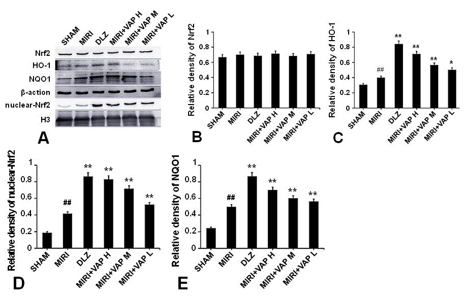
Fig. 5.
Effects of VAP on the expression of Nrf2 (A, B, D), HO-1 (A, C), NQO1 (A, E) in MIRI rats by Western blotting.
Note: Compared with Control group, ## p<0.01; Compared with MIRI group, * p<0.05, ** p<0.01.


