

Tetrandrine Induces Apoptosis in HepG2 by Modulating Hippo Signalling Pathway


Tetrandrine Induces Apoptosis in HepG2 by Modulating Hippo Signalling Pathway
Fuling Wang1,2, Changlin Yue2, Hong Li2, Wenjing Yu1, Wenlan Li1* and Guosong Xin1*
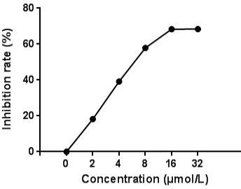
Inhibitory effect of TET treatment on cell proliferation of 24 h old HepG2 cells.
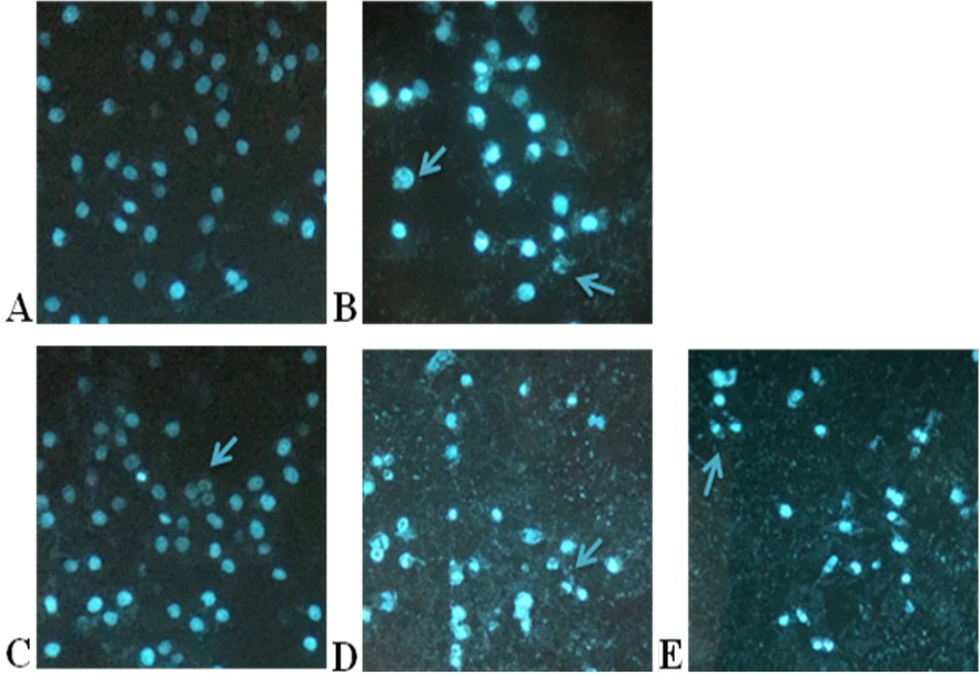
Fluorescent micrographs showing TET induced morphological changes in HepG2 cells visualized after Hoechst 33258 staining (200× magnification): (A) Control; (B) HCPT (7 μmol/L); (C) TET (3.75 μmol/L); (D) TET (7.5 μmol/L); and (E) TET (15 μmol/L).
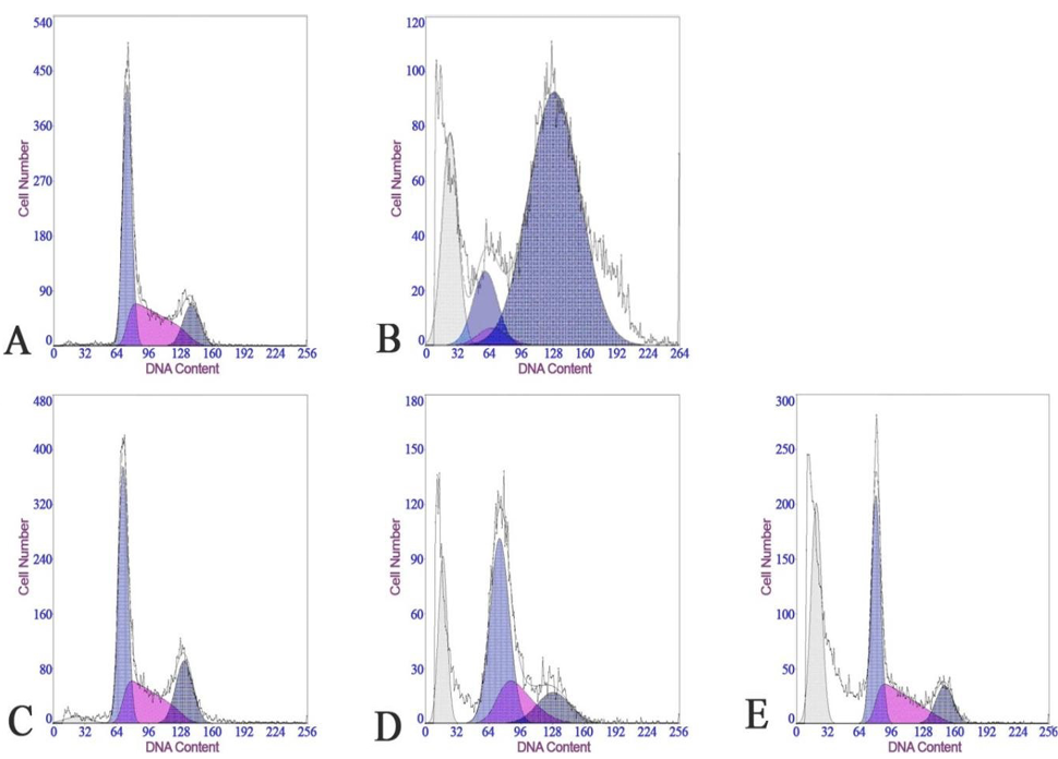
Analysis of apoptosis of HepG2 cells upon treatment with TET or HCPT followed by PI staining, using a flow cytometer (A) Control; (B) HCPT (7 μmol/L); (C) TET (3.75 μmol/L); (D) TET (7.5 μmol/L); and (E) TET (15 μmol/L).
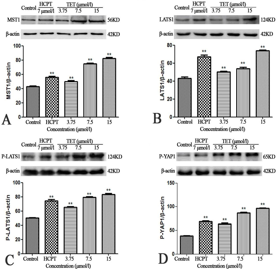
Protein expression profile after 48 h TET treatment (3.75, 7.5, or 15 μmol/L) HepG2 cells. Western blots represent (A) MST1, (B) LATS1, (C) P-LATS1, and (D) P-YAP1. (**P<0.01, vs control).
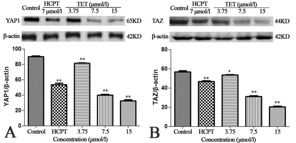
Expression profile proteins participating in Hippo signalling pathway in HepG2 cells upon 48 h TET treatment (3.75, 7.5, or 15 μmol/L). Western blots represent (A) YAP1 and (B) TAZ. (**P<0.01, *P<0.05).

