A Novel Gap Junction Alpha 8 (GJA8) Mutation Associated with a Congenital Cataract Patient in Pakistan
A Novel Gap Junction Alpha 8 (GJA8) Mutation Associated with a Congenital Cataract Patient in Pakistan
Ayesha Zahid,Ammara Muazzam, Sidra Mustafa, Saba Irshad*,Malik Siddique Mahmood and Rehman Shahzad
SSCP banding pattern of PCR products of GJA8 exon 2 of patient samples affected 10 to affected 17, with primer set 4. M, 1Kb DNA ladder; Lane 1, A10; Lane 2, A11; Lane 3, A12; Lane 4, A13; Lane 5, A14; Lane 6, A14; Lane 7, A15; Lane 8, A16; Lane 9, A17. White arrowheads indicate an extra band in Lane 5 and 6.
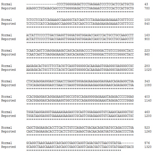
Sequence alignment of reported sequence of GJA8 exon 2 retrieved from NCBI (GenBank NG_016242.1) and normal sequence. Star indicates the matches.
Sequence Chromatogram of patient sample A14, at reference position 330 to 340. Red arrowhead indicates the position of mutation.
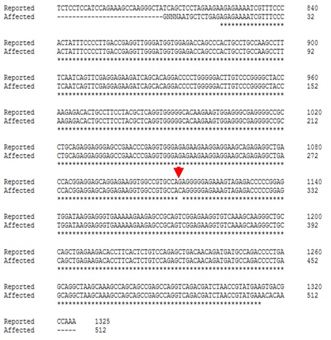
Sequence alignment of Patient sample with the reported sequence of GJA8 exon 2 submitted in NCBI (GenBank KY556641) and normal sequence retrieved from NCBI (GenBank NG_016242.1). Star indicates the matches. Red arrowhead indicates the substitution in coding region of GJA8 exon 2 at nucleotide 1104.
Amino acid sequence alignment of the patient sample with reported sequence. Difference is highlighted in yellow color which indicates the substitution of glutamic acid to glutamine at codon position 368.
Secondary structure of wild type GJA8 from amino acid 360 to 380 by I-Tasser. In wild type protein glutamic acid at position 368 as indicated by arrow head is involved in coil formation with a total conf. score of 7. Greater score indicates maximum accuracy of this secondary structure.
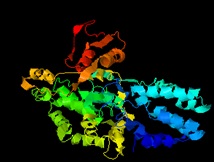
3-D structure of wild type GJA8 proposed by I-Tasser with reference to 10 most significant threading templates selected on the basis of their Z-score, with a total quality factor of 89.880 calculated by ERRAT.
Ramachandran plot of wild type GJA8 protein, showing most of the amino acid residues fall in the permitted regions, whereas few fall in the prohibited regions of plot.
Superimposed 3-D structure of wild type and mutant GJA8 proteins proposed by SWISS MODEL. Green arrow indicates glutamic acid at position 368 in wild type protein whereas red arrow indicate the position of glutamine in mutant GJA8 protein.


