

Alpha-2-Macroglobulin and Alpha-2-HS Glycoprotein are Potential Markers of Renal Cell Carcinoma: An Insight from Proteome Profile of Cancer Tissues


Alpha-2-Macroglobulin and Alpha-2-HS Glycoprotein are Potential Markers of Renal Cell Carcinoma: An Insight from Proteome Profile of Cancer Tissues
Safa Akhtar1,3, Shahzadi Noreen2, Anna E. Lokshin3 and Muhammad Waheed Akhtar1*
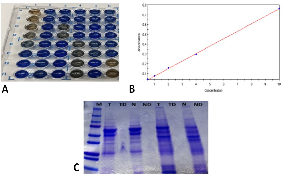
(A) Bradford plate assay. (B) Estimation of protein concentration of tryptic digested and non digested lysates by microplate quantification assay. (C) SDS-PAGE analysis of tryptic digests and non digested tissue lysates M symbolizes the ladder T and TD represent tumor and tumor digest, respectively whereas N and ND symbolize Normal and normal digest.
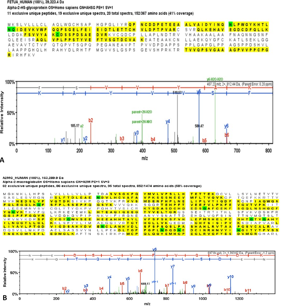
Mass spectrometric analysis of proteins with 99% Minimum 2 Min # Peptides 0.9% Decoy FDR; Spectra at 85.0 % Minimum 0.07% Decoy FDR for (A)- Alpha-2 HS glycoprotein (FETUA), (B)- Alpha-2 macroglobulin.
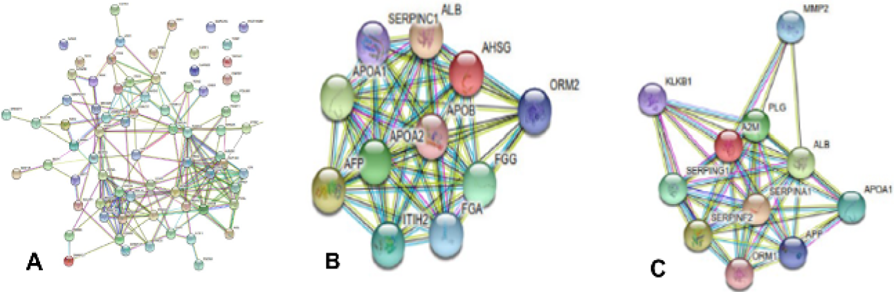
(A) STRING analysis of differentially expressed proteins that were identified through LC-MS/MS in RCC. UniProt ID of 78 proteins were added in the category of multiple protein. The viewed evidence was shown in different coloured lines to indicate type of evidence in order to support each interaction. STRING analysis showed more interaction among set of protein than expected indicating that proteins were partially connected biologically. Protein interaction maps of B)- AHSG (FetuA).and C)- A2M.

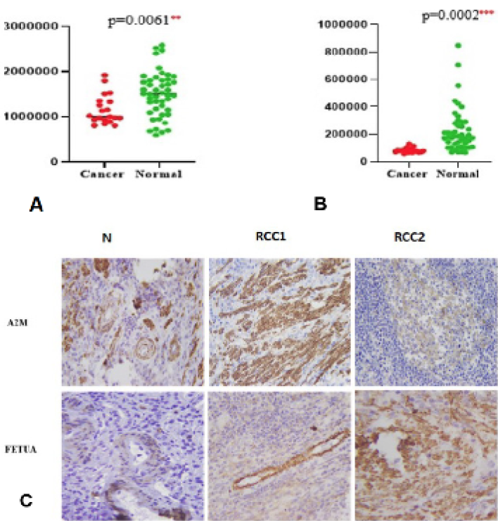
Dot plots of Luminex for A2M (A) and FetuA (B). C, IHC analysis of upregulated proteins A2M and FetuA in RCC and normal tissue.

