

CPEB3 Targets E-Cadherin mRNAs in a Post-Transcriptional Regulation Manner and Inhibits the Invasiveness of Ovarian Cancer Cells


CPEB3 Targets E-Cadherin mRNAs in a Post-Transcriptional Regulation Manner and Inhibits the Invasiveness of Ovarian Cancer Cells
Yanhua Zhang1, Hui Li1, Xinya Li1, Yong Zhang1, Rongqing Li1* and Chen Qing2*
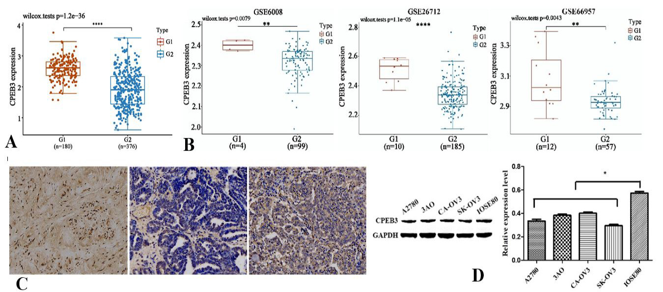
Fig. 1.
The expression of CPEB3 is reduced in ovarian cancer.
A, CPEB3 expression in ovarian cancer (OC) tissues (G2) from the TCGA database was significantly lower than that in normal ovarian samples (G1) from GTEx. B, CPEB3 expression in OC was significantly lower in three GEO datasets than that in normal ovarian tissue. C, Immunohistochemical (IHC) analysis of OC tissue chip showed that 75 OC cases were positive and 5 cases were negatively expressed, resulting in a positive rate of 75/80 (93.6%). In normal ovarian tissue, 7 cases were positive and 0 cases were negative, with a positive rate of 7/7 (100%). D, Western blot was used to detect the expression level of CPEB3 protein in various OC cell lines.
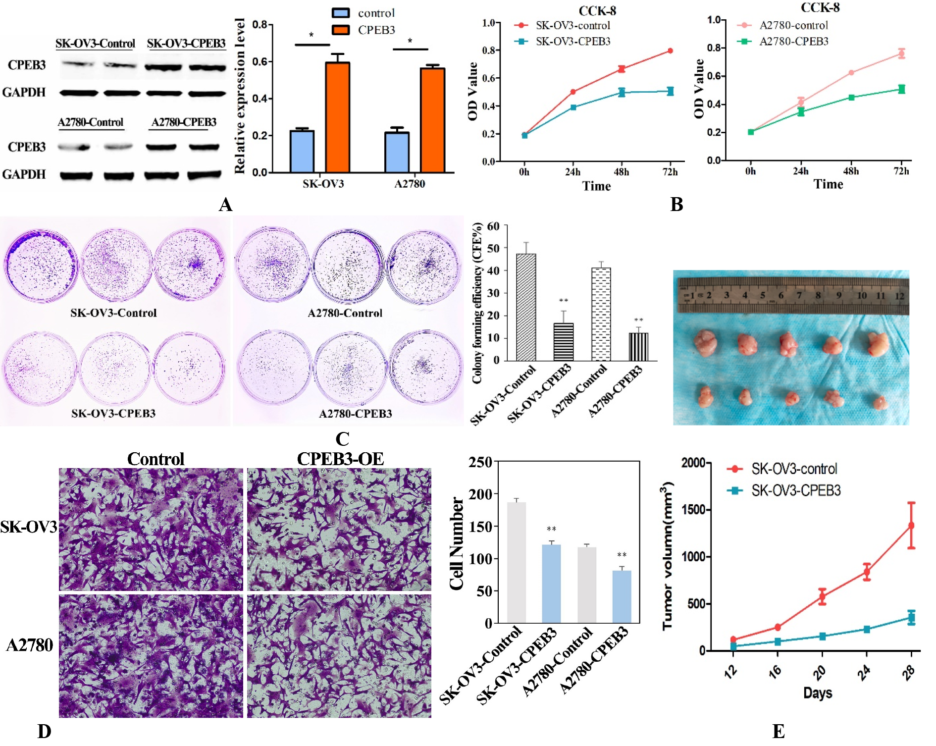
Fig. 2.
CPEB3 significantly inhibited the ability of proliferation and invasion of ovarian cancer in vitro and in vivo.
A, Construction of CPEB3-stabilized OC cell lines using lentiviral vectors in SK-OV3 and A2780 cells. B, CPEB3 overexpression resulted in a reduction in OC cell proliferation through a CCK-8 assay. C, Clonogenic growth experiments revealed that CPEB3 overexpression significantly reduced the clonal formation ability of OC cells. D, In the transwell experiment, the ability of OC cells overexpressing CPEB3 to penetrate cell membranes was significantly weakened. E, In vivo experiments involving nude mice subcutaneously inoculated with SK-OV3 cells overexpressing CPEB3 showed a significant reduction in tumor volume compared to the control group.

Fig. 3.
Functional enrichment analysis and expression analysis of key genes in the CPEB3 regulatory network of ovarian cancer.
A, B, GO terms and KEGG pathway enriched in differentially expressed CPEB3-realated genes in ovarian cancer. C, Immunohistochemical analysis showing the expression of E-cadherin, EGFR and BCL2 in ovarian cancer tissue chips (Bars represent 20 µm). D, Western blotting showing the expression of related proteins in SK-OV3 and A2780 cell lines overexpressing CPEB3 (CON: blank control group, and OE: overexpression CPEB3; p <0.01, compared to control group). E, Western blotting analysis of related proteins in tumor tissues from nude mice overexpressing SK-OV3-CPEB3 (p <0.05, compared to control group).
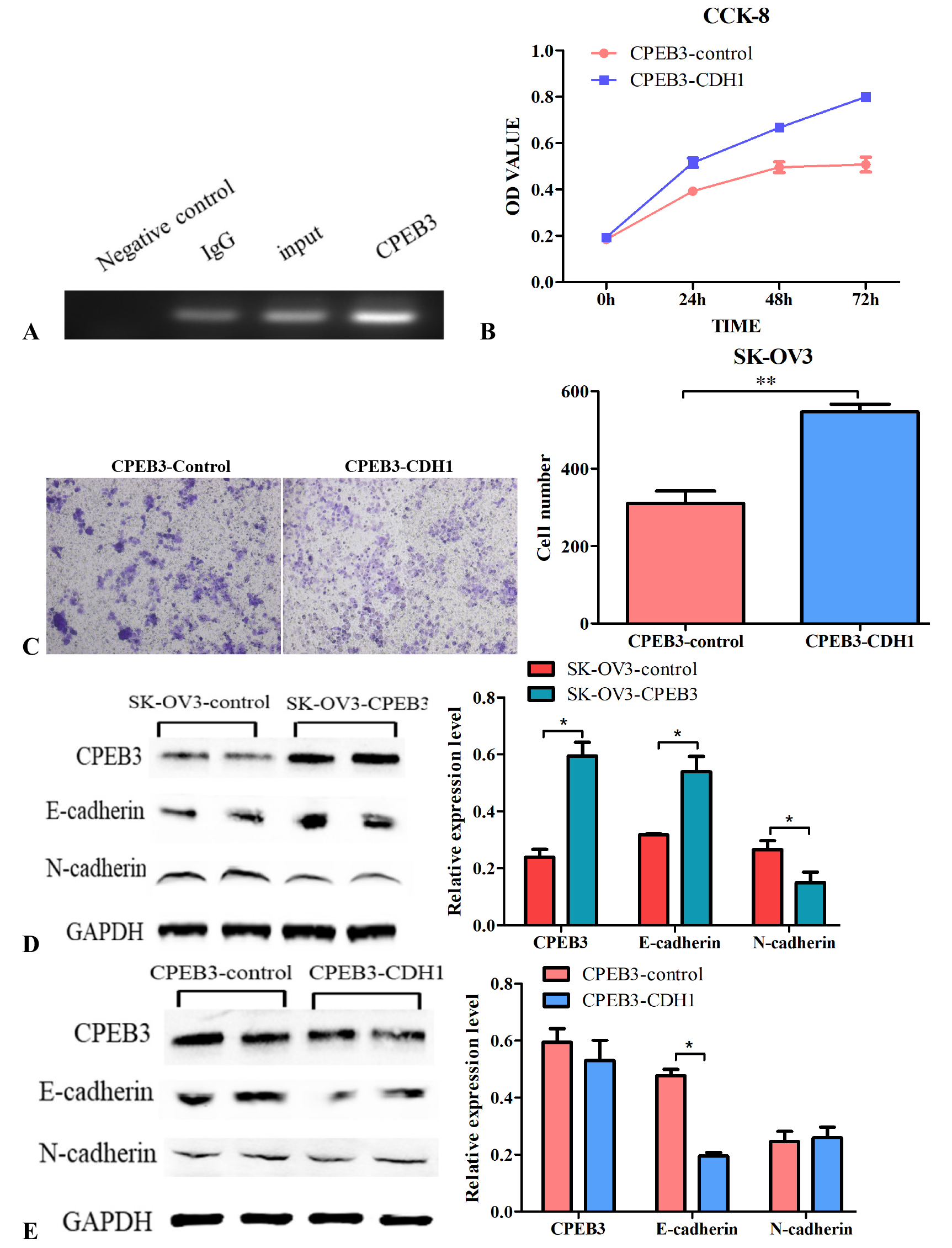
Fig. 4.
CPEB3 inhibits invasion by regulating E-cadherin in ovarian cancer.
A, RIP experiment was carried out to verify CPEB3 directly interact with E-cadherin in ovarian cancer cell line SK-OV3. B, C, after interfering CDH1 with siRNA in SK-OV3 ovarian cancer cells expressing CPEB3, cell proliferation and invasion abilities were detected using CCK-8 assay and Transwell assay, respectively. D, Western blotting showing the changes in E-cadherin and N-cadherin expression levels after overexpression of CPEB3 in SK-OV3 cells. E, Western blotting displaying E-cadherin and N-cadherin expression levels after interfering CDH1 with siRNA in SK-OV3 ovarian cancer cells expressing CPEB3.

November 2024
Pakistan J. Zool., Vol. 56
