

Epidemiological Study of Lumpy Skin Disease Outbreaks in Egypt Based on Viral Isolation and Molecular Detection


Epidemiological Study of Lumpy Skin Disease Outbreaks in Egypt Based on Viral Isolation and Molecular Detection
Hend E.M. Elsheikh1*, Mamdouh F. El-Mekkawi1, A.A. Abou-Zaid1 and
Amal M. Abd El Raof2
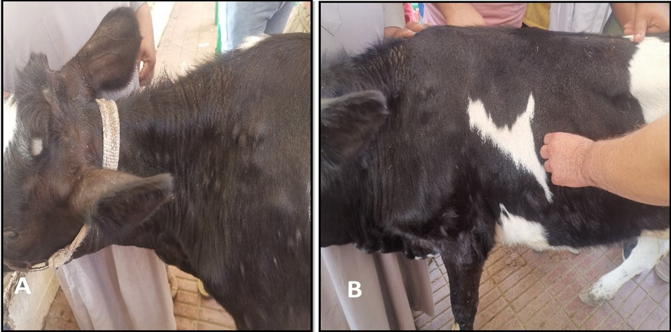
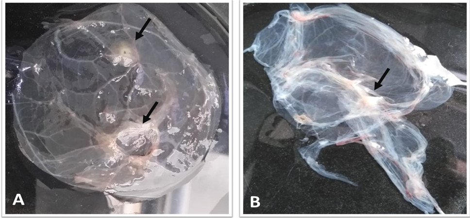
Mild form of LSDV revealed few numbers of closed nodules as shown in A and B.
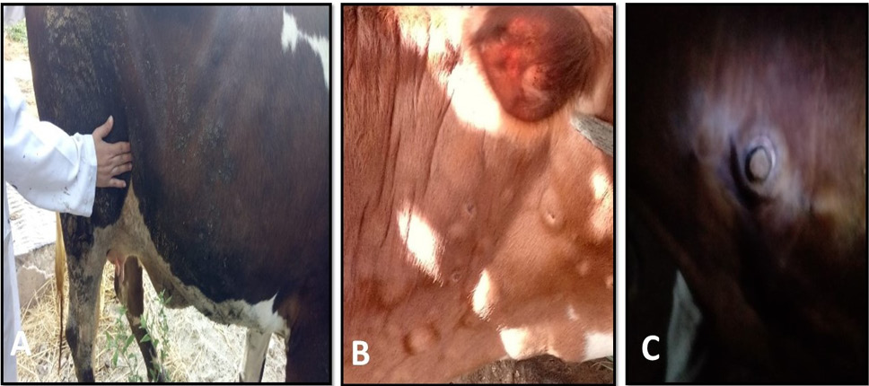
Moderate form of LSDV. (A) Swollen prefemoral lymph node. (B, C) distinctive feature lesions of LSDV sit fast.
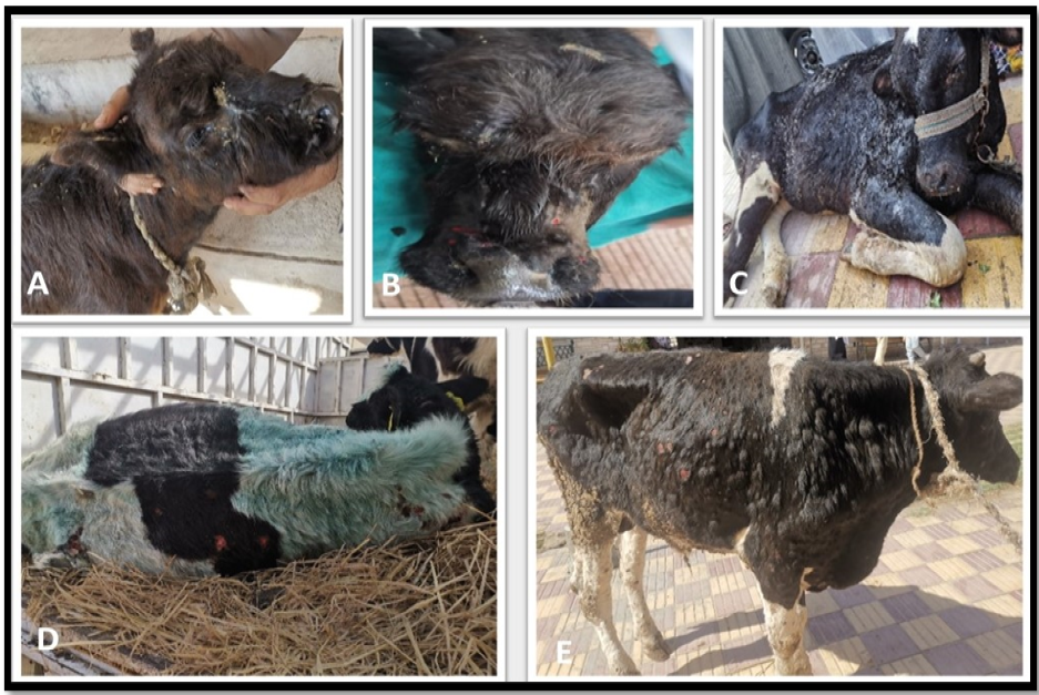
Severe form of LSDV. (A) Diseased calf showed, extreme dyspnea, nasal, ocular discharge, and respiratory manifestations. (B) Nodules present on the muzzle, as well as within the nasal and buccal mucosa. (C) Edema in legs and brisket and infected calf unable to stand. (D, E) The cutaneous lesion coalesced and large area of skin is sloughed creating deep ulcers.
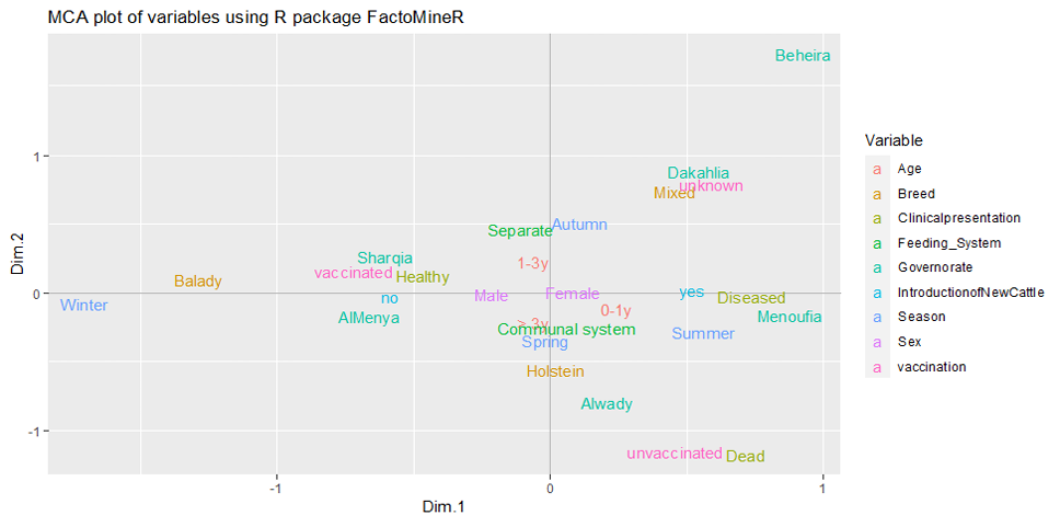
Two-dimensional plot of multiple correspondence analyses representing relationship between predictor’s categories.
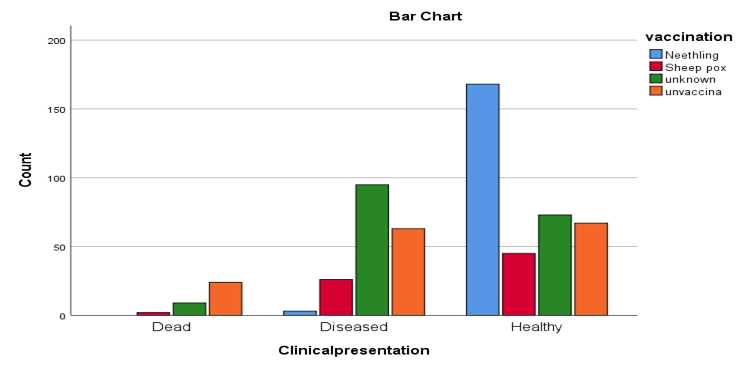
Relationship between vaccination type and clinical case of LSDV: Neethling strain vaccine did not show any deaths of LSD, and the lower number of diseased animals compared by sheep pox vaccine. Unvaccinated and animals with unknown condition of vaccination recorded the highest diseases and deaths of LSD.

