Histopathological Effect of Traces for Uranium on Cow Kidneys
Histopathological Effect of Traces for Uranium on Cow Kidneys
Oula E. Hadi*, E.H. Altaee
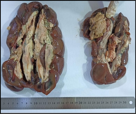
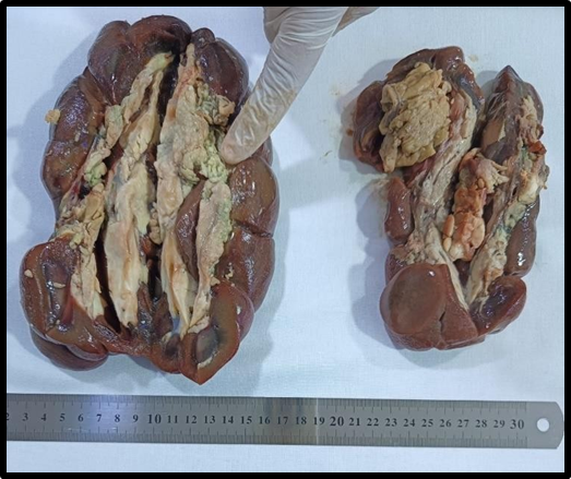
Clear cell carcinoma.
Sarcomatoied renal cell carcinoma.
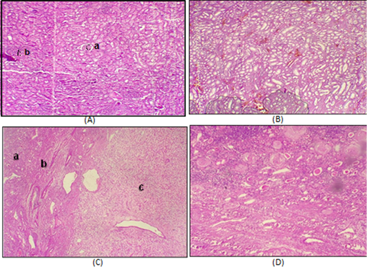
A: Normal Kidney tissue (a) glomerulus (b) renal tubules 4X, B: Nephritis of Kidney with inflammatory cell aggregation in low concentration of uranium 4X. C: (a) Normal kidney tissue. (b) area of fibrosis. (c) renal cell carcinoma 4X, D: Abnormal kidney with sclerosis of glomeruli renal tubules atrophy with collection of protein material in renal tubules in low concentration of uranium 4X.
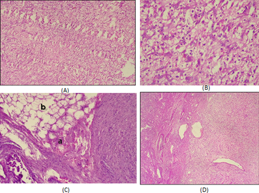
A: Clear cell carcinoma in kidney in high concentration of uranium 4X. B: renal cell carcinoma (a) clear cell cytoplasm with minimal pleomorphism (not high grade with thin (b) fibrovascular stroma and (c)infiltration of inflammatory cells in high concentration of uranium 40X. C: Fatty tissue invaded by renal cell carcinoma (a) tumor cell (b) fatty cell 4X. D: Renal cell carcinoma with sarcomatoied component (differentiation cell int connective tissue tumor sarcoma like) 4X.
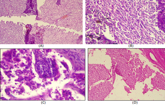
A: (a) clear cell carcinoma. (b) sarcomatoied carcinoma. B: Sarcomatoied renal cell carcinoma with (a) spindle cell with (b)hyperchromatic nuclei 10X. C: Sarcomatoied renal cell carcinoma with spindle cell with hyperchromatic nuclei 40X. D: Clear cell carcinoma of kidney in high concentration of uranium 4X.
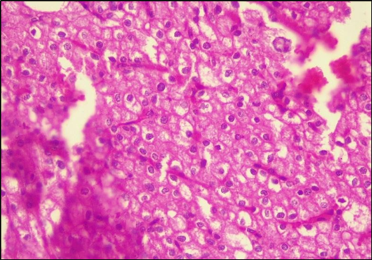
Clear cell carcinoma of kidney in high concentration of uranium 40X.


