
Histophysiological Effect of Rosuvastatin on the Kidney in Male Albino Rats


Histophysiological Effect of Rosuvastatin on the Kidney in Male Albino Rats
Haneen Imad Al-Sultani1*, Ahmed Obaid Hussain1, Hazar Shakir Saleh2


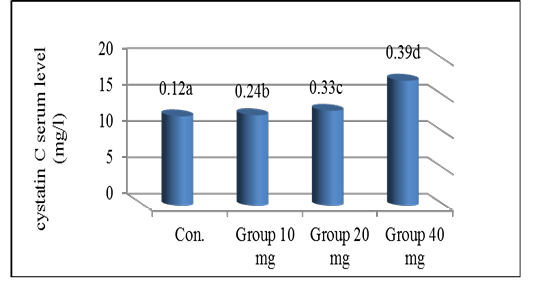
The mean (n=8) serum cystatin C level (mg/l) in the study groups.
a-d Different letters indicate standard deviation at (P≤0.05)

The mean (n=8) serum vitamin D3 level (ng/ml) in the study groups.
a-d Different letters indicate standard deviation (P≤0.05).

Photomicrograph of rat kidney of the control group showing: kidney normal histology, stained with H&E, Scale bar=100 µm.

Photomicrograph of rat kidney of 10 gm group showing, (A): hemorrhage (yellow arrow), degeneration in the epithelial cells (black arrow), (B): irregularity in the normal form of the renal glomerulus (red arrow), stained with H&E, (A) Scale bar=100 µm, (B) Scale bar=50 µm.

Photomicrograph of rat kidney of 20 gm group showing, (A): glomerulus damage (black arrow), necrosis (blue arrow), dilation of the renal tubules (yellow arrow), lymphocytic infiltration (red arrow), (B): congestion (yellow arrow), hemorrhage (black arrow), fatty changes (red arrow) stained with H&E, Scale bar=100 µm.
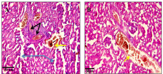
Photomicrograph of rat kidney of 40 gm group showing: sever congestion (black arrow), sever hemorrhage (yellow arrow), sever glomerular degeneration (blue arrow), stained with H&E, Scale bar=50 µm.
The mean (n=8) serum cystatin C level (mg/l) in the study groups.
a-d Different letters indicate standard deviation at (P≤0.05)
