
In Vivo Anti-Trypanosomal Activity of Basil Extract on Trypanosoma evansi


In Vivo Anti-Trypanosomal Activity of Basil Extract on Trypanosoma evansi
Esam A Razin1, Hassan Sobhy2, Tarek R. AboElnaga1, Asmaa A. Darwish1*, Rasha S. Mohammed1
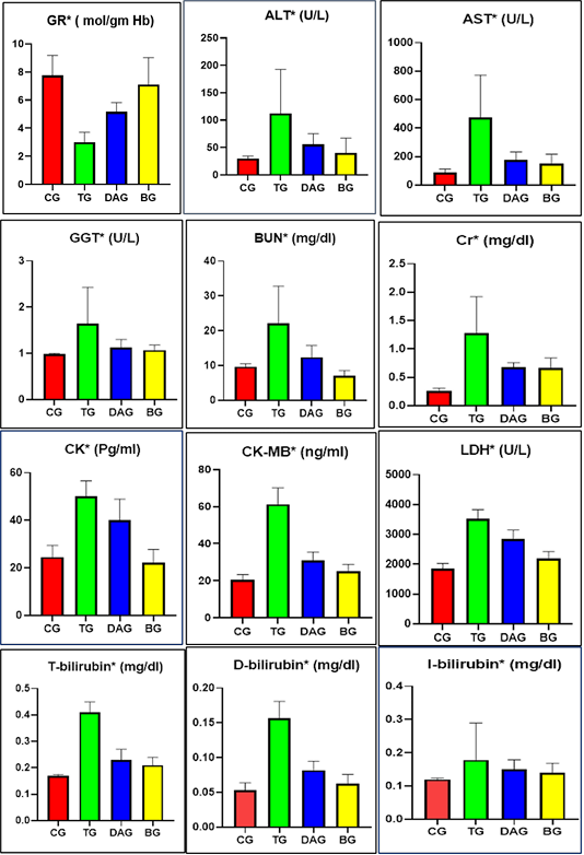
Comparison of oxidative stress and organs function tests between the studied groups, differences between groups were considered significant when P ˂ 0.05 and indicated by (*).
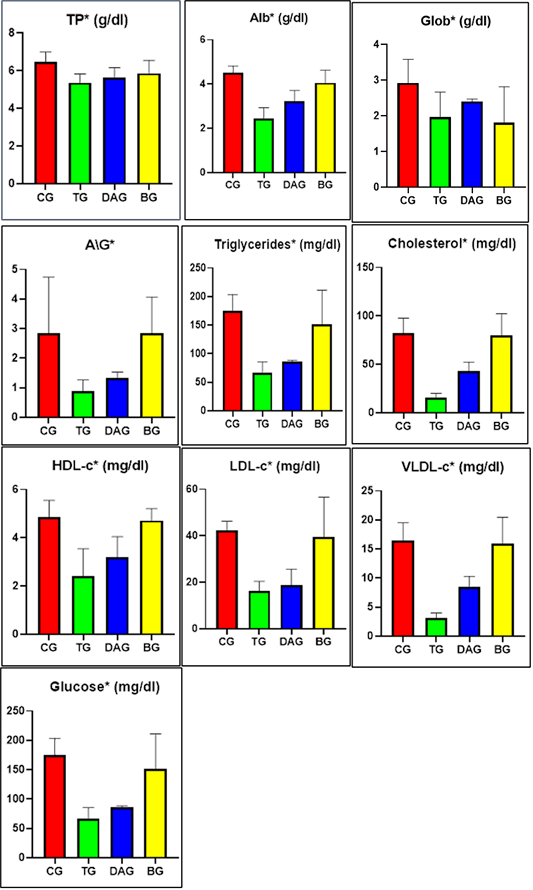
Comparison of glucose, protein, and lipid profiles between the studied groups, differences between groups were considered significant when P ˂ 0.05, and indicated by (*).
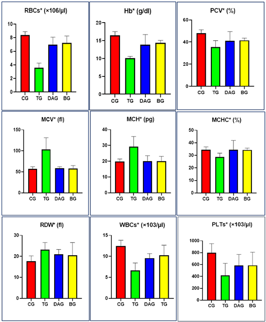
Comparison of hematological parameters between the studied groups, differences between groups considered significant when P ˂ 0.05 and indicated by (*).
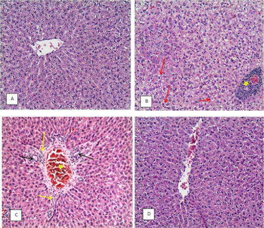
(H&E X20). A: liver of CG, B: liver of TG showing congestion, area of coagulative necrosis with inflammatory cell infiltration (star) and vacuolar degeneration of hepatocytes (red arrow), C: liver of DAG showing congestion and edema (star), infiltration of inflammatory cells (yellow arrow), newly formed bile duct (black arrow) and dilatation of hepatic sinusoids, D: liver of BG more or less to normal with slight congestion.
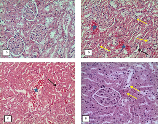
(H&E X20), A: kidney of CG, B: kidney of TG showing congestion (star), tubular necrosis (yellow arrow) and increase space of Bowman’s capsule (black arrow), C: kidney of DAG showing congestion and edema (arrow), necrosis of tubules and atrophy of glomeruli (black arrow), D: kidney of BG more or less to normal with slight congestion and necrosis of tubules (yellow arrow).
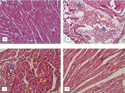
(H&E X20), A: heart of CG, B: heart of TG showing large area of hemorrhage and edema, myocardiolysis (star) and necrosis of muscle (black arrow), C: heart of DAG showing myocardiolysis, hemorrhage and edema (star), swelling of muscles and muscle necrosis (yellow arrow), D: heart of BG more or less to normal with slight myocardiolysis.
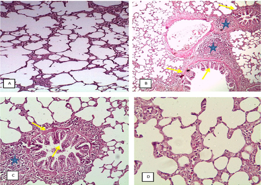
(H&E X4), A: lung of CG showing alveolar emphysema, B: lung of TG showing bronchitis and bronchiolitis, peribronchial edema, hyperplasia of epithelial lining (yellow arrow) and sever peribronchial infiltration of chronic inflammatory cells (star) and alveolar emphysema, C: lung of DAG showing catarrhal bronchiolitis, hyperplasia of epithelial lining, edema (yellow arrow) with massive infiltration of chronic inflammatory cells, (star) and alveolar emphysema, GD: lung of BG beginning to return to normal showing alveolar emphysema.
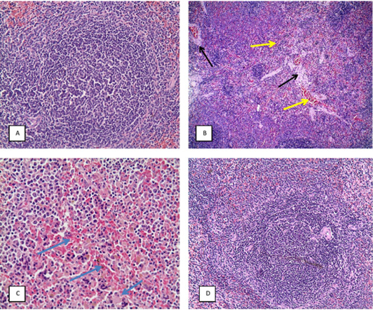
(H&E X20), A: spleen of CG, B: spleen of TG showing splenic hemorrhage and congestion (yellow arrow), vascular and perivascular edema (black arrow) and depletion of white pulp, C: spleen of DAG showing splenic hemorrhage (blue arrow), edema and depletion of white pulp, D: spleen of BG showing more or less to normal.
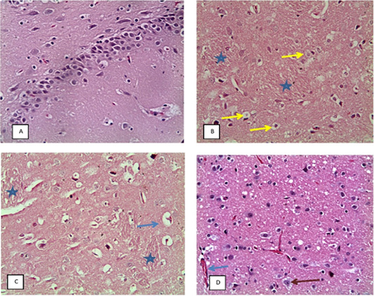
(H&E X20), A: showing CG brain, B: brain of TG showing demyelination (star), chromatolysis of neuron, neuronal edema (yellow arrow) and perivascular edema. C: brain of DAG showing demyelination of brain (star) and perivascular edema (blue arrow), D: brain of BG showing decreased lesion severity, chromatolysis of neuron (red arrow) and congested blood vessels (blue arrow).
