Joint Action of Trichoderma hamatum and Difenoconazole on Growth of a Phytopathogen Sclerotinia sclerotiorum under Laboratory Conditions
Joint Action of Trichoderma hamatum and Difenoconazole on Growth of a Phytopathogen Sclerotinia sclerotiorum under Laboratory Conditions
Dan Yü, Chuchu Li, Yü Huang and Zhen Huang*
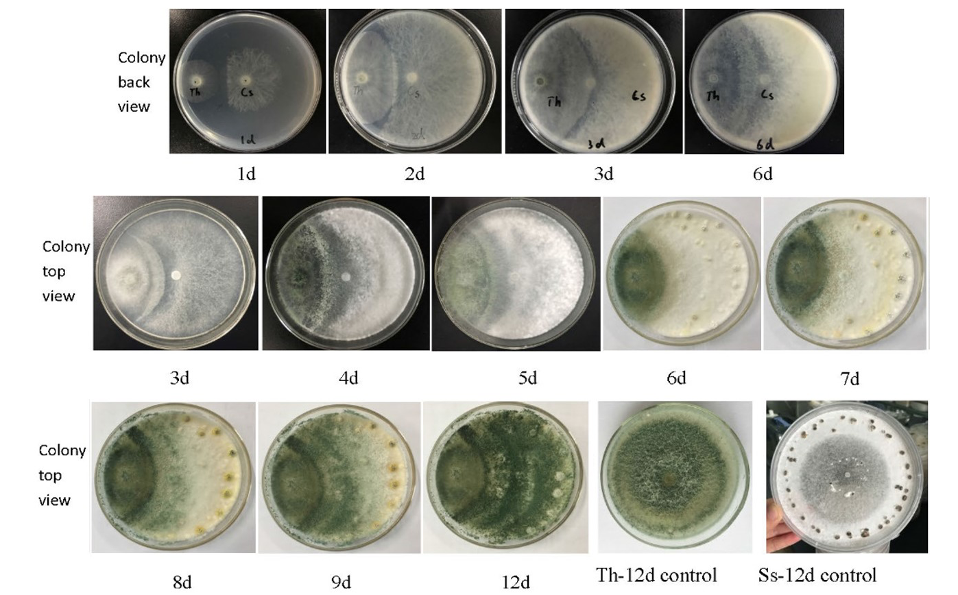
Inhibition of Trichderma hamatum on mycelial growth and Sclerotia formation of Sclerotinia sclerotiorum.
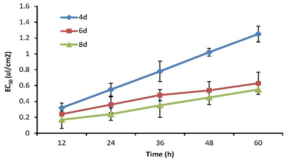
The inhibition of EC50 of T. hamatum cell-free culture supernatant on mycelial growth of S. sclerotiorum.
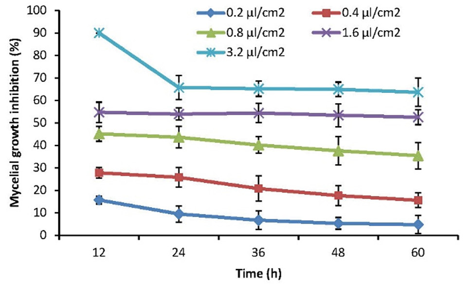
The inhibition of T. hamatum cell-free culture supernatant on mycelial growth inhibition of S. sclerotiorum after treated in 70°C for 10 min. Data on mean (±SE) inhibitory percentage were subjected to arcsine transformation prior to computation
The inhibition of T. hamatum cell-free culture supernatant on mycelial growth inhibition of S. sclerotiorum after treated in 70°C for 0, 10, 20 and 30 min. Data on mean (±SE) inhibitory percentage were subjected to arcsine transformation prior to computation. Means±SE in time dots (the column) marked with different letters are significantly different (Tukey’s HSD, a=0.05).
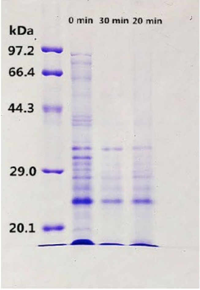
SDS-PAGE of protein from T. hamatum cell-free culture supernatant after treated in 70°C for 0, 20 and 30 min.
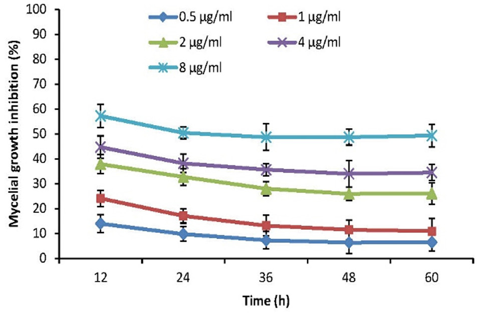
The inhibition of T. hamatum ethylacetate on mycelial growth of S. sclerotiorum. Data on mean (±SE) inhibitory percentage were subjected to arcsine transformation prior to computation.
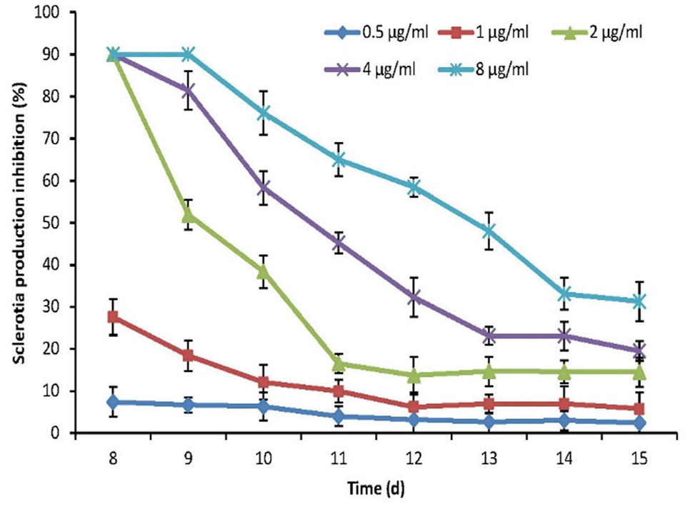
The inhibition of T. hamatum ethylacetate extracts on sclerotia production of S. sclerotiorum. Data on mean (±SE) inhibitory percentage were subjected to arcsine transformation prior to computation.
The inhibition of difenoconazole on mycelial growth of S. sclerotiorum. Data on mean (±SE) inhibitory percentage were subjected to arcsine transformation prior to computation.


